The Central Nervous System: The Brain and Spinal Cord
Summary
TLDRThis script offers an introductory exploration of the human brain's structure and function, highlighting the central nervous system's complexity. It details the brain's development from a neural tube into primary and secondary vesicles, leading to the adult brain's distinct regions. The focus is on the cerebral cortex's role in conscious thought, and the brain's various sensory and motor areas. The summary also touches on the diencephalon, brainstem, cerebellum, and protective structures like meninges, concluding with a brief mention of the spinal cord and its functions.
Takeaways
- 🧠 The human brain is considered the most complex object in the known universe, with its structure and function, especially consciousness, not yet fully understood.
- 🌱 The development of the brain begins with the neural tube in an embryo, which differentiates into primary and secondary brain vesicles, eventually forming the adult brain regions.
- 🏰 The central nervous system (CNS) includes the brain and spinal cord, with the brain being divided into the cerebrum, diencephalon, brainstem, and cerebellum.
- 🧬 Neurons and various types of neuroglia, such as astrocytes, microglial cells, and oligodendrocytes, play crucial roles within the CNS.
- 🧐 The cerebral cortex, part of the cerebrum, is responsible for conscious mind functions and is divided into motor, sensory, and association areas.
- 🤲 The motor homunculus illustrates the body parts' size in proportion to the number of neurons controlling them, highlighting the complexity of motor control.
- 👁️ The sensory areas of the brain process information from various senses, including touch, vision, hearing, smell, taste, and balance.
- 🔄 The diencephalon, located at the brain's center, includes the thalamus and hypothalamus, which are vital for learning, memory, and autonomic functions.
- 🌐 The brainstem, consisting of the midbrain, pons, and medulla oblongata, connects to the spinal cord and manages essential life functions.
- 🏋️♂️ The cerebellum is responsible for muscle coordination and smooth motion, playing a key role in motor control.
- 🛡️ The brain is protected by the meninges, including the dura mater, arachnoid mater, and pia mater, which provide a protective layer between the brain and skull.
Q & A
What are the two main divisions of the nervous system?
-The two main divisions of the nervous system are the central nervous system (CNS), which consists of the brain and spinal cord, and the peripheral nervous system (PNS).
What is the human brain considered to be in terms of complexity?
-The human brain is considered to be the single most complex object in the known universe due to its vast number of neuronal connections and the not yet fully understood mechanisms of consciousness.
What is the process called that allows the brain to occupy the available space within the skull?
-The process is called gyrification, which involves the formation of many folds in the brain to best occupy the available space within the skull.
What are the four main regions of the brain?
-The four main regions of the brain are the cerebral hemispheres, diencephalon, brain stem, and cerebellum.
What is the function of the ventricles in the brain?
-The ventricles are hollow cavities in the brain filled with cerebrospinal fluid and lined with ependymal cells, which help circulate the fluid.
What are the roles of the different types of neuroglia in the central nervous system?
-Neuroglia in the CNS include astrocytes, which perform regulatory functions; microglial cells, which monitor neuron health; and oligodendrocytes, which form myelin sheaths.
What is the primary function of the cerebral cortex?
-The cerebral cortex, made of gray matter, is the most recently evolved section of the animal brain and is where the conscious mind is found. It is responsible for higher mental functions such as memory and language.
What does the term 'motor homunculus' represent?
-The term 'motor homunculus' represents an image of the human body where each body part is depicted in a size proportional to the quantity of neurons that control it, showing the disproportionate amount of cortex devoted to fine motor control areas like the face, tongue, and hands.
What are the main functions of the diencephalon in the brain?
-The diencephalon, consisting of the thalamus, hypothalamus, and epithalamus, is involved in directing information to the cerebral cortex, controlling the autonomic nervous system, regulating body temperature, hunger, thirst, sleep cycles, emotional responses, and housing the pituitary and pineal glands.
What is the cerebellum responsible for in terms of motor control?
-The cerebellum is responsible for regulating muscle contraction to generate smooth and coordinated motion.
What are the protective structures for the brain and spinal cord called?
-The protective structures for the brain and spinal cord are called meninges, which include the dura mater, arachnoid mater, and pia mater.
Outlines

Esta sección está disponible solo para usuarios con suscripción. Por favor, mejora tu plan para acceder a esta parte.
Mejorar ahoraMindmap

Esta sección está disponible solo para usuarios con suscripción. Por favor, mejora tu plan para acceder a esta parte.
Mejorar ahoraKeywords

Esta sección está disponible solo para usuarios con suscripción. Por favor, mejora tu plan para acceder a esta parte.
Mejorar ahoraHighlights

Esta sección está disponible solo para usuarios con suscripción. Por favor, mejora tu plan para acceder a esta parte.
Mejorar ahoraTranscripts

Esta sección está disponible solo para usuarios con suscripción. Por favor, mejora tu plan para acceder a esta parte.
Mejorar ahoraVer Más Videos Relacionados
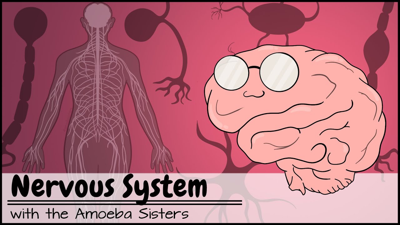
Nervous System

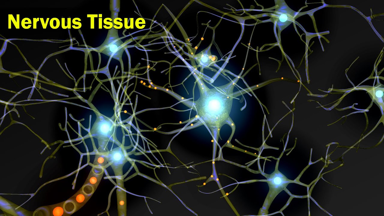
Types of Tissue Part 4: Nervous Tissue

SISTEM KOORDINASI, REPRODUKSI, DAN HOMEOSTATIS PADA MANUSIA (PART 1) - IPA KELAS 9 SMP
Nervous Tissue | Structural Organization in Animals | Anatomy | Inter 2nd year Class 11 Biology

The Human Body | Facts About the Parts of the Human Body System

Parts of the Brain Song
5.0 / 5 (0 votes)
