Labelled skull bones
Summary
TLDRThis educational video script delves into the intricate anatomy of the human skull, highlighting the evolution of the presenter's teaching style with enhanced 3D visualization and dynamic labeling. It covers individual bones, their fusion points, and sutures, with a focus on the frontal, parietal, temporal, and occipital bones, as well as the maxilla and zygomatic bones. The script promises further exploration of the skull's complex structures, including foramina and sutures, in subsequent sessions, aiming to captivate viewers with the fascinating world of human anatomy.
Takeaways
- π The video is focused on the anatomy of the human skull, revisiting the topic with an updated presentation style.
- π The speaker plans to cover the individual bones of the skull, examining them on both colored and white models for better understanding.
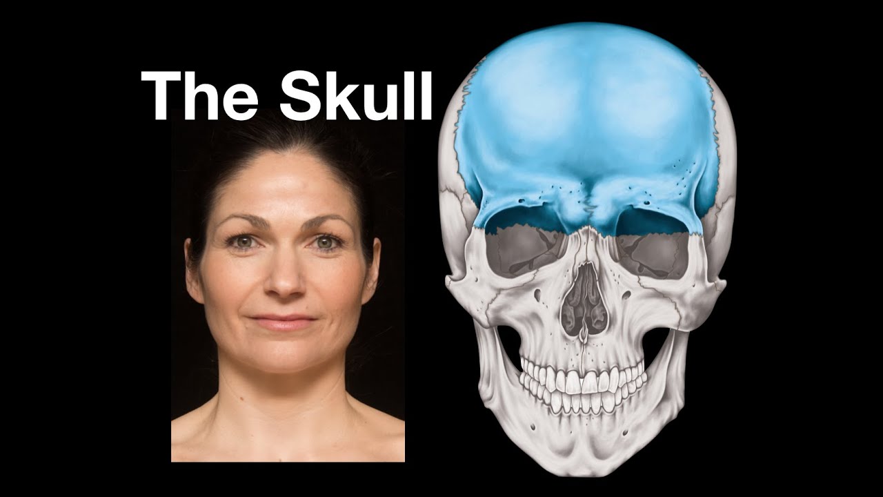
- π§ The frontal bone is described as originally forming from two bones that fuse together to become a single midline bone.
- ποΈ The parietal bones are referred to as 'walls', forming a significant part of the skull's structure, and are paired on each side.
- π°οΈ The temporal bone is highlighted for its role in the aging process, as it's where graying of hair typically begins.
- π The temporal bone is complex, with multiple parts including the squamous, petrous, and mastoid processes, and is involved in hearing and balance.
- π The occipital bone is at the back of the head, with a thick and solid structure, and features the foramen magnum for the spinal cord.
- π¦· The maxilla is the major bone forming the upper jaw and is responsible for housing the upper teeth.
- ποΈ The zygomatic bone, or cheekbone, is a prominent bone that connects the maxilla, temporal, and frontal bones and contributes to the orbit's structure.
- 𦴠The sphenoid bone is central and complex, linking various other bones and housing the pituitary gland, with important foramina for structure passage.
- π The ethmoid bone forms part of the nasal cavity and the medial wall of the orbit, with a role in air warming and humidification.
- ποΈ The vomer is a midline bone forming part of the nasal septum, separating the nasal cavity into left and right sides.
- π’ The lacrimal bone is associated with tear production, housing the nasolacrimal duct that links the eye to the nasal cavity.
Q & A
What is the main focus of the video script?
-The main focus of the video script is to discuss the anatomy of the human skull, specifically the bones that make up the skull, their locations, and their functions.
How has the video creator's style evolved over the years according to the script?
-The video creator's style has evolved to include dynamic labeling, better 3D lighting, and 4K resolution, enhancing the presentation of the anatomy of the skull.
What is the frontal bone and how does it develop?
-The frontal bone is a single central bone of the skull that originally forms as two separate bones, left and right. It fuses together early in life, eliminating the frontal suture, resulting in a single frontal bone.
What does the term 'parietal bone' refer to and how many are there?
-The term 'parietal bone' refers to the wall-like bones of the skull, and there are two parietal bones, one on each side of the head.
Why is the temporal bone called 'temporal'?
-The temporal bone is called so because 'temporal' refers to the passing of time, and this is where people typically go gray first, showing the passing of time.
What are the main parts of the temporal bone?
-The main parts of the temporal bone include the squamous part (flat part laterally), the petrous part (rocky ridge containing structures of the inner ear and middle ear), and the mastoid process.
What is the occipital bone and what significant feature does it have?
-The occipital bone is the bone at the back of the head, which is thick and solid. It has a significant feature called the foramen magnum, the large hole through which the spinal cord passes.
What is the maxilla and what does it contribute to?
-The maxilla is a single midline bone that forms the upper jaw and houses the upper teeth. It also contributes to the floor of the orbit, the socket for the eye.
What is the sphenoid bone and what is its significance?
-The sphenoid bone is a central, single bone that links the temporal, parietal, frontal, and zygomatic bones. It is butterfly-shaped, forms part of the posterior orbit, and houses the pituitary gland. It also has important foramina for structures to pass through.
What is the function of the zygomatic bone?
-The zygomatic bone, also known as the cheekbone, is a prominent bone that joins the maxilla, temporal, and frontal bones. It forms part of the lateral wall of the orbit.
What is the role of the ethmoid bone in the skull?
-The ethmoid bone is a single central bone that forms the upper part of the nasal cavity and is part of the medial wall of the orbit. It also separates the cranial cavity from the nasal cavity.
What are the two bones that form the lower jaw?
-The mandible is the bone that forms the lower jaw. It is a single bone, unlike the upper jaw (maxilla), which starts as two bones that fuse together.
What is the vomer bone and what does it form?
-The vomer is a single midline sheet of bone that forms part of the nasal septum, separating the nasal cavity into left and right sides.
What is the lacrimal bone and its relation to the tear duct?
-The lacrimal bone houses the nasolacrimal duct, a bony tube that links the orbit with the nasal cavity, allowing tears to drain from the eye into the nose.
What are the palatine bones and their contribution to the skull?
-The palatine bones are small, paired bones that form part of the hard palate at the back and extend superiorly to contribute to the formation of the nasal cavity.
What are the remaining bones in the skull that the script does not detail?
-The remaining bones not detailed in the script are likely the small bones of the ear ossicles and possibly the hyoid bone, which are very small and not easily pointed out on a skull model.
Outlines
π Skull Anatomy Overview
The script begins with an introduction to the anatomy of the skull, highlighting the evolution of the video style over the years to include dynamic labeling and 4K resolution. The focus is on the individual bones of the skull, their locations, and their 3D characteristics. The speaker expresses a deep fascination with the human skull, especially the sutures, joints, and foramina, and promises to explore these in future videos. The frontal bone is introduced as the first bone, which originally forms as two separate bones that fuse together in early life.
ποΈ Bones and Structures of the Skull
This paragraph delves into the specific bones of the skull, including the parietal bone, which forms a significant part of the neurocranium, and the temporal bone, known for its association with aging as it's the first area where hair turns gray. The temporal bone's structure is complex, with the squamous part visible on the skull's side and the petrous part housing the inner ear and middle ear structures. The mastoid process and zygomatic arch are also discussed as part of the temporal bone's anatomy. The occipital bone, with its thick and solid structure and the foramen magnum, is also covered, along with the maxilla and its role in forming the upper jaw and the floor of the orbit.
𦴠Detailed Examination of Skull Bones
The script continues with an in-depth look at the bones of the skull, focusing on the sphenoid bone, which is central and links various other bones. It forms part of the orbit and houses the pituitary gland, with several important foramina for structures to pass through. The pterygoid plates of the sphenoid bone and their attachment points for muscles are also mentioned. The hard palate, formed by the maxilla and palatine bones, and the mandible, which forms the lower jaw, are discussed. The temporomandibular joint, where the mandible articulates with the temporal bone, is highlighted. The orbit's formation from various bones, including the frontal, maxilla, sphenoid, and zygomatic bones, is also covered, with a special mention of the ethmoid bone's role in the medial wall of the orbit and the nasal cavity.
π Further Exploration of Skull Bones and Structures
The final paragraph explores additional bones and structures within the skull. The ethmoid bone's role in the nasal cavity and its function in warming and humidifying the air is explained. The vomer bone, which forms part of the nasal septum, and the inferior nasal concha, a separate bone, are also discussed. The nasal bones, which make up the external nose, and the lacrimal bone, which contains the nasolacrimal duct connecting the eye to the nasal cavity, are highlighted. The speaker invites viewers to search for more detailed videos on specific bones and structures, acknowledging the complexity of the skull's anatomy and the limitations of memory, even for a professional in the field.
Mindmap
Keywords
π‘Anatomy
π‘Skull
π‘Frontal Bone
π‘Parietal Bone
π‘Temporal Bone
π‘Occipital Bone
π‘Maxilla
π‘Zygomatic Bone
π‘Sphenoid Bone
π‘Ethmoid Bone
π‘Mandible
π‘Foramina
Highlights
Introduction to revisiting the anatomy of the skull with updated video techniques for better understanding.
Exploration of the skull's bones individually, emphasizing their 3D structure and anatomical relationships.
Explanation of the frontal bone's development from two separate bones that fuse together.
Description of the parietal bone's role in forming a significant part of the neurocranium.
Discussion on the temporal bone, its parts, and its relation to aging and graying of hair.
Identification of the temporal bone's complex structure including the squamous part, petrous part, and mastoid process.
Introduction of the occipital bone, its solid structure, and the foramen magnum's significance.
Clarification on the maxilla's role in forming the upper jaw and its contribution to the orbit's floor.
Highlighting the zygomatic bone's importance in connecting the maxilla, temporal, and frontal bones.
Detailed examination of the sphenoid bone's central position and its various parts.
Explanation of the pterygoid plates' function and their attachment to muscles.
Discussion on the hard palate's formation by the maxilla and the contribution of the palatine bones.
Introduction of the mandible and its articulation with the temporal bone at the temporomandibular joint.
Identification of the ethmoid bone's role in the nasal cavity and its parts within the orbit and cranial cavity.
Description of the vomer bone's function in forming part of the nasal septum.
Highlighting the nasal bones' composition and their distinction from the cartilaginous external nose.
Explanation of the lacrimal bone's involvement in tear duct function and its connection to nasal congestion.
Invitation to use YouTube's search function to find more detailed videos on specific skull bones.
Conclusion summarizing the overview of the skull's bones andι’ε of future discussions on sutures and foramina.
Transcripts
[Music]
this is going to really
confuse my auto focus in it oh it's
doing it it's doing all right
all right look at these
look at these beautiful things
so
i think we're going to go back to the
anatomy of the skull for a few weeks
um i recorded the anatomy of videos
about the anatomy of the skull
five years ago i think and i think my my
video style has evolved over the years
now we've got you know dynamic labeling
we've got better kind of more 3d
lighting good we've got the 4k goodness
right
so
i think what we're going to do is we're
going to go back today and look at the
bones of the skull each one individually
and we'll look at it on the colored
skull we'll look at it on the white
skull we'll get an idea of where it is
it's 3dness what's near it and you know
we'll get some labeling on there all the
good things right so
maybe
maybe i just love the human skull
anatomy so much i want to talk about it
for a few weeks is that all right
[Music]
um skull anatomy i mean is there
anything more human
i love it but skull anatomy we're
interested in the bones we're interested
in the sutures the joints between the
bones we're interested in the the
foramina the holes in the bones and what
passes through them but today we'll just
look at the bones and then we'll build
on that in the future
this
is the frontal bone
it it forms
originally as two bones left and right
in the two halves and then early on in
life
it fuses there is a frontal suture to
start with as this bone is forming and
then that frontal suture um
disappears the two bones fuse together
so we have a single
central midline
frontal bone
so
this is the
frontal bone
[Music]
posterior to the frontal bone we have
the parietal bone so parietal means wall
right so we have two parietal bones one
on the right one on the left
and the pro the frontal bone meets the
parietal bone and you can see that the
parietal bone is forming a great deal of
the
neurocranium the the skull
the
parietal bone
here
[Music]
inferior to the parietal bone we have
the temporal bone long time viewers will
know that every time i talk about the
temporal bone i say and it's called the
temporal bone because this is where we
see the passing of time that's what
temporal refers to as we we go gray this
is where we go gray first well i am so
my hair is going gray here showing the
passing of time this is the temporal
bone and the temporal region by the way
the lobes of the brain are largely named
because they are deep to these bones so
it's easy to say this bone this bone
this bone the lobes of the brain you
know not always quite so clearly
demarcated so the lobes of the brain are
deep they're named because of the bones
they're deep to so the temporal bone now
this is interesting it has multiple
parts this is the flat part laterally
the the squamous part
um
so that's what we're seeing here the
squamous part of the temporal bone now
if i take
the skull cap off
so we're taking off parietal and frontal
bone bits there
that's also
the temporal bone in here and this is
the petrous part of the temporal bone
the rocky ridge that's in there and that
rocky ridge exists because these
structures of the inner ear are in there
and the middle ear is is in here so
the
the temporal bone has the external
auditory meatus that is the ear oil
and the petrus part has internal
structures in there
this lumpy bit here this chunk that's
the mastoid process of the temporal bone
you can palpate that here it's a very
obvious lump
the mastoid process of the temporal bone
and also
this here is the zygomatic arch and look
the temporal bone is contributing to
that
all right temporal bone
squamous part and parietal part a
squamous part and
peter's part the rocky bit inside
if we
look posteriorly
this is the occipital bone here so the
occipital bone is at the back of your
head and if you palpate the back of your
head you feel a nice solid lump these
lumpy bits here these are all parts of
the occipital bone the occipital bone is
pretty thick and solid and if i tilt
this
we can see that this hole here this is
foramen magnum the big hole that's what
the spinal cord goes through that is
part of the occipital bone
all right
put this one back together
again
that is the occipital bone
here
uh the other thing about the acceptable
bone is we can see these articulating
processes here these are the acceptable
condyles
that is where the skull sits upon the
vertebral column
difficult thing to balance right
so the occipital bone is a single
midline bone
like the frontal bone is whereas the
parietal
and temporal bones
there are one on either side of the body
so they are paired
turning the skull around to look at the
face
the face also gets known as the viscero
cranium which is the neurocranium
surrounds the brain and we see this
major bone here this is the maxilla
and the maxilla again is a single
midline bone but it starts off as two
bones
and then they fuse in the midline
like the frontal bone does so we have a
single maxilla or maxillary bone and the
maxilla is forming the upper jaw the
teeth the upper teeth are embedded into
the maxilla
and if we look into the orbit you can
see that the maxilla
is contributing to
the floor of the orbit
that is the hole that the eye lives in
so
this is
the maxilla here
[Music]
so the other bone then is prominent here
is we already hinted towards it is the
the zygomatic bone or the zygoma the the
cheekbone very very prominent the
cheekbone
and
this is a separate bone
but look it joins
the maxilla
and
the temporal bone
and the frontal bone
they all are joined by the zomatic
zygomatic bone and look the zygomatic
bone is also forming
the lateral wall or part of the lateral
wall of the orbit so the zygomatic bone
but the zygomatic arch is a complex made
up of multiple bones look
[Music]
and then deep to the zygomatic arch we
have
this
space we have the
infra-temporal fossa
so then
in the infra-temporal fossa we see this
red bone this is the sphenoid bone this
is a tricky one to work out where it is
the sphenoid bone is a very very central
bone so we see it here the sphenoid bone
is a single central bone
um it is but it's linking the temporal
bone the parietal bone the frontal bone
the zygomatic bone and if we look into
the orbit it's also forming the
posterior part of the orbit
deep within there
so the sphenoid bone
is if we
[Music]
so the sphenoid bone is forming the
posterior orbit if we take the skull cap
off
and we consider where the orbit is
this is the sphenoid bone here
it's a central single bone it is kind of
butterfly shaped
and it is
extending out and around here
the pituitary gland sits within it
and it has a number of important holes
in it important foramina that structures
pass through it is a bone that links to
the the orbit and the face and so on so
the sphenoid bone very very central
that central sphenoid bone then also has
if we look inferiorly
these
pterygoid plates
these kind of winged structures
and these paragoid plates are part of
the sphenoid bone and we have a number
of muscles and what have you attaching
there
so the pterygoid terry oy terry wing oid
shaped so wing shaped bits of bone
part of the sphenoid bone
down here
and while we're down here
the
hard palate the roof of the mouth there
that is formed by
the maxilla
maxilla
see the purple
maxilla
the hard palate the roof of the mouth is
formed by the maxilla now on this
colorful model we can see that the
posterior part of the
of the hard palate is red that's not the
sphenoid bone these are the palatine
bones there's one on either side
and the palatine bones are forming
they're little bones they're kind of we
see the base of them there the flat base
they're kind of tall and pointy
so they are forming part of the hard
palate the posterior part of the hard
palate but they're also extending
superiorly so they're going to
contribute to forming part of the nasal
cavity and the link between the sphenoid
bone and the the maxilla palatine bones
little diddy guys difficult to visualize
where they are and what they do
okay so we've built up quite a lot of
the skull so far
this is the mandible down here forming
the lower jaw
and the lower teeth are part of the
mandible and the mandible then is
articulating with
the temporal bone at the tempuro
temporomandibular
joint
in the orbit
we can see that the orbit is formed from
frontal bone
maxilla
sphenoid bone
zygomatic bone now
there's another yellow bone
in there in the medial wall of the orbit
and i think this is the hardest bone in
the skull to visualize where it is and
what shape it is that is the ethmoid
bone so the ethmoid bone is again a
single
central
unpaired bone
it's forming the at the top of the the
nasal cavity in there
um and it's kind of you know it's almost
like u-shaped but it's more complicated
than that
and what we can see inside the nasal
cavity is we can see lots of curvy bits
with these other country these curved
bits of bone
they cover the mucous membrane they help
warm and humidify the air when we
breathe it in through our noses and into
our respiratory tracts so
the ethmoid bone is the upper part of
the nasal cavity
so the ethmoid bone
is part of the medial wall of the orbit
and also part of the
superior part of the nasal cavity and if
we look
inside the cranial cavity that's the
ethmoid bone there
right so sphenoid bone is here
ethmoid bone is kind of this
rectangular strip anteriorly so it is
also separating the cranial cavity from
the nasal cavity so that's the ethmoid
bone
that's almost all of them
if we think about the nasal cavity
inside here inferiorly
i said the superior and middle nasal
country are part of the ethmoid bone but
there are inferior conchi as well an
inferior curved shell-like bone and that
is a bone on its own so the inferior
concha there's a left one in the right
one that's a separate bone the other
thing we can see here is we can see voma
vomer
is a single midline sheet of bone and it
is forming part of the nasal septum so
the nasal septum is separating our nasal
cavity into left and right sides part of
it is cartilaginous part of it is bone
and the bony bit is voma and that's what
we can see here because this is a bony
skeleton
with the cartilage removed so the nasal
cavity has got a number of bones within
it
and in fact the remaining two bones are
here as well
these
are the nasal bones so while much of the
nose is cartilaginous the external nose
up here it's bone so these are the nasal
bones they're two separate bones one on
either side
and
here
this orange bone this is the lacrimal
bone so the lacrimal glands are actually
up here they're laterally and superior
in the eye lacrimal fluid that is tears
wash across the eye
and in the nasal lacrum well in the
lacrimal bone we have the nasolacrimal
duct there's a bony tube there which
links the orbit with the nasal cavity
which is why when you cry when you make
lots of tears you get a snotty nose
because your tears go across the eye
through the nasolacrimal duct into the
nose and they come out through your nose
or you swallow them or whatever
you do a lot of sniffling
so that's the lacrimal bone there
on the white skull
you have to look really carefully to see
the sutures the edges of the bone but
that's the
nasal bone there
and that's the lacrimal bone in there
so those are the bones of the skull
frontal parietal occipital
temporal
maxilla mandible
sphenoid bone is central
sphenoid uh
nasal
ethmoid lacrimal
palatine
vomer
inferior nasal concha is that it
there are some other bones in the skull
that i have not mentioned
can you think what they are
clue they're very very small
i can't point at them on this i have
probably looked at some of these bones
in more detail i probably looked at the
bones of the orbit and the nose in way
more detail um i've done so many videos
i can't and i get paid
to talk anatomy every day so i can't
remember what i've done i will just like
you will have to search the youtube use
the youtube search function to find out
if i've done a video on any of these if
you'd like to know more but
this is a this is another start we've
looked at the bones of the skull and
then we'll look at the sutures and the
framing and stuff like that in the
coming weeks i think i hope this has
been
illustrative
illustrated illustrative useful
uh see you next week
see you next week
[Music]
5.0 / 5 (0 votes)