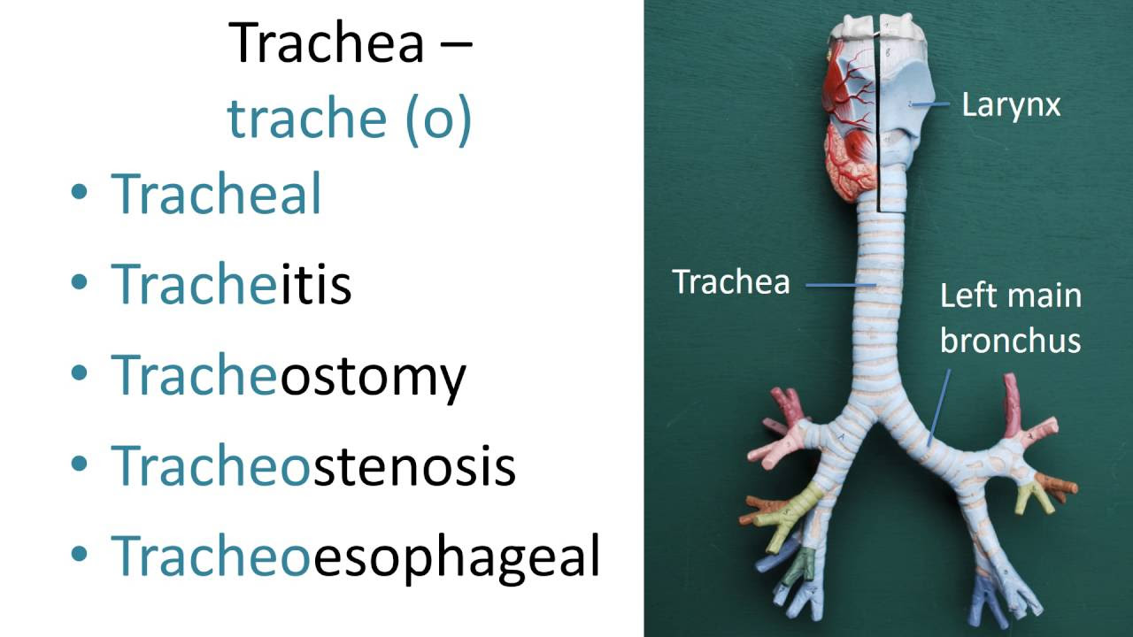
Shotgun Histology Trachea
Summary
TLDRThis transcript provides a detailed description of the anatomical structure and features of the trachea. It highlights the cartilage surrounding the air-filled space, the presence of ciliated pseudostratified columnar epithelium with goblet cells, and submucosal glands. The video also points out the smooth muscle in the posterior part of the trachea, called the trachealis muscle, as well as the adjacent esophagus. The discussion emphasizes the connective tissues, blood vessels, and cartilage, offering an in-depth look at the histological layers of the trachea.
Takeaways
- 🫁 The structure being examined is the trachea, part of the upper respiratory tract.
- 🦴 The trachea has a space filled with air, surrounded by cartilage, with a small absence of cartilage in certain areas.
- 🔬 The lining of the trachea consists of pseudostratified columnar epithelium, with ciliated cells and occasional goblet cells.
- 🧬 The mucosa includes a layer of connective tissue known as the lamina propria, composed of loose connective tissue.
- 🩸 Below the mucosa is the submucosa, which contains blood vessels and submucosal glands responsible for secreting mucus.
- 🛡️ The cartilage in the trachea is hyaline cartilage, but can resemble elastic cartilage unless a special stain is used to differentiate them.
- 💪 At the posterior part of the trachea, where cartilage is absent, there is smooth muscle called the trachealis muscle.
- 🔄 The trachea connects closely to the esophagus, which lies posterior to it and has a different type of lining: stratified squamous epithelium.
- 📏 The adventitia of the trachea contains a mix of connective tissue, cartilage, and smooth muscle, providing structure and flexibility.
- 🧪 The entire trachea structure is described as having a classical appearance, with all key components like mucosa, submucosa, cartilage, and smooth muscle clearly visible.
Q & A
What is the primary function of the trachea?
-The primary function of the trachea is to provide a passage for air to move between the throat (pharynx) and the lungs. It also has ciliated pseudostratified columnar epithelium to help filter particles and mucous-producing goblet cells to trap debris.
What type of cartilage surrounds the trachea, and what is its role?
-The trachea is surrounded by hyaline cartilage, which forms C-shaped rings. These rings provide structural support to keep the airway open, allowing air to flow easily while still allowing flexibility for movement during breathing.
What is the mucosa layer of the trachea composed of?
-The mucosa layer of the trachea is composed of pseudostratified ciliated columnar epithelium, with occasional goblet cells that secrete mucus. This layer helps trap foreign particles and moves mucus upward towards the throat.
What is the role of goblet cells in the trachea?
-Goblet cells in the trachea secrete mucus, which helps trap dust, pathogens, and other particles. The mucus is then moved by the cilia towards the throat, where it can be swallowed or expelled.
What is the function of the cilia in the pseudostratified epithelium of the trachea?
-The cilia in the pseudostratified epithelium beat in a coordinated manner to move mucus and trapped particles upward toward the pharynx, where they can be swallowed or expelled, keeping the respiratory tract clear of debris.
What is the lamina propria, and where is it located in the trachea?
-The lamina propria is a layer of loose connective tissue found directly beneath the mucosa in the trachea. It provides structural support and houses blood vessels that nourish the epithelial cells.
What is the submucosa, and what structures does it contain?
-The submucosa is a layer beneath the lamina propria in the trachea that contains rich blood vessels and submucosal glands, which secrete mucus to help keep the airway moist and trap particles.
What is the trachealis muscle, and where is it located?
-The trachealis muscle is a band of smooth muscle located in the posterior part of the trachea, where there is no cartilage. It helps control the diameter of the trachea, allowing it to constrict or expand during breathing or coughing.
How can elastic cartilage be distinguished from hyaline cartilage in the trachea?
-Elastic cartilage can be distinguished from hyaline cartilage by performing a stain for elastic fibers. Elastic cartilage contains more elastic fibers, whereas hyaline cartilage does not. In the trachea, hyaline cartilage is more commonly found.
What tissue is found posterior to the trachea, and what is its relationship with the trachea?
-The esophagus is located posterior to the trachea. The two structures are separated by a band of smooth muscle, allowing the esophagus to expand when swallowing food without constricting the airflow in the trachea.
Outlines

This section is available to paid users only. Please upgrade to access this part.
Upgrade NowMindmap

This section is available to paid users only. Please upgrade to access this part.
Upgrade NowKeywords

This section is available to paid users only. Please upgrade to access this part.
Upgrade NowHighlights

This section is available to paid users only. Please upgrade to access this part.
Upgrade NowTranscripts

This section is available to paid users only. Please upgrade to access this part.
Upgrade NowBrowse More Related Video

Clavicle Anatomy Animation | General features, Osteology, Attachments, Development, clinical anatomy

Female Pelvis Sagittal Section
Medical terms 7, Respiratory system

Oral Cavity Proper (Palate & Tongue) - Oral Cavity Anatomy

Airway Anatomy - 3D Tutorial

Fetal Pig Dissection - Anatomy of the Respiratory System
5.0 / 5 (0 votes)