DNA Sequencing Techniques | An Overview
Summary
TLDRThis lecture offers an in-depth overview of DNA sequencing techniques, explaining the process of determining the order of base pairs in DNA molecules. It covers the significance of DNA sequencing in detecting mutations, distinguishing organisms, and identifying human haplotypes. The video explores various sequencing methods, including direct sequencing techniques like Sanger sequencing and chemical sequencing, as well as next-generation sequencing technologies such as ion conductance, reversible dye terminator, sequencing by ligation, and nanopore sequencing. Each method is briefly explained, highlighting its unique approach to DNA analysis.
Takeaways
- 𧬠DNA sequencing is the process of determining the order of base pairs in a DNA molecule, crucial for understanding genetic information.
- π There are four bases in DNA: adenine (A), cytosine (C), guanine (G), and thymine (T), which are the building blocks of genetic code.
- π DNA sequencing helps in detecting mutations, distinguishing organisms, and identifying human haplotypes and polymorphisms.
- π§ͺ Direct sequencing methods like chemical sequencing (Maxim Gilbert) and Sanger sequencing allow for direct reading of DNA base sequences.
- π¬ Chemical sequencing uses chemical agents to cleave DNA at specific base pairs and involves electrophoresis to visualize and read the sequence.
- π Sanger sequencing is a modification of the DNA replication process using dideoxynucleotides (ddNTPs) to terminate DNA strand growth at specific bases.
- π Pyrosequencing is an indirect method that detects DNA sequence by monitoring light generation as nucleotides are incorporated into a growing DNA strand.
- 𧡠Bisulfite DNA sequencing is used to detect methylated cytosines, which play a role in gene regulation and chromatin structure.
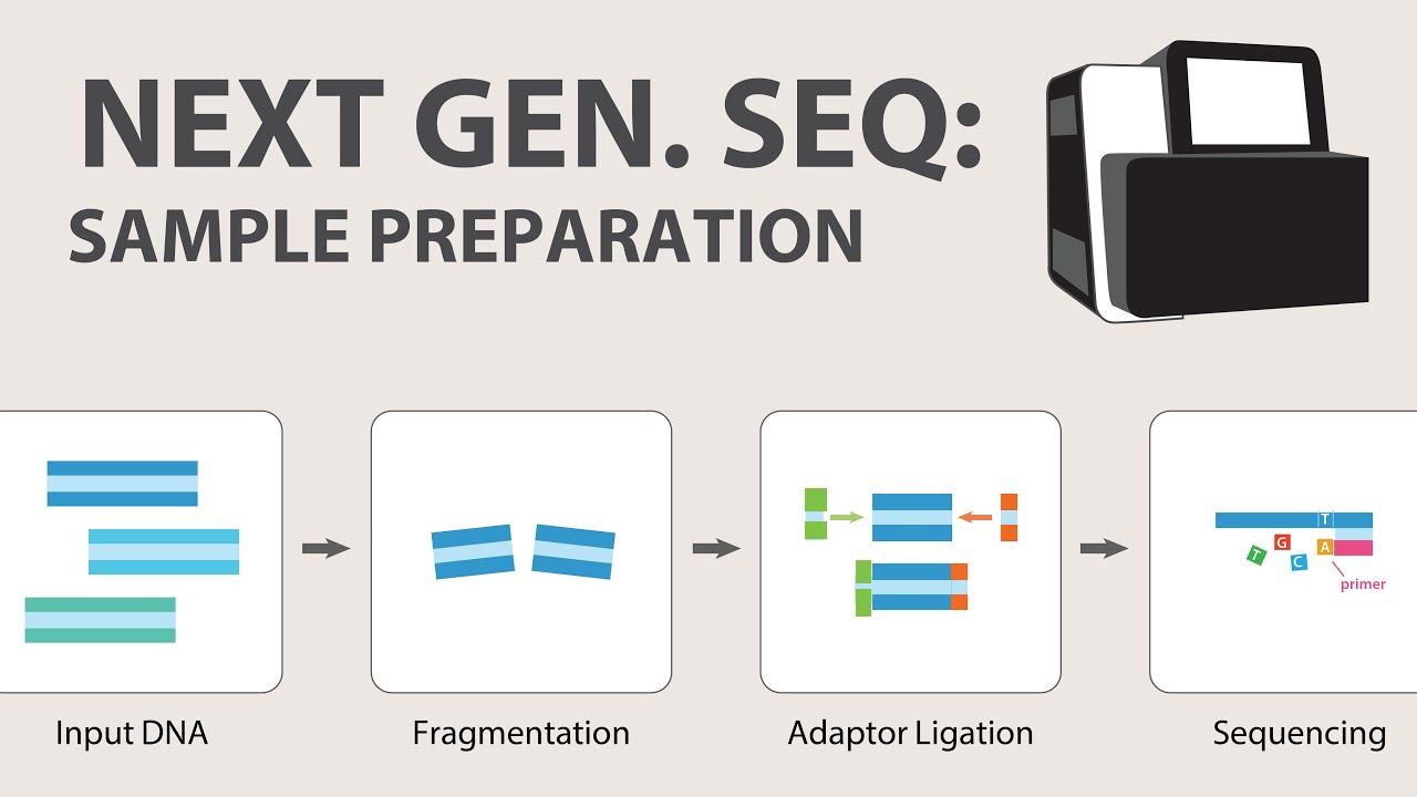
- π Next Generation Sequencing (NGS) technologies like ion conductance, reversible dye terminator, and nanopore sequencing allow for rapid, high-throughput DNA sequencing.
- 𧬠NGS techniques are used for large-scale genomic studies and require powerful computing for data analysis and assembly of sequenced libraries.
Q & A
What is DNA sequencing and why is it important?
-DNA sequencing is the process of determining the order of base pairs in a DNA molecule. It's important because it can help detect mutations in genes, distinguish between different organisms by analyzing specific gene sequences, and identify human haplotypes and polymorphisms, which provide insights into gene inheritance and function.
What are the four bases found in DNA and how are they abbreviated?
-The four bases found in DNA are adenine (A), cytosine (C), guanine (G), and thymine (T).
How does DNA sequencing help in identifying mutations?
-DNA sequencing helps identify mutations by determining the exact sequence of bases in a gene. A change in just one base can lead to a change in the gene's function, which can have significant effects on an individual.
What is the purpose of using the 16s rRNA gene in distinguishing microorganisms?
-The 16s rRNA gene is used to distinguish microorganisms because it contains universal sequences that are conserved across different species, allowing for the identification of specific differences that can differentiate one microorganism from another.
What are the two main types of direct DNA sequencing techniques mentioned in the script?
-The two main types of direct DNA sequencing techniques mentioned are chemical sequencing (Maxim Gilbert sequencing) and Sanger sequencing.
How does chemical sequencing differ from Sanger sequencing?
-Chemical sequencing uses chemical agents to cleave DNA at specific base pairs, while Sanger sequencing is a modification of the DNA replication process that uses modified dNTPs (ddNTPs) to create different lengths of DNA fragments.
What is the role of electrophoresis in DNA sequencing?
-Electrophoresis is used in DNA sequencing to separate DNA fragments based on their size. The separated fragments are then visualized and the sequence is read from the pattern they form on the gel or in the capillary.
Why is Sanger sequencing still widely used today?
-Sanger sequencing is still widely used because it can be adapted for automation, which allows for faster and more efficient sequencing. It uses fluorescent dyes instead of radioactive labels, making it safer and more convenient.
What is pyrosequencing and how does it determine the DNA sequence?
-Pyrosequencing is a DNA sequencing method that relies on the generation of light through luminescence whenever nucleotides are added to a growing strand of DNA. It determines the sequence by detecting which nucleotide generates a light signal when added.
What is bisulfite DNA sequencing and why is it used?
-Bisulfite DNA sequencing is used to detect methylated cytosines, which play a role in gene expression regulation and chromatin structure. It involves treating DNA with bisulfite to convert cytosines to uracil, leaving methylated cytosines unchanged, and then sequencing to identify the methylated sites.
What are the key features of Next Generation Sequencing (NGS) technologies?
-NGS technologies are characterized by their ability to sequence large numbers of templates carrying millions of bases in a short time. They use target libraries, require powerful computers and bioinformatics for data analysis, and include methods like ion conductance sequencing, reversible dye terminator sequencing, sequencing by ligation, and nanopore sequencing.
Outlines
𧬠DNA Sequencing Techniques Overview
This paragraph introduces the concept of DNA sequencing, which is the process of determining the order of base pairs in a DNA molecule. It explains the significance of knowing the DNA sequence, including detecting mutations, distinguishing between organisms, and identifying human haplotypes. The paragraph outlines two main types of DNA sequencing: direct sequencing, which includes Maxam Gilbert sequencing and Sanger sequencing, and their applications in genetic analysis.
π¬ Direct Sequencing: Chemical and Sanger Methods
The paragraph delves into the details of direct sequencing methods, starting with chemical sequencing or Maxam Gilbert sequencing. It describes the process of labeling DNA with radioactive isotopes and using chemical agents to cleave DNA at specific bases. The resulting fragments are separated by size through electrophoresis to determine the sequence. The paragraph then contrasts this with Sanger sequencing, which is a modified DNA replication process using dideoxynucleotides (ddNTPs) to terminate DNA strand growth at specific bases, creating fragments that can be sequenced by gel or capillary electrophoresis.
π Sanger Sequencing and Automation
This section focuses on the Sanger sequencing method, emphasizing its role in automated DNA sequencing. It explains the use of fluorescent dyes instead of radioactive labels for automated reading of sequences. The paragraph outlines two types of automated Sanger sequencing: dye primer and dye terminator sequencing, each with its method of electrophoresis. It also mentions the advantages of capillary electrophoresis in automated sequencing and how the resulting data is interpreted to determine DNA sequences.
π Indirect Sequencing: Pyrosequencing and Bisulfite Sequencing
The paragraph introduces indirect sequencing methods, starting with pyrosequencing, which detects DNA sequence by monitoring light generation upon nucleotide incorporation. It details the steps involved, from the addition of dNTPs to the detection of light signals and the interpretation of these signals to determine the DNA sequence. The paragraph also covers bisulfite DNA sequencing, a method used to detect methylated cytosines, which involves treating DNA with bisulfite to convert unmethylated cytosines to uracil, followed by sequencing to identify methylation patterns.
π Next Generation Sequencing (NGS) Technologies
This section discusses Next Generation Sequencing (NGS) technologies, which include ion conductance sequencing, reversible dye terminator sequencing, sequencing by ligation, and nanopore sequencing. It highlights the massive parallel sequencing capabilities of NGS, allowing for the analysis of millions of bases in a short time. The paragraph describes each NGS method, explaining how they work, the type of libraries they use, and the equipment and bioinformatics required for data processing and analysis.
π§ͺ Sequencing by Ligation and Nanopore Sequencing
The final paragraph focuses on sequencing by ligation, which uses oligomers that hybridize and ligate to the DNA template, allowing for the detection of two bases at a time. It also covers nanopore sequencing, a unique method that sequences a single long DNA molecule by identifying each nucleotide as it passes through a protein pore, causing a disruption in the current. The paragraph provides insights into the technical aspects of these advanced sequencing techniques and their applications in genomic research.
Mindmap
Keywords
π‘DNA Sequencing
π‘Base Pairs
π‘Mutations
π‘Direct Sequencing
π‘Chemical Sequencing
π‘Sanger Sequencing
π‘Pyrosequencing
π‘Bisulfite DNA Sequencing
π‘Next Generation Sequencing (NGS)
π‘Ion Conductance Sequencing
π‘Sequencing by Ligation
Highlights
Introduction to DNA sequencing, the process of determining the order of base pairs in DNA.
Explanation of the four DNA bases: adenine (A), cytosine (C), guanine (G), and thymine (T).
Overview of the 16s rRNA gene in E. coli and its 1500 base pairs.
Applications of DNA sequencing include mutation detection and organism identification.
Direct sequencing methods, such as chemical sequencing and Sanger sequencing, are introduced.
Description of chemical sequencing, including the use of radioactive labels and chemical agents.
Process of Sanger sequencing, involving the use of ddNTPs and electrophoresis.
Advantages of Sanger sequencing, including its adaptability for automation.
Introduction to indirect sequencing methods like pyrosequencing, which relies on luminescence.
Bisulfite DNA sequencing for detecting methylated cytosines and its role in gene regulation.
Overview of Next Generation Sequencing (NGS) technologies, including their high-throughput capabilities.
Ion conductance sequencing and its detection of hydrogen ions released during DNA synthesis.
Reversible dye terminator sequencing and the formation of DNA colonies on a flow cell.
Sequencing by ligation, which detects two bases at a time using fluorescently labeled oligomers.
Nanopore sequencing, a single-molecule sequencing method that identifies nucleotides by current disruption.
Practical applications and the importance of bioinformatics in reassembling sequenced libraries.
Transcripts
hi everyone
in this lecture we will have an overview
of the different dna sequencing
techniques
now what is dna sequencing this is the
process of determining the sequence or
order of base pairs in a given molecule
of dna
as a recap there are four bases
in your dna and these are adenine
abbreviated by a
cytosine which is abbreviated by c
guanine which is abbreviated by g and
thymine which is abbreviated by t
in this example you can see the sequence
of the 16s rrna gene in e coli and you
can see there are about 1500 base pairs
and we know this sequence because of dna
sequencing
so why do we need to know the sequence
of dna or certain genes
here are just some applications first
it can help us detect mutations in
specific genes of interest
in this example you can see that this
gene is composed of around 1 500 bases
but just change to one of these bases
can lead to more function of the gene
and even detrimental effects to an
individual
next
knowing the sequence of a specific gene
can help us distinguish one organism
from another this is especially useful
in identifying microorganisms which can
be differentiated from each other by
looking at specific differences
in universal genes like in this example
the 16s rna gene
it can also be used to help us identify
human haplotypes and designate
polymorphisms
this gives us information on how genes
are inherited and how these variations
might affect a gene's function
now let's go into
the different types of dna sequencing
techniques the first
grouping of dna sequencing is called
direct sequencing
and here there are two examples the
maxim gilbert sequencing and sanger
sequencing
so what is direct sequencing
this is the most definitive molecular
method to identify genetic lesions and
to know the sequence of dna
here the base sequences are read
directly and usually direct sequencing
methods involve some sort of
electrophoresis in which you can
visualize bands that correspond to
whatever base is in the sequence
this is especially useful in detecting
small variations in a specific sequence
the first type of sequencing we have is
called chemical sequencing or maxim
gilbert sequencing
this type of technique requires single
single-stranded dna or double-stranded
dna with a radioactive label at one end
usually this is the 5-prime end
this type of sequencing is called
chemical sequencing because it uses
chemical agents to cleave dna
at specific base pairs
different fragments are then separated
based on size to identify the sequence
the first step in chemical sequencing is
the addition of a radioactive label
the label being used is a radioactive
phosphorus and usually this is
conjugated to atp
this is added to the five prime end of a
dna fragment using your polynucleotide
kinase enzyme
if you wanted to add
this radioactive phosphorus to the three
prime end the usual method is by using
terminal transferase plus alkaline
hydrolysis to remove excess adenylic
acid residues
in this figure you can see that you take
our template dna and after this step
they will now be labeled with a
radioactive label now the radioactive
label here is very important in order
for us to visualize the dna fragments
later on
the next step is to use chemical agents
to break apart the fragment
so our template dna which has already
been labeled is divided into four
each aliquot is treated with a different
chemical and there are four being used
first we have dimethyl sulfate
abbreviated by dms
formic acid which is abbreviated by fa
hydrazine which is abbreviated by h and
a combination of hydrazine and a salt
abbreviated here with h plus s
in this figure you can see our four
tubes each containing
one of our different chemical agents
so we add our template dna to each tube
and incubate it for a period of time
then a strong reducing agent such as 10
percent pipiridine is added which breaks
the strand at specific nucleotides what
you are left with
is different fragments of different
sizes each ending in a specific
nucleotide depending on which
compound or which chemical it was
incubated with
in this table we can see the different
chemical agents are base modifiers being
used in chemical sequencing
so these base modifiers break apart
your dna fragment at specific
nucleotides and it does these using
these different actions
we can also see here the time it takes
or the incubation time for each of these
base modifiers to completely break apart
your template dna
so first we have dimethyl sulfate or dms
and this breaks the dna fragment
whenever it encounters a guanine
formic acid breaks apart the dna
fragment whenever it encounters a
guanine and an adenine then we have
hydrazine which breaks apart the chain
whenever it encounters a thymine and a
cytosine and we have hydrazine salt
which
breaks apart the chain whenever it
encounters a cytosine
the last step in chemical sequencing is
the separation of different fragments
and the reading of the sequence
in the previous step we are left with
different fragments and depending on the
chemical agents being used we are sure
that these fragments end in a certain
nucleotide
now these fragments are loaded into a
gel and separated based on their size
using electrophoresis the different
fragments are separated by size on a
denaturing polyacrylamide gel by
electrophoresis
the radioactive label which was added in
step one is used to visualize the bands
either using autoradiography or by
exposing the gel to an x-ray film
the sequence is read from the bottom to
the top and this is from the five prime
end all the way to the three prime end
in this figure you can see an example of
a gel being used in chemical sequencing
so at the bottom you will find the
shortest fragments and this is closer to
the five prime end this is a recap we
added the primer in the five prime end
okay and in the top here you have much
longer fragments
now the lane in which the band appears
is used to identify the nucleotide so
for example if you have a band in the g
and the g plus a lane then the
nucleotide in that sequence is a guanine
if you have a band both in the c plus t
lane and the c lane then the nucleotide
is a cytosine
if you have a band only in the c plus t
lane the nucleotide is a thymine and if
you have a band only in the g plus a
lane then the nucleotide is an adenine
so reading this gel from the bottom this
is the five prime end you can see that
the first band we can see is found in
the g plus a lane only this indicates
that the first nucleotide in the
sequence
is an a
then the following
bands are found in the c plus t
and the c lane and we have two bands so
this tells us the next basis in the
sequence is a c and a c
and we just go through this gel until
you reach the top so in the top you can
see here that
there is a band in the c plus t lane
only and this tells us that
our
next base in the sequence is a t
chemical sequencing also comes with some
disadvantages it can only be used for
sequencing short lengths of dna
and it uses hazardous materials like
your hydrazine and your pipiridine and
this means that you need special
equipment and specially trained
individuals in order to perform our
chemical sequencing
the next type of direct sequencing is
called your die deoxy chain termination
or sanger sequencing
this is a modification of the dna
replication process
it uses single stranded template dna and
a single stranded primer
in this type of sequencing a modified
dntp called
dioxynucleotide or ddntp is used to
create different lengths of dna
fragments
and the fragments are run through a gel
or capillary electrophoresis in order to
tell the sequence of the dna fragment
the first step in your sanger sequencing
is the addition of the different
reactants here are the different
reactants being used first we have your
template dna this will be your pcr
product then you have your primer so the
primer binds to the three prime end of
the template and it creates copies of
the template from the five prime end to
the three prime end
sometimes the primer can be conjugated
with a radioactive phosphorus or a
fluorescent dye as a label
other reactants that we use
are the dntps just as a recap dntp
stands for
deoxyribonucleoside triphosphate but we
just call them dntps for short
these are the building blocks of our dna
whenever we do
amplification
we have four dntps one each for adenine
guanine cytosine and thymine
then in sanger sequencing we have these
ddntps
which are like your regular dntps but
they lack the hydroxyl group found on
the third ribose carbon
of the deoxynucleotide so here we have
our regular dntp and our d dntp
and here you have
the missing hydroxyl group
whenever our ddntps are added to a
growing sequence of dna
this terminates the replication process
of course you also need the other
components for dna replication like your
polymerase and other
substrates that it may use
the next step in sanger sequencing is
the dna replication so the reactions
occur in four different tubes and each
tube has the four dntps and one specific
ddntp at a lower concentration
whenever a ddntp is added to a growing
dna strand the replication stops and
this results in different length strands
each ending with a specific dntp
in this figure you can see our four
different tubes
again each tube has
a specific ddntp and all tubes have the
four dntps which are essential building
blocks
for dna replication
it should also be noted that the ratio
between ddntps and dntp in each tube
must be optimized if there is too much
dntp then you will be left with very
very short fragments and if there is not
enough ddndp you will end up with very
long fragments
so you need an optimized ratio of the
two in order to get a variety of
different fragments which will allow you
to easily sequence your dna fragment
the last step in our sanger sequencing
is the separation of fragments and the
actual sequencing
to do this a sequencing ladder is first
created here you can see an example
of a sequencing ladder
this is still a polyacrylamide gel
electrophoresis similar to our chemical
sequencing
each reaction is drawn on a different
lane so we have four lanes here
and since we know that each reaction
contains a specific ddntp we can
associate bands in each lane with a
specific nucleotide base so we have one
for a c b and g
the sequence is read based on the
position and the lane of the band
so the lane in which the band can be
found will tell us the base identity
and
the position of the band or the
migration length will tell us the
nucleotide sequence
so shorter fragments have a faster
migration
bands that migrate farther tell us that
the nucleotide is closer to the five
prime end
where we have our primer and
the larger bands
will migrate less which tells us that
these nucleotides are much farther away
from our primer
so when we read this gel for example you
read it from the bottom to the top that
is the five prime end to the three prime
n
the first band we see here is found in
the lane for a
so this means that the first nucleotide
in our sequence is a
here you have a g
the next in the sequence is a c
followed by another g and by a t so you
just read all of these bands until you
get to the top band which is your g
sanger sequencing is still being widely
used today mainly because it can be
adapted for automation
in an automated sequencing instead of
using radioactive dyes we use different
fluorescent dyes corresponding to hd
ddntp to allow for automated reading of
the sequencing ladder
there are two types of automated sanger
sequencing
first we have die primer sequencing
in this sequencing the dye is attached
to our primer as the name suggests and
we have diet terminator sequencing here
the dye is being attached or conjugated
through the dntp
automated science sequencing can also
employ two different types of
electrophoresis first you have the more
traditional gel electrophoresis which
can either use dye primer sequencing or
die terminator sequencing
and the more popular capillary
electrophoresis which can only use diet
terminator sequencing
capillary electrophoresis is more widely
used because it allows for all four
sequencing reactions to be performed
in the same tube
each band has its own corresponding
fluorescent color and this is caused by
the ddntp which terminates each fragment
after the machine
reads the fluorescence it comes up with
this electro ferrogram
here you can see a variety of peaks and
these indicate the fluorescence which
was detected by your machine
each peak is a different color of
fluorescence and this corresponds to a
specific nucleotide for example this
first peak
is corresponding to our g nucleotide a
and then followed by two peaks which
correspond to two t's and this continues
until the entire sequence is red
now depending on the reagents and the
gels being used the number of bases per
sequence read averages from 300 to 500
bases per read
next we'll talk about the indirect
methods for sequencing dna the first is
pyrosequencing
unlike your direct sequencing methods
which
directly show us the different bands
which correspond to the different
nucleotides pyrosequencing relies on the
generation of light through luminescence
whenever nucleotides are added to a
growing strand of dna
this is designed to determine a dna
sequence without having to make a
sequencing ladder here you can see an
example of a machine by illumina which
is one of the most common machines being
used for pyrosequencing in fact one of
the common names for pyrosequencing is
also called illumina sequencing
here you can see the different
components in a sequencing reaction
these include our single stranded dna
templates
our sequencing primer and some enzymes
which include sulfurylase and luciferase
and their substrates adenosine 5
phosphosulfate or aps and luciferin
the first step in pyrosequencing is the
addition of individual dntps we have our
template dna which is usually our pcr
product and this is immobilized in
individual flow cells or wells
then these wells are flooded with only
one type of dntp at a time
if the added dntp is complementary to
the template it is added to the strand
and pyrophosphate is released through
this
reaction
so this is your pyrophosphate
the next step in pyrosequencing is
luminescence and detection
previously we were left with our
pyrophosphate now sulfurolase combines
pyrophosphate and aps into atp
next this adp is used by luciferase to
convert luciferin into oxyluciferin and
in this reaction also this produces
light
now the light here is detected by our
luminometer
so our nucleotides are only added one at
a time to each well and the sequence is
determined by which the four nucleotides
generates a light signal so for example
if the light signal was detected when we
added
a guanine dntp then we can tell that the
next base in the sequence is guanine if
the light was seen when we added our
cytosine dntp then we can tell that the
next base in the sequence is your
cytosine
the last step in pyrosequencing is
resetting the system
here aparase is used to remove excess
free dntp and datp so that another dntp
can be added and here you can see the
reactions
that api race does in order to remove
these excess reactants
pyrosequencing machines produce
pyrograms this is a graphical
representation of what the machine was
able to observe during the reaction
this consists of peaks of luminescence
associated with the addition of
complementary nucleotide here at the
bottom you can see each instance where a
nucleotide was added and whenever a
luminescence was observed you can see a
peak so for example here when the g and
the c nucleotides were added the machine
was able to observe some luminescence
corresponding to this peak right here
so the machine will add g and c to the
nucleotide sequence however when we
added the t nucleotide no luminescence
was observed so
no peak is formed and the machine
foregoes this nucleotide in the sequence
or it skips it
repeated nucleotides show larger
luminescence peak height for example
here when the g nucleotide was added
a larger luminescence was observed and
this indicates that
at this
part of the sequence there are two g's
and the same can also be seen here with
two c's
next we have bisulfite dna sequencing
this type of dna sequencing is also
known as methylation-specific sequencing
and it's used to detect methylated
cytosines these methylated cytosines are
an important player in the regulation of
gene expression and chromatin structure
so sometimes although our dna may not
have any mutations the presence of these
different cytosines might still cause
some genes to be downregulated or even
inactivated
the first step in bisulfite dna
sequencing is bisulfite conversion
here dna is fragmented and purified then
the different fragments are denatured
with heat at approximately 97 degrees
for five minutes and exposed to a
bisulfite solution this solution is
composed of sodium bisulfite sodium
hydroxide and hydrokenone for 16 to 20
hours
what this reaction does it converts
regular cytosines into uracil however if
those cytosines are already methylated
then this reaction will leave them
unchanged
the next step is sequencing the treated
fragments are sequenced using sanger
sequencing or pyrosequencing and the
degree of methylation is determined by
comparing our bisulfite treated setups
with our untreated dna fragments
here you can see an example of a
pyrogram showing you the different
converted cytosines they are
denoted here by the t
which stands for your uracil
and you also have your unchanged
cytosines
which are here seen as c with an
underscore
now we compare these with the untreated
dna fragments in order to find the
degree of methylation
next we will talk about next generation
sequencing or ngs
these include methods like your ion
conductance sequencing reversible die
terminator sequencing sequencing by
ligation and nanopore sequencing
now what is next generation sequencing
they are also known as massive parallel
sequencing techniques and they were
designed in recent years to sequence
large numbers of templates carrying
millions of bases in a short time period
they are usually used for genomic or
gene panel studies and they have these
common characteristics first they use
target libraries which are a collection
of dna fragments to be sequenced
usually these fragments may be labeled
or indexed
they also need to use very powerful
computers and bioinformatics to
reassemble the sequenced libraries and
give us information about the gene or
genome of interest
the first ngs technique we'll be
discussing is called ion conductance
sequencing this technique uses indexed
libraries otherwise known as gene panels
and they are amplified using primers
immobilized on micro particles so here
you can see a microtube containing a
variety of microparticles or beads
and our template dna attaches to these
beads and are amplified using epcr or
emulsion pcr
next
these beads carrying the amplicons or
our sequence templates are placed on a
solid surface gene chip
next nucleotides are added in a
predetermined order or one at a time
and when a complementary dntp is added a
hydrogen ion is released and you can see
that in this figure right here so aside
from pyrophosphate which was discussed
in our pyrosequencing hydrogen ions are
also released whenever a
new nucleotide is added to a growing
sequence
the hydrogen ion will lower the ph of
the reaction by a specific amount and
this is recorded by a sequencer so
whenever a change in ph is detected
whatever dntp was added
to the plate is recorded as what is next
in the sequence
the next ngs technique we'll be
discussing is called reversible die
terminator sequencing
here amplified fragments are hybridized
to immobilize primers on a solid surface
called a flow cell
the fragments hybridized to the
immobilized primers and are amplified
using a special pcr called branch pcr
and this forms clusters of products
called colonies
so this is what a
flow cell looks like and a microscopic
look at the flow cell will show you
these different primers so we have a
primer for reverse and a forward primer
so through our branch pcr these clusters
of
dna are formed
called our colonies
so once these different colonies are
created they are sequenced in place by
the sequential addition of fluorescently
labeled nucleotides
so here you can see
an example of our different colonies and
we add a variety of nucleotides to them
these nucleotides have a specific
fluorescent label so whenever we detect
the light coming from a specific
nucleotide that nucleotide is then added
to the sequence of this colony
next we'll talk about sequencing by
ligation
unlike the other techniques which use
individual dntps sequencing by ligation
uses short fluorescently labeled
oligomers that hybridize in short
increments if they are complementary to
the dna template when we say oligomers
these refer to short chains of nucleic
acid
template dna anchored to a glass slide
is flooded with a fluorescent labeled
oligonucleotide
and if the oligonucleotide is
complementary to the template it is
ligated by dna ligase
using this method two bases are detected
at a time
oligonucleotide is cleaved followed by
the next round of ligation each time two
new nucleotides are detected
so in this figure you can see a
graphical representation of our
sequencing by ligation here you have our
different oligomers and each type of
oligomer has a specific fluorescent
probe attached
once the oligomer attaches to the
template strand ligation is done
followed by
detection
then
cleavage of the unused oligomer so that
a new oligomer can bind to the template
lastly we will talk about nanopore
sequencing
unlike other methods nanopore sequencing
is unique because it does not require
fragmentation and amplification of the
template dna instead it uses one long
double-stranded dna molecule up to 1
megabase pairs long this is equivalent
to 1 million base pairs and this is
drawn through a protein pore
each nucleotide is identified by a
disruption in the current as it passes
through the pore so here we have an
example of our different protein pores
and as the dna passes through this pore
each
base pair causes a change in the current
which is then detected by your sequencer
here you can see the different changes
in the current by our different
nucleotide bases
if you wanted to learn more about the
things we just discussed
please check out this
reference
thank you for listening make sure to
subscribe for more videos like this
5.0 / 5 (0 votes)