Chem 1307 Ch 19.3 Proteins - Secondary Structure
Summary
TLDRThis script delves into the secondary structures of proteins, focusing on the alpha helix and beta pleated sheets, which are stabilized by hydrogen bonds. It explains how these structures are crucial for protein function, with the alpha helix being more polar and the beta sheet accommodating hydrophobic amino acids. The script also introduces the triple helix found in structural proteins like collagen. It connects the misfolding of beta-amyloid proteins from alpha helices to beta pleated sheets in Alzheimer's disease, illustrating the critical role of protein structure in health and disease.
Takeaways
- 🧬 The secondary structure of proteins refers to the local folding patterns that occur due to hydrogen bonds between amino acids within the same or different peptide chains.
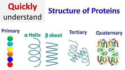
- 🌀 Two common types of secondary structures are the alpha helix and beta pleated sheets, which are crucial for the protein's function and stability.
- 🌈 The alpha helix is characterized by hydrogen bonds between the carbonyl oxygen and the amide hydrogen of the amino acids in the next turn of the helix, forming a spiral staircase shape.
- 📜 Beta pleated sheets form when hydrogen bonds occur between carbonyl oxygen atoms and amide hydrogen atoms of different amino acids, creating a sheet-like structure.
- 🌿 Hydrophobic amino acids are more common in beta pleated sheets, while hydrophilic or polar amino acids are more exposed in alpha helices due to their structural differences.
- 📊 Ribbon diagrams are used to represent the secondary structures of proteins, simplifying the visualization by showing the overall shape rather than individual atoms.
- 🔗 Hydrogen bonds are the primary force maintaining the secondary structures of proteins, such as in alpha helices and beta pleated sheets.
- 🌟 Collagen, a structural protein, contains a triple helix, where three polypeptide chains are intertwined and stabilized by hydrogen bonds, providing strength to tissues like skin, tendons, and cartilage.
- 🧠 Protein misfolding and aggregation, such as the conversion of beta amyloid proteins from alpha helices to beta pleated sheets in Alzheimer's disease, can lead to the formation of insoluble plaques and neuronal dysfunction.
- 💡 The diagnosis of Alzheimer's disease is typically confirmed post-mortem by examining brain slices for atrophy and the presence of neurofibrillary tangles affecting nerve cell transmission.
- 🛑 Neurons do not regenerate quickly, and the death of nerve cells due to protein aggregation and plaque formation in Alzheimer's leads to memory loss and loss of motor function.
Q & A
What is the secondary structure of a protein?
-The secondary structure of a protein refers to the local folding patterns of the peptide chain, which are stabilized by hydrogen bonds between the backbone atoms. It includes common structures such as the alpha helix and beta pleated sheets.
What are the two most common types of secondary structures in proteins?
-The two most common types of secondary structures in proteins are the alpha helix and beta pleated sheets.
How are alpha helices formed in proteins?
-Alpha helices are formed by hydrogen bonds between the oxygen of the carbonyl groups and the hydrogen of the NH groups of the amide bonds in the next turn of the helix, which gives the peptide chain a helical shape.
What interactions are responsible for the formation of beta pleated sheets in proteins?
-Beta pleated sheets are formed by hydrogen bonds between the carbonyl oxygen atoms and the hydrogen atoms in the amide group, which bend the polypeptide chain into a sheet-like structure.
What is the difference between alpha helices and beta pleated sheets in terms of amino acid exposure?
-Alpha helices tend to have more polar or hydrophilic amino acids exposed due to their tight interaction, while beta pleated sheets may have more hydrophobic amino acids tucked away from the aqueous environment.
What is a triple helix and where is it commonly found?
-A triple helix is a secondary structure found in structural proteins like collagen, where three polypeptide chains are woven together with hydrogen bonds holding the chains together, providing additional strength.
How does the representation of proteins change when using a ribbon diagram?
-In a ribbon diagram, the protein is represented with a general structure rather than individual atoms, showing the secondary structures like alpha helices and beta pleated sheets as ribbons, which simplifies the visualization of large proteins.
What is the significance of the N-terminus and C-terminus in the context of protein structure?
-The N-terminus and C-terminus are the beginning and end of a protein chain, respectively. They can be identified in protein diagrams to understand the directionality of the amino acid chain and are important for the overall three-dimensional structure.
How can changes in protein secondary structure lead to disease?
-Changes in protein secondary structure, such as the conversion of alpha helices into beta pleated sheets in Alzheimer's disease, can cause proteins to misfold and aggregate, leading to the formation of insoluble plaques that disrupt normal cellular functions and contribute to disease pathology.
What is the role of beta amyloid proteins in Alzheimer's disease?
-In Alzheimer's disease, beta amyloid proteins change from their normal alpha helical shape to beta pleated sheets, forming insoluble aggregates that accumulate as plaques, which are implicated in the disruption of neuronal signaling and contribute to the disease's progression.
How can the diagnosis of Alzheimer's disease be confirmed?
-The official diagnosis of Alzheimer's disease can only be done post-mortem by examining brain slices for atrophy and the presence of neurofibrillary tangles. However, symptoms like memory loss and loss of motor function can be used for an unofficial diagnosis.
Outlines

Cette section est réservée aux utilisateurs payants. Améliorez votre compte pour accéder à cette section.
Améliorer maintenantMindmap

Cette section est réservée aux utilisateurs payants. Améliorez votre compte pour accéder à cette section.
Améliorer maintenantKeywords

Cette section est réservée aux utilisateurs payants. Améliorez votre compte pour accéder à cette section.
Améliorer maintenantHighlights

Cette section est réservée aux utilisateurs payants. Améliorez votre compte pour accéder à cette section.
Améliorer maintenantTranscripts

Cette section est réservée aux utilisateurs payants. Améliorez votre compte pour accéder à cette section.
Améliorer maintenantVoir Plus de Vidéos Connexes

Tertiary structure of proteins | Macromolecules | Biology | Khan Academy

Protein structure | Primary | Secondary | Tertiary | Quaternary

Struktur Sekunder, Tersier, Kuartener Protein | Fungsi dan Struktur Protein

Proteins: Primary and Secondary Structure | A-level Biology | OCR, AQA, Edexcel

Beta sheet structure of proteins

Protein Structure - Primary, Secondary, Tertiary, & Quarternary - Biology
5.0 / 5 (0 votes)
