Onion Peel Under the Microscope | How to Prepare Stained Temporary Mount of Onion Peel
Summary
TLDRThis educational video guides viewers through the process of preparing and observing onion epidermal cells under a microscope. It details the necessary equipment, the staining process with methylene blue, and the creation of a slide to prevent cell dehydration. The video demonstrates the use of both low and high magnification to reveal the distinct boundaries, rectangular shape, and nucleus location of plant cells, offering insights into basic cellular structures and their functions.
Takeaways
- 🧐 The video demonstrates the process of preparing and observing onion cells under a compound microscope.
- 🔍 The necessary tools for the demonstration include glass slides, cover slips, a needle, a scalpel, forceps, scissors, onions, glycerin, and a staining agent like methylene blue.
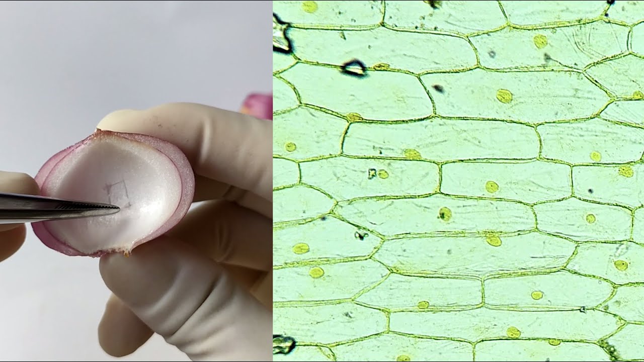
- 🧑🍳 The presenter peels an onion and carefully removes the inner epidermal layer to use as a specimen for staining.
- 🎨 Methylene blue is used to stain the onion cells, which helps in observing the cell structure more clearly under the microscope.
- ⏳ The staining process should be quick to prevent the cells from drying out, and the stained specimen is placed on a slide with glycerin to keep it moist.
- 🔬 The use of a coverslip is crucial to protect the specimen and to avoid air bubbles that could distort the view under the microscope.
- 🔬 The presenter uses both low (10x) and high (45x) magnification to observe the onion cells, highlighting the differences in the number of cells visible at each magnification level.
- 📐 The cells observed are expected to be nucleated, have a distinct boundary due to the cell wall, and be rectangular or brick-like in shape.
- 🔵 The cytoplasm of the cells is stained light blue, while the nucleus takes up a darker shade of blue, indicating successful staining.
- 🔍 Higher magnification allows for a closer look at the nucleus and cytoplasm, but cell organelles are not visible without an electron microscope.
- 📚 The video concludes with an invitation to subscribe to the channel and visit the website for more educational content on various subjects.
Q & A
What is the main subject of the video?
-The main subject of the video is the preparation and observation of onion peel cells under a compound microscope.
What materials are needed to prepare a slide of onion peel?
-The materials needed include clean glass slides, cover slips, a needle, a scalpel, forceps, a pair of scissors, onions, glycerin, and a staining agent like methylene blue.
Why is glycerin used on the slide before placing the specimen?
-Glycerin is used to keep the specimen moist and prevent it from drying out while being observed under the microscope.
How does one remove the inner lining of the onion peel?
-The inner lining of the onion peel is removed by breaking the fleshy leaf into small portions and teasing out the epidermal layer using forceps or a scalpel.
What is the purpose of staining the onion peel specimen?
-Staining the onion peel specimen enhances the visibility of cellular structures, such as the nucleus and cytoplasm, under the microscope.
Why is it important to avoid air bubbles under the coverslip?
-Air bubbles can interfere with the clarity of the microscopic image, making it difficult to observe the specimen's details.
What technique is used to place the coverslip without trapping air bubbles?
-The technique involves touching the edge of the coverslip to the slide, balancing it with a needle, and slowly removing the needle to allow the coverslip to settle without air bubbles.
What magnifications are used to observe the onion peel cells in the video?
-The video uses both 10x magnification (low power) and 45x magnification (high power) to observe the onion peel cells.
What are the characteristics of the onion peel cells observed under the microscope?
-The onion peel cells are observed to have a distinct boundary, a nucleus located towards the periphery, and are rectangular in shape with closely packed cytoplasm.
Why can't cell organelles be seen with the compound microscope used in the video?
-Cell organelles require a much higher magnification than what a compound microscope can provide; an electron microscope would be needed for such detailed observation.
Where can viewers find more courses like the one presented in the video?
-Viewers can find more courses on physics, chemistry, biology, mathematics, and computer coding by subscribing to the channel and visiting the website www.manojacademy.com.
Outlines
🔬 Preparing and Observing Onion Epidermal Cells
In this segment, the video script details the process of preparing a slide of onion peel cells for microscopic observation. The narrator guides the audience through the necessary materials, including glass slides, cover slips, a scalpel, forceps, onions, glycerin, and a staining agent like methylene blue. The process involves cutting the onion, peeling off the outer layer, teasing out the epidermal layer, staining it, and placing it on a slide with glycerin to prevent drying. A coverslip is carefully applied to avoid air bubbles, ensuring clear visibility under the microscope.
🌿 Analyzing Plant Cells Under the Microscope
This paragraph describes the observation of onion epidermal cells using a compound microscope. The narrator starts with a low power objective to get an overview of the cells, noting their distinct boundaries, rectangular shape, and closely packed arrangement, which is typical for plant epidermal cells. The cells' nucleus is stained dark blue and is located at the periphery, while the cytoplasm appears light blue. Switching to a high power objective reveals fewer cells due to the reduced field of view but provides a more detailed view of the nucleus and cytoplasm. The narrator mentions that to observe cell organelles, an electron microscope would be required. The video concludes with an invitation to subscribe to the channel and visit the academy's website for more courses.
Mindmap
Keywords
💡Onion
💡Compound Microscope
💡Epidermal Layer
💡Dissection Box
💡Glycerin
💡Methylene Blue Stain
💡Coverslip
💡Air Bubbles
💡Nucleus
💡Cytoplasm
💡Cell Wall
Highlights
Introduction to observing onion cells under a compound microscope.
List of materials required for preparing an onion peel slide.
Process of cutting an onion in half and peeling out a fleshy leaf.
Kneading the inner lining of the onion peel to obtain the epidermal layer.
Use of methylene blue stain to color the slide for better observation.
Importance of working quickly to prevent cells from drying out.
Application of glycerin to keep the specimen moist during observation.
Technique for placing the specimen on the slide without rolling.
Method to avoid air bubbles when placing the coverslip.
Preparation of the specimen slide ready for microscope observation.
Use of a compound microscope with different magnifications.
Expectation of observing nucleated, rectangular, closely packed epidermal cells.
Characteristic features of onion peel cells under low power magnification.
Observation of the nucleus and cytoplasm staining under high power magnification.
Limitation of microscope magnification for observing cell organelles.
Conclusion of the virtual practical class and invitation to subscribe for more.
Promotion of Manoj Academy's courses in various subjects.
Transcripts
i'm sure you have all seen and eaten an
onion but have you ever wondered what
the cells of an onion look like
in this video we are going to do exactly
that
we are going to prepare a slide of an
onion peel and observe it under a
compound microscope
for that these are the following things
that you would require from your
dissection box
we will require clean glass lights cover
slips
needle
a scalpel
forceps
a pair of scissors
onions
a little bit of glycerine and
i am using methylene blue stain to stain
the slide you may use safranin or any
other stain that is available now let us
start with the work
so first of all i have taken an onion
and cut it into halves
and then i will have to peel out one of
the fleshy leaves of onion
so you see i will just take out the
leaves in such a manner that i get rid
of
this dry
scaly leaf which is present
outside
and i only have this fleshy part that i
need
now after this we are going to
knead the inner lining of this fleshy
leaf
which is actually an epidermal layer
how will i take it out
so i break this
in a small
into a small portion
and then
with the help of a pair of forceps
i will just tease the inner lining of
the onion peel with the forceps or you
can here use a scalpel
but a forceps is always very convenient
you just have to tease it and you will
see the inner lining is coming out
like
a thin strip
all right so this is the layer of
epidermis that we require
however we do not require so much so
what we will do is
we will quickly and everything has to be
done very quickly because you do not
want the cells to dry up so very quickly
i will take a small piece
of this
peel
and this much is enough in fact this is
more than enough
i will put this in the methylene blue
stain that i have taken
on a watch glass
so now you can see
that the specimen is inside the stain
and depending on the strength of the
stain you might have to keep it for a
couple of minutes to five minutes
after this we will take a clean glass
light
we will take the stained
specimen
once it has been stained you can take it
and we will have to place it on the
glass light but before that
we will use a little bit of glycerin and
put it on the slide
why are we using glycerin we are using
glycerin because we do not want the
specimen to dry out while we are
observing it under the microscope the
glycerin will keep the specimen moist
okay now
we will take
the specimen out of the stain
place it on the slide
make sure that this
section that you have taken does not
roll so if you find that it is rolling
anywhere you can simply hold it with
your forceps and tease it with the
needles so that it is straight and it is
laid out on the slide
to make sure that this specimen does not
dry out we will have to cover it with a
coverslip
now this is a coverslip here i am using
a square coverslip you can use a round
cover slip if you have one but while we
put down the coverslip we have to be
very sure that there is no air bubble
trapped inside because if there is air
bubble inside then it will not be
clearly visible the specimen will not be
clearly visible under the microscope so
there is a certain technique for laying
down the coverslip such that you do not
have air bubbles inside let me show you
how
so we will just touch the coverslip
onto the slide
and
balance it
support it with the needle
and we will slowly take the needle out
very slowly
and you will see that there is no air
bubble left
now under this coverslip now we have the
specimen ready so now what do we have to
do
we will have to simply put the spec
slide under the microscope and observe
it so here i am using a compound
microscope we have already seen the
parts of a compound microscope first i
will be using a 10x magnification which
is low power and then i will be using a
45x magnification which is high power to
observe the cells
what kind of cells do you expect to see
in the onion peel
since these are epidermal cells and
these are plant cells we expect to see
nucleated cells we expect to to see
cells which have a very strong boundary
or a very distinct boundary and usually
the epidermal cells are rectangular in
shape so let us see how the cells will
look like and then we can
write down the characteristic features
so now i will just focus the microscope
i am using the 10x magnification i will
focus the slide
so here we can see the cells in low
power as you can see we have
seen that these cells have a distinct
boundary because these are plant cells
so they have a cell wall
they are living cells so you can see a
clear blue dot inside which is the
nucleus and what is the location of the
nucleus the nucleus is located towards
the periphery of the cell
you also see that these cells are
rectangular in shape brick like and are
absolutely closely packed because you
want an epidermal layer to be closely
packed there should be no gap so that it
can provide enough protection
the cytoplasm has been stained light
blue whereas the nucleus has taken up a
dark blue stain
let us bring this under high power and
see if we can magnify the nucleus and
the cytoplasm a little further
so you see under high power you see less
number of cells why because when you are
magnifying the tissue in a microscopic
field
in low power you see a large area and
therefore you can see a larger number of
cells
the moment you use a higher
magnification you see a smaller area of
the specimen
although the image is magnified you see
lesser number of cells
but the nucleus is prominent here so is
the cytoplasm
we cannot see the cell organelles
because for that we need a much higher
magnification which is possible with the
help of an electron microscope
so that was all for today's class i'm
sure you enjoyed it we will be back with
more such virtual practical classes from
manoj academy thank you and see you soon
subscribe to our channel
and go to our website
www.manojacademy.com
courses on physics chemistry biology
mathematics and computer coding
hope you will have fun
happy learning
Ver Más Videos Relacionados
Onion Skin Epidermal Cells: How to Prepare a Wet Mount Microscope Slide
Biology Lab || Mitosis
Microscopy - How to use a microscope - GCSE Science Required Practical
Mitosis in Onion Root tip Experiment
GCSE Biology Revision "Required Practical 1: Microscopes"
Comparing animal and plant cells | Cells and organisms | Middle school biology | Khan Academy
5.0 / 5 (0 votes)