Praktikum Biologi SMP - Pengamatan Sel Tumbuhan
Summary
TLDRThis educational video demonstrates the process of observing plant cells under a microscope, focusing on onion and corn cells. The tutorial explains the necessary materials, such as a light microscope, pipette, and glass slides, and the steps for preparing wet and dry preparations. The video walks viewers through placing a thin onion skin onto a slide, adding a drop of water, and adjusting the microscope for optimal magnification. It also covers using preserved dry preparations, like corn cells, to study plant structure. The video aims to enhance understanding of plant cell anatomy through clear, step-by-step instructions.
Takeaways
- 😀 The experiment focuses on observing plant cells, specifically onion cells, under a microscope.
- 😀 Essential materials include a light microscope, pipette, glass slides, cover slips, and preserved or dried specimens.
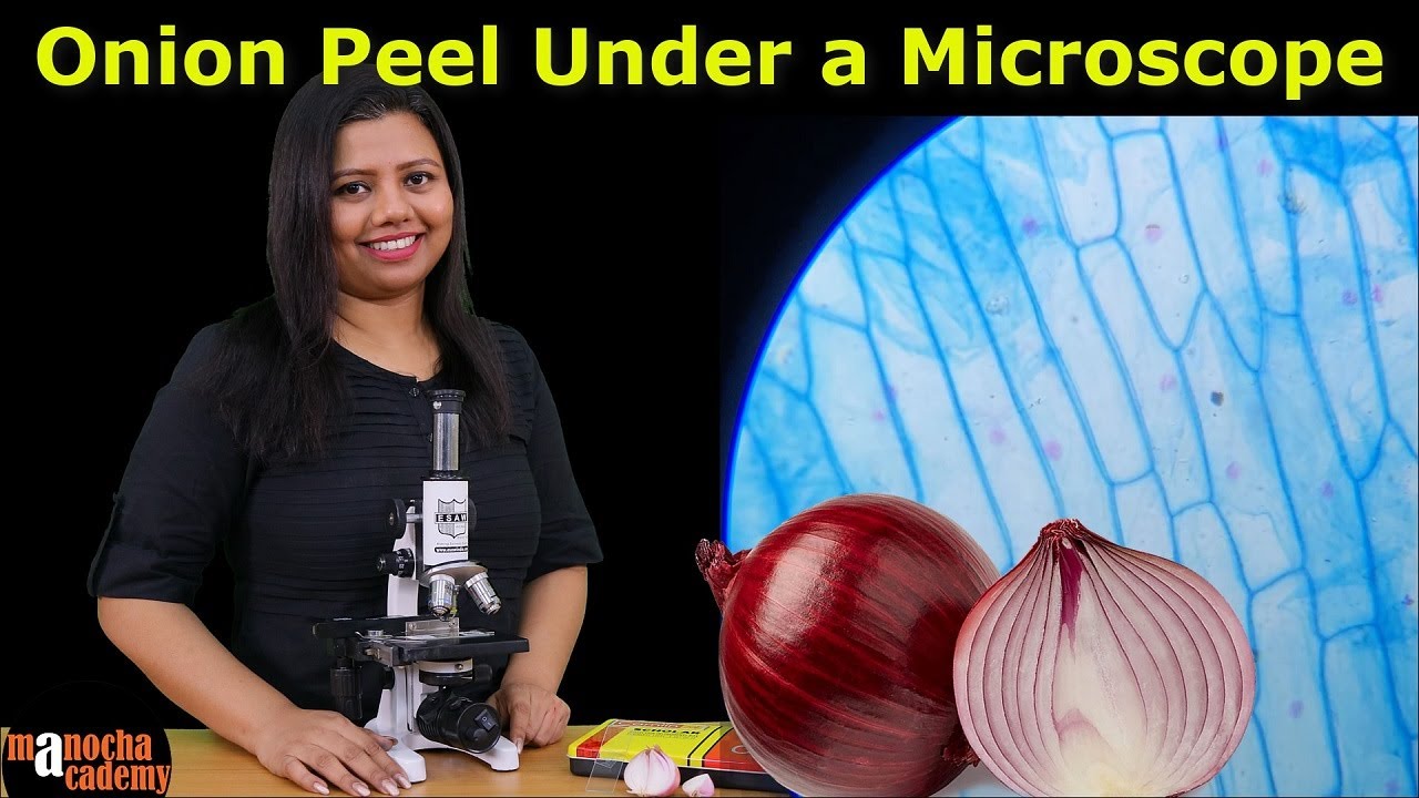
- 😀 The first step is to peel a thin layer of onion skin and place it on a glass slide.
- 😀 A few drops of water are added to the onion skin before covering it with a cover slip for clearer observation.
- 😀 The slide is then placed on the microscope stage, and a clamp is used to secure it in position.
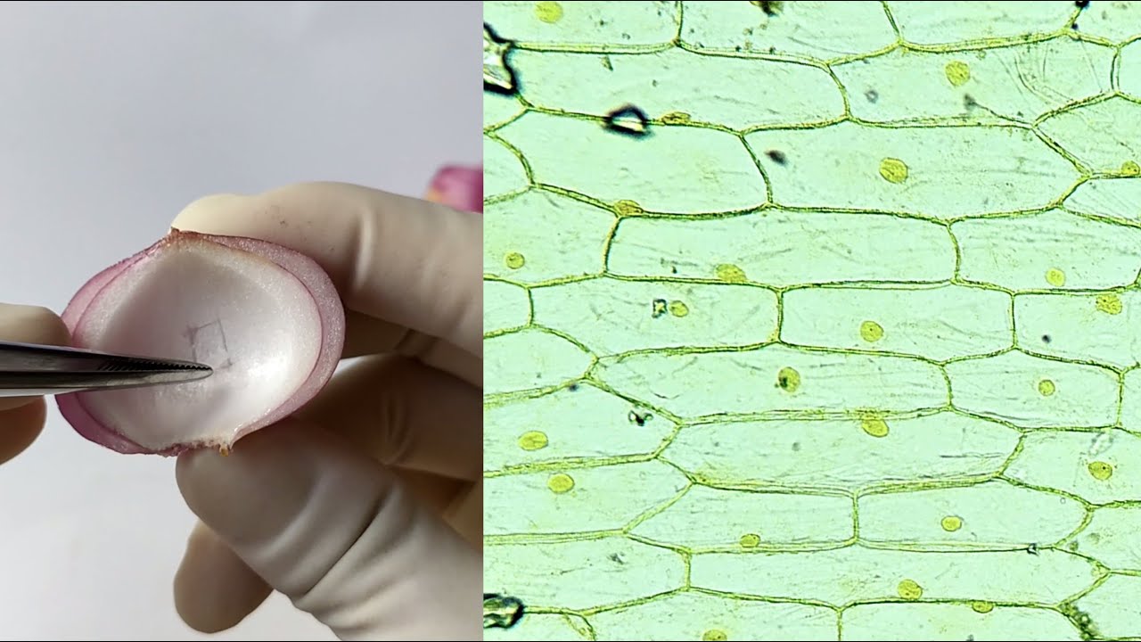
- 😀 The magnification is set to 100x (10x ocular lens and 10x objective lens) to observe the cell structure.
- 😀 Adjusting the light and fine-tuning the microscope’s focus ensures clear visibility of the onion cell structure.
- 😀 The same procedure can be used to observe dry or preserved plant specimens, such as corn cells.
- 😀 For dry specimens like corn, no water is used, and the specimen is directly observed under the microscope.
- 😀 The process is concluded with the observation of both onion and corn plant cells, showing different structures.
- 😀 The lesson aims to provide a hands-on understanding of plant cell structure through direct observation using a microscope.
Q & A
What is the main purpose of the video?
-The main purpose of the video is to demonstrate how to observe plant cells under a microscope, specifically focusing on onion cells and using both wet and dry mount techniques.
What materials are required for the observation of plant cells in the video?
-The materials required include a light microscope, chemical pipette, Petri dish, aquades (distilled water), object glass, cover glass, prepared or dried specimens, scalpel, tissue, and onion and garlic.
What is the first step in preparing the onion cells for observation?
-The first step is to peel a thin layer of onion skin and place it on an object glass. A few drops of water are added to cover the specimen before placing a cover glass on top.
Why is water added to the onion skin sample before placing the cover glass?
-Water is added to ensure the specimen is properly hydrated and to help make the onion cells clearer for observation under the microscope.
How is the specimen positioned on the microscope for observation?
-The specimen is moved to the microscope stage and held in place using clips to ensure that it stays in position during observation.
What magnification is used during the observation with the microscope?
-A total magnification of 100x is used, which is achieved by combining a 10x ocular lens with a 10x objective lens.
How is the light adjusted to improve visibility during the observation?
-The light mirror is adjusted to direct the light toward the specimen, enhancing its visibility and clarity for observation.
What is the difference between wet mount and dry mount preparations?
-In a wet mount, water is used to keep the specimen hydrated and help with the observation, while in a dry mount, the specimen is prepared without any water, typically using a prepared or dried specimen.
What alternative specimen is used for the dry mount method?
-For the dry mount, corn plant cells are used, which do not require any water droplets and can be observed directly under the microscope.
What is the expected outcome when observing corn plant cells under the microscope?
-When observing corn plant cells under the microscope using a 10x ocular lens and a 40x objective lens, the cells are clearly visible and can be examined in detail.
Outlines

This section is available to paid users only. Please upgrade to access this part.
Upgrade NowMindmap

This section is available to paid users only. Please upgrade to access this part.
Upgrade NowKeywords

This section is available to paid users only. Please upgrade to access this part.
Upgrade NowHighlights

This section is available to paid users only. Please upgrade to access this part.
Upgrade NowTranscripts

This section is available to paid users only. Please upgrade to access this part.
Upgrade NowBrowse More Related Video
Onion Peel Under the Microscope | How to Prepare Stained Temporary Mount of Onion Peel
Onion Epidermal Cell Peel Slide Preparation Practical Experiment

Onion Skin Epidermal Cells: How to Prepare a Wet Mount Microscope Slide

Staining of Nucleic Acid by Acetocarmine
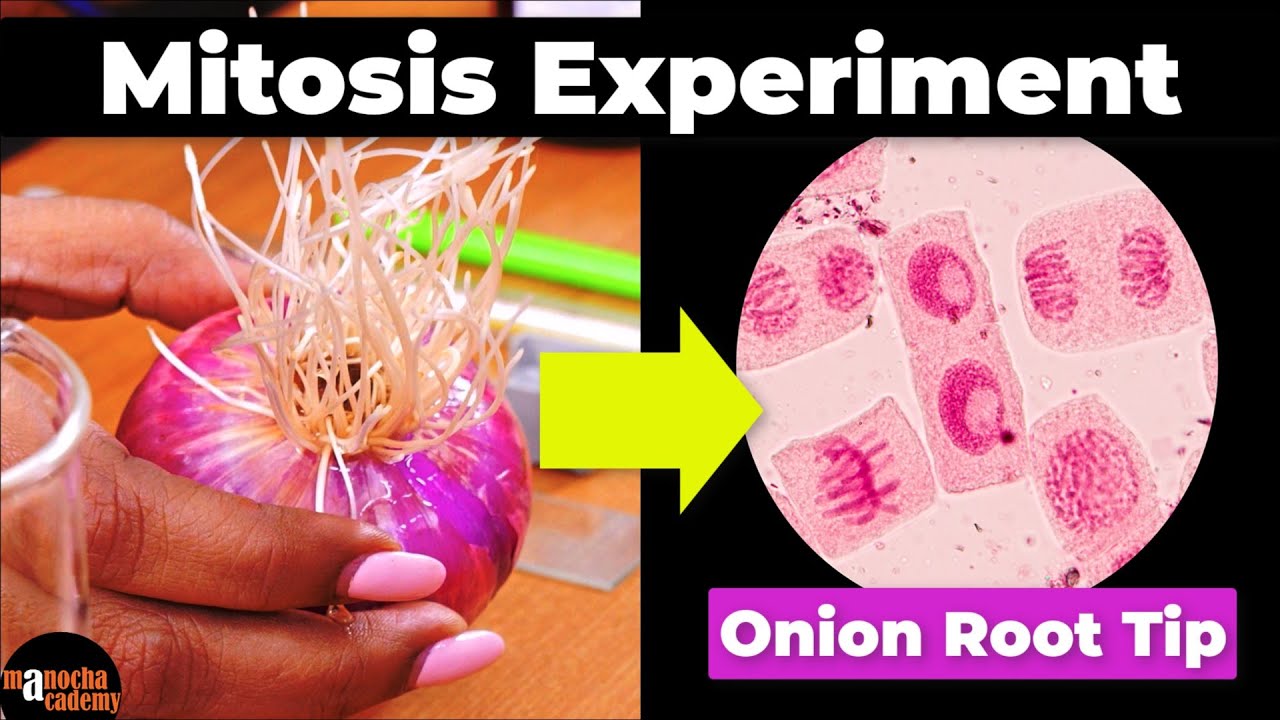
Mitosis Experiment Onion Root Tip Procedure
Preparação de lâminas para observação de células vegetais
5.0 / 5 (0 votes)