Onion Skin Epidermal Cells: How to Prepare a Wet Mount Microscope Slide

Tami Guy, MS, CPPS
15 Oct 201206:51

Summary

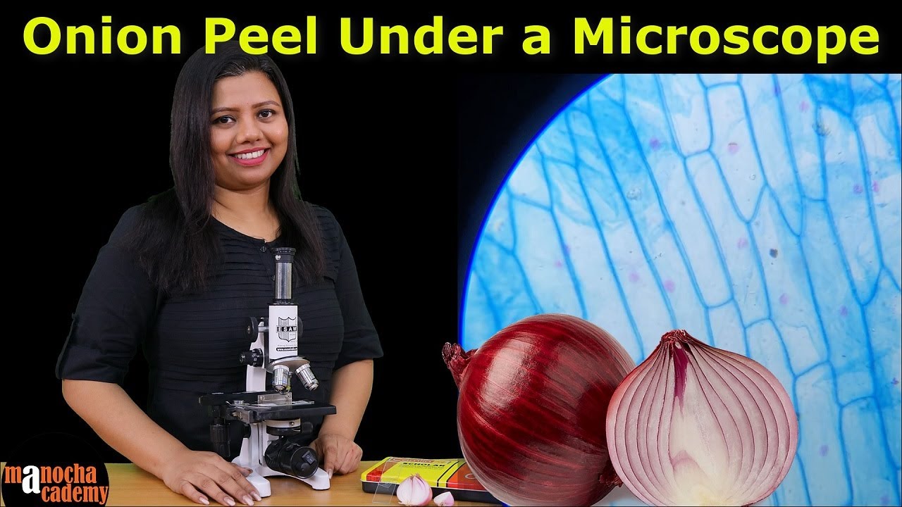
TLDRThis educational video demonstrates the process of preparing a wet mount slide of stained onion epidermal cells. The presenter explains how to select and peel the thin, transparent layer of cells from an onion bulb, transfer it onto a microscope slide, and apply iodine for staining. The video also covers techniques for placing a cover slip to minimize air bubbles and adjusting the microscope's light levels for optimal viewing. Finally, the presenter shows the cells magnified under different objective lenses, highlighting the cell walls, nuclei, and the importance of staining for visibility.

Takeaways

  • πŸ”¬ The video demonstrates how to prepare a wet mount slide of stained onion epidermal cells.
  • 🧐 The process begins with selecting a smooth and shiny layer of onion bulb for obtaining the specimen.
  • πŸ”Ž A thin, transparent layer of cells is peeled off the onion layer using tweezers for use as the specimen.
  • πŸ“ An alternate technique mentioned is peeling the onion skin directly onto the slide for a larger, flatter specimen.
  • πŸ’§ Iodine is used as the wet mount solution, which also serves to stain the cells for better visibility.
  • 🏷️ A cover slip is applied by holding it at a right angle and allowing it to drop onto the slide, minimizing air bubbles.
  • πŸ”­ The specimen is first examined under a 4X objective lens, magnifying the cells 40 times actual size.
  • πŸ” The video explains that a compound microscope is parfocal, allowing for easy transition between different magnification levels.
  • πŸŒ‘ Adjusting the light level by using the dimmer dial or iris diaphragm can improve contrast and visibility of the specimen.
  • πŸ”¬ The final examination is done under a high dry lens at 400 times actual size, revealing detailed cell structures.

Q & A

  • What is the purpose of the video?

    -The purpose of the video is to demonstrate the process of preparing a wet mount slide of stained onion epidermal cells.

  • What type of specimen is used in the video for the wet mount slide?

    -The specimen used is a thin, transparent layer of onion epidermal cells obtained from the inner layer of an onion bulb.

  • How does one identify the correct layer of onion epidermal cells?

    -The correct layer is identified by its smooth and shiny appearance compared to the rough and matte side of the onion layer.

  • What technique is shown for obtaining the onion epidermal cells?

    -The technique involves breaking the onion layer towards the shiny side and gently peeling apart the two pieces to obtain the thin layer of cells.

  • How should one handle the thin layer of onion epidermis to place it on the slide?

    -The thin layer should be torn off with tweezers and then carefully spread out on the slide to avoid it folding over on itself.

  • What alternative method is suggested for transferring the onion skin to the slide?

    -The alternative method is to peel the layer of onion skin directly onto the slide, allowing it to wrap around the slide like Saran Wrap.

  • What staining agent is used in the video for the wet mount?

    -Iodine is used as the staining agent, which provides both the wet component and stains the cells for better visibility.

  • How is a cover slip applied to the specimen on the slide?

    -The cover slip is held at a right angle to the slide and then dropped, allowing it to fall and push the air out, creating fewer air bubbles.

  • What magnification is used to initially examine the onion epidermal cells?

    -The initial examination uses a 4X objective lens, magnifying the specimen 40 times actual size.

  • How can the contrast of the specimen be improved if the stain is not taken up well?

    -The contrast can be improved by adjusting the light level, either by using the dimmer dial on the microscope base or by opening or closing the iris diaphragm.

  • What is the highest magnification used in the video to view the onion epidermal cells?

    -The highest magnification used is with the high dry power objective lens, which magnifies the specimen 400 times actual size.

Outlines

00:00

πŸ”¬ Preparing a Wet Mount Slide of Onion Epidermal Cells

This paragraph describes the process of preparing a wet mount slide of stained onion epidermal cells. The presenter starts by selecting a microscope slide and obtaining a specimen from the onion bulb. The bulb has a rough matte side and a smooth shiny side, and the goal is to peel off the thin transparent layer of cells from the shiny side. The presenter uses tweezers to tear off a piece of the onion epidermis and places it on the slide. The delicate layer of cells may fold over, so it needs to be spread out. The presenter also mentions an alternative technique for transferring the onion skin directly onto the slide. For staining, iodine is used, which serves as both the wet mount and staining agent. A cover slip is applied by holding it at a right angle and letting it drop onto the slide to minimize air bubbles. The specimen is then examined under a 4X objective lens, which magnifies the cells 40 times. The cells are described as misshapen bricks, and the video shows a microscopic view of the onion epidermal cells.

05:03

πŸ“Έ Enhancing Microscopic Image Contrast and Viewing Techniques

In this paragraph, the focus is on enhancing the contrast of the specimen when viewing under a microscope and adjusting light levels to improve visibility. The presenter demonstrates how to adjust the light by using the dimmer dial on the microscope base or by opening and closing the iris diaphragm, which controls the amount of light shining through the specimen. After focusing the specimen at low power, the presenter switches to a high dry power lens, which magnifies the specimen 400 times. The video shows examples of onion epidermal cells viewed at this higher magnification, where the cell walls and nuclei are more clearly visible. The presenter also suggests visiting Science Prof Online for more educational content.

Mindmap

Keywords

πŸ’‘Wet Mount Slide

A wet mount slide is a type of microscope slide preparation where a small piece of biological material is placed in a drop of liquid on a slide. In the context of the video, the liquid is iodine, which serves to both wet the specimen and stain it for easier viewing under a microscope. The process involves placing a thin layer of onion epidermal cells onto the slide and then covering it with a drop of iodine to prepare for microscopic examination.

πŸ’‘Onion Epidermal Cells

Onion epidermal cells refer to the outermost layer of cells of an onion bulb, which are used as a specimen in this video. These cells are thin, transparent, and serve as a model system for demonstrating cell structure under a microscope. The video describes how to carefully peel these cells from the onion layer to prepare a wet mount slide, highlighting their importance in the process.

πŸ’‘Microscope Slide

A microscope slide is a small glass or plastic plate used to hold a specimen for examination under a microscope. In the video, the microscope slide is where the onion epidermal cells are placed and prepared for staining and viewing. The slide is an essential tool in microscopy, providing a stable platform for the specimen.

πŸ’‘Iodine

Iodine is a chemical element used in the video as a staining agent for the onion epidermal cells. It not only provides the 'wet' part of the wet mount slide by adding moisture but also colors the cells, enhancing their visibility under the microscope. The use of iodine is crucial for making the cell structures stand out clearly.

πŸ’‘Cover Slip

A cover slip is a small, thin piece of glass placed over the specimen on a microscope slide to protect the sample and provide a smooth surface for viewing. In the video, the cover slip is carefully applied to the iodine-wetted onion epidermal cells to prepare the wet mount slide for microscopic examination. Proper application of the cover slip is demonstrated to minimize air bubbles.

πŸ’‘Objective Lens

The objective lens is the lens of a microscope that is closest to the specimen. It is used to magnify the image of the specimen. In the video, the presenter uses a 4X objective lens to initially examine the onion epidermal cells, providing a lower magnification view before moving to higher magnifications. The objective lens is a key component in determining the level of detail visible in the microscope.

πŸ’‘Magnification

Magnification in microscopy refers to the increase in the apparent size of an object due to the use of a microscope. The video mentions magnification levels of 40 times and 100 times actual size, achieved by using different objective lenses. Magnification is essential for observing the fine details of the onion epidermal cells that are not visible to the naked eye.

πŸ’‘Compound Microscope

A compound microscope is a type of microscope that uses multiple lenses to achieve higher magnification and resolution. The video discusses the use of a compound microscope to view the onion epidermal cells at different magnifications. The compound microscope allows for the observation of cellular structures that are not possible with the naked eye.

πŸ’‘Parfocal

Parfocal is a term used to describe the ability of a microscope to maintain focus when switching between different objective lenses. In the video, the presenter mentions that the microscope is parfocal, which means once the specimen is in focus with one objective, it will remain relatively in focus when switching to another, requiring only minor adjustments.

πŸ’‘Light Adjustment

Light adjustment refers to the process of modifying the intensity and distribution of light to optimize the viewing conditions in microscopy. The video demonstrates how adjusting the light level, using the dimmer dial and iris diaphragm, can improve the contrast and visibility of the onion epidermal cells under the microscope. Proper lighting is crucial for achieving clear and detailed images.

πŸ’‘Cell Wall

The cell wall is a rigid outer layer that provides support and protection to plant cells, including onion epidermal cells. In the video, the presenter notes the presence of thick cell walls in the onion epidermal cells when viewed under higher magnification. The cell wall is an important structural component of plant cells, contributing to their shape and function.

Highlights

Introduction to preparing a wet mount slide of stained onion epidermal cells.

Selection of a microscope slide for the specimen preparation.

Obtaining a specimen from the smooth, shiny side of an onion bulb layer.

Technique to gently peel apart the onion layer to access the epidermal cells.

Method to tear off a thin layer of onion epidermis using tweezers.

Spreading the delicate onion epidermis layer on the slide.

Alternate technique for transferring onion skin directly onto the slide.

Use of iodine as a staining agent for the wet mount.

Procedure for applying a cover slip to the specimen to avoid air bubbles.

Examination of the specimen under a 4X objective lens for initial magnification.

Description of onion epidermal cells appearing like misshapen bricks under the microscope.

Explanation of the compound microscope's parfocal property for ease of focus adjustment.

Observation of onion epidermal cells at 100x magnification for a closer look.

Technique to improve contrast by adjusting light levels for better visibility.

Demonstration of using the dimmer dial and iris diaphragm to control light.

Switching to high dry power for 400x magnification to observe cell details.

Observation of thick cell walls and cell nuclei in the onion epidermal cells at high magnification.

Promotion of additional educational resources available at Science Prof online.

Transcripts

play00:01

in this video I'm going to show you how

play00:03

to prepare a wet Mount slide of stained

play00:06

onion epidermal

play00:09

cells here's my microscope slide I want

play00:12

to have that out and

play00:18

ready I can obtain my specimen from one

play00:21

layer of onion bulb if I look at this

play00:24

layer one side is kind of rough and

play00:27

matte and the other side is smooth

play00:30

smooth and

play00:32

shiny I need to break my onion layer

play00:35

towards this shiny side and then very

play00:38

gently

play00:40

peel the two pieces apart there's a thin

play00:44

transparent layer of cells holding these

play00:47

two pieces of the onion layer together

play00:50

that's the layer of epidermal cells that

play00:52

I'm looking for for my

play00:57

specimen now I need to obtain a piece of

play01:00

that onion epidermis to place on my

play01:03

slide I can do that by tearing it off

play01:05

with a pair of

play01:09

tweezers this is a very delicate thin

play01:13

layer of ss and they'll fold over on

play01:16

themselves uh so when you place that

play01:18

onto the slide once you get it off your

play01:21

tweezers you're going to need to spread

play01:23

it out a little bit the little piece of

play01:25

onion epidermis can easily get all

play01:27

watted up and that makes it very

play01:29

difficult to see this

play01:43

sounds while filming in the lab I forgot

play01:46

to include an easier alternate technique

play01:48

for getting that onion skin onto the

play01:50

slide so I'm going to show it to you

play01:52

here in my dining room with my two

play01:56

helpers the best way to get a large flat

play01:59

sheet of these onion epidermal cells is

play02:03

to peel that layer of onion skin off

play02:06

directly onto the slide it's very thin

play02:09

it'll wrap right around the slide just

play02:11

like Saran

play02:16

Wrap so apparently I've lost my helpers

play02:19

because onions are way too stinky so we

play02:22

might as well head back to the lab

play02:31

for this wet Mount I'm just going to use

play02:33

iodine this will provide the wet part of

play02:35

the wet Mount and it will also stain the

play02:38

cells and make them easier to

play02:53

see now I'm going to obtain a cover slip

play02:56

a clear square that I'm going going to

play03:00

place on top of my specimen on the

play03:09

slide the best way to apply that cover

play03:11

slip is to hold it at a right angle to

play03:14

the slide at one side of your specimen

play03:17

and then let it drop let it fall as it

play03:20

falls it will push the air out and

play03:22

you'll have fewer air bubbles this way

play03:25

here's my finished prepared specimen

play03:31

here's a closer look at the application

play03:34

of that cover slip again I hold it at a

play03:36

right angle to the slide I let it drop

play03:39

that pushes the air out sometimes I'll

play03:41

give it a little tap and now I'm all

play03:51

set I'm now going to examine my specimen

play03:55

using the 4X objective lens that will

play03:57

magnify the specimen 40 times actual

play04:02

size here's my first look

play04:04

microscopically at the onion epidermal

play04:07

cells magnified 40 times these cells

play04:10

look kind of like misshapen bricks a

play04:13

whole bunch of

play04:14

them the compound microscope is par

play04:17

focal what that means is once I get my

play04:20

specimen in focus at one power in this

play04:23

case the scanning power I can switch to

play04:26

the next higher objective the low power

play04:29

in this case and my specimen will still

play04:31

pretty much be in Focus I'll only have

play04:33

to adjust the focus a little bit to get

play04:35

it

play04:37

crisp here are my onion epidermal cells

play04:40

magnified 100 times actual

play04:43

size so this is a closer look at that

play04:46

specimen we just looked at under 40

play04:49

total magnification the specimen that

play04:52

did not stain so

play04:54

well we stained the specimen to increase

play04:57

the contrast to make it more visible and

play05:00

since the stain wasn't taken up that

play05:02

well there are other ways to improve the

play05:04

contrast of our specimen when viewing

play05:07

this specimen and taking this picture I

play05:10

adjusted the light level I turned it way

play05:12

down and that helped me see the cells

play05:14

much more distinctly and

play05:17

clearly here's just a reminder of the

play05:19

different ways you can adjust the light

play05:21

level shining up through your

play05:24

specimen the first way is to use the

play05:28

dimmer dial on the base of your scope

play05:31

that dial will make the light shining up

play05:34

from the base brighter or

play05:38

dimmer you can also adjust the amount of

play05:41

light shining up through your specimen

play05:44

by opening or closing the iris diaphragm

play05:48

that's directly beneath the stage it

play05:50

looks and feels like a camera lens you

play05:53

twist it and you can let more or less

play05:55

light through the

play05:58

stage once the specimen is clearly in

play06:01

focus at low power that yellow banded

play06:04

lens I can switch the objective to the

play06:08

high dry power that's the blue banded

play06:10

lens and it will magnify my specimen a

play06:13

total of 400 times actual size here are

play06:17

a couple examples of onion epidermal

play06:19

cells viewed at 400 times actual size

play06:22

through that high dry lens these samples

play06:25

have stained a little better than the

play06:27

ones I showed you previously I can see

play06:29

the thick cell walls and these elongated

play06:32

cells in this picture I can see a cell

play06:35

nucleus in the second cell below the

play06:43

pointer more seriously nerdy amazing

play06:46

free stuff at science Prof online.com go

play06:50

there

Rate This
β˜…
β˜…
β˜…
β˜…
β˜…

5.0 / 5 (0 votes)

Related Tags
MicroscopyBiologyLab TechniqueEducationalOnion CellsWet MountStainingMicroscope UseScience TutorialCell Structure