IMAT Biology Lesson 6.1 | Anatomy and Physiology | Animal Tissues Part I
Summary
TLDRIn this educational video, Andy from Med School EU explores the physiology of animals and humans, focusing on the four primary types of tissues: epithelial, connective, muscle, and nervous. He delves into the functions, locations, and specific roles of each tissue type, highlighting the importance of epithelial tissue in protection, absorption, and sensation, and connective tissue in support, binding, storage, and immune response.
Takeaways
- 📚 The video is an educational lecture on the physiology of animals and humans, focusing on the different types of tissues in the human body.
- 🔍 The classification of biological systems is reviewed, starting from cells to tissues, organs, organ systems, and finally the entire organism.
- 🧬 Four primary types of tissues are discussed: epithelial, connective, muscle, and nervous tissues, each with specific functions and locations in the body.
- 💪 Epithelial tissue's main function is physical protection, but it also plays roles in absorption, sensation, and secretion.
- 👤 Different types of epithelial tissues include simple squamous, simple cuboidal, simple columnar, stratified, and pseudostratified columnar, each with unique characteristics and locations.
- 🧠 Merkel cells in the skin are responsible for receiving sensations of touch and passing them on to the nerves.
- 🦴 Connective tissue has various functions including support, binding, storage, transport, protection, and immune response.
- 🦾 There are four major types of connective tissue: connective tissue proper, cartilage, bone, and blood, each with specific roles in the body.
- 🧬 Mesenchyme is an embryological connective tissue that develops into other types of connective tissue and is considered the beginning of all connective tissues.
- 🦴 Cartilage comes in three types: hyaline, elastic, and fibrocartilage, each with distinct properties and locations in the body.
- 🦴 Bone anatomy is detailed, including the epiphyseal plate, epiphyseal line, red bone marrow, periosteum, and the roles of fibroblasts and osteoclasts in bone repair and growth.
Q & A
What is the main topic of the video script?
-The main topic of the video script is the physiology of animals and humans, specifically focusing on the different types of animal tissues.
How many primary types of tissues are discussed in the script?
-The script discusses four primary types of tissues: epithelial, connective, muscle, and nervous tissue.
What is the fundamental function of epithelial tissue?
-The fundamental function of epithelial tissue is physical protection, both internally and externally, from mechanical, chemical, and physical injury, as well as dehydration.
What are the different types of epithelial tissues mentioned in the script?
-The different types of epithelial tissues mentioned are simple squamous, simple cuboidal, simple columnar, stratified, and pseudostratified columnar.
What is the primary function of simple squamous epithelium?
-The primary function of simple squamous epithelium is to allow the movement of molecules across the cell, and it is typically found in the alveoli and the inner lining of blood vessels.
What is the main function of simple cuboidal epithelium?
-The main function of simple cuboidal epithelium is to absorb fluids across both surfaces of the tissue, such as in the nephron of the kidney.
What is the primary function of simple columnar epithelium?
-The primary function of simple columnar epithelium is absorption and secretion, which is prominent in the digestive system.
What are the four major types of connective tissue?
-The four major types of connective tissue are connective tissue proper, cartilage, bone, and blood.
What is the role of fibroblasts in connective tissue?
-Fibroblasts in connective tissue produce protein fibers and ground substance, including collagen, elastic fibers, and reticular fibers, which provide support and protection to cells.
What are the three types of cartilage mentioned in the script?
-The three types of cartilage mentioned are hyaline cartilage, elastic cartilage, and fibrocartilage.
What is the primary function of the periosteum in bone anatomy?
-The periosteum is the outer surface of the bone and plays a role in the growth and repair of the bone by providing a source of cells and nutrients.
What are the functions of red bone marrow?
-The functions of red bone marrow include the production of red blood cells, platelets, and white blood cells, as it contains stem cells that differentiate into various blood cells.
What is the role of fibroblasts and osteoclasts in bone repair?
-Fibroblasts and osteoclasts play a crucial role in bone repair. Fibroblasts lay down bone tissue, while osteoclasts break down bone tissue, maintaining a balance that is essential for the healing and remodeling of bones.
Outlines

Esta sección está disponible solo para usuarios con suscripción. Por favor, mejora tu plan para acceder a esta parte.
Mejorar ahoraMindmap

Esta sección está disponible solo para usuarios con suscripción. Por favor, mejora tu plan para acceder a esta parte.
Mejorar ahoraKeywords

Esta sección está disponible solo para usuarios con suscripción. Por favor, mejora tu plan para acceder a esta parte.
Mejorar ahoraHighlights

Esta sección está disponible solo para usuarios con suscripción. Por favor, mejora tu plan para acceder a esta parte.
Mejorar ahoraTranscripts

Esta sección está disponible solo para usuarios con suscripción. Por favor, mejora tu plan para acceder a esta parte.
Mejorar ahora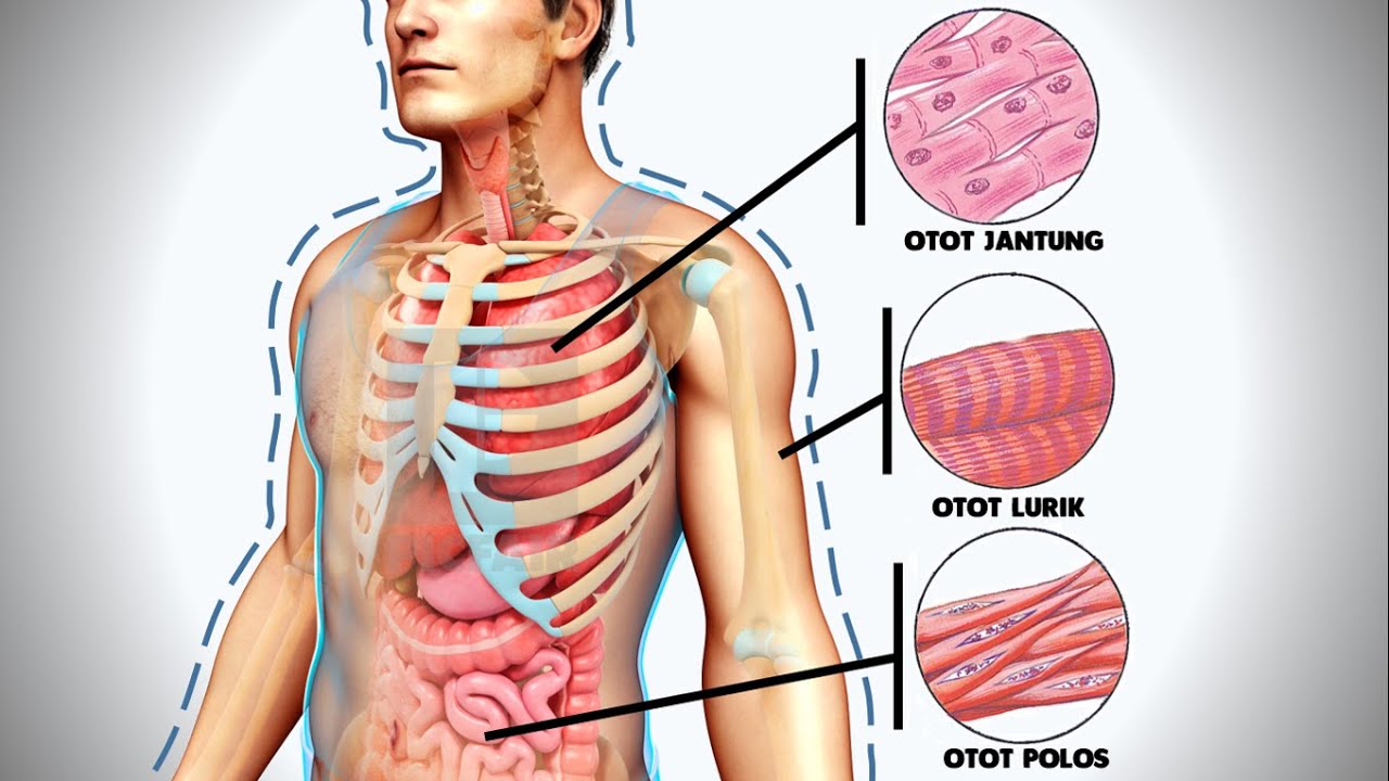


5.0 / 5 (0 votes)
