El CUERPO HUMANO, los huesos y músculos (para estudiar)🦴
Summary
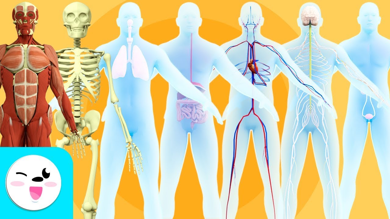
TLDREste vídeo ofrece una visión detallada del cuerpo humano, enfocándose en los sistemas esquelético y muscular. Se explica cómo los huesos y los músculos, junto con otros tejidos y órganos, trabajan en conjunto para permitir el movimiento y mantener la estructura corporal. Se detallan las funciones de los 206 huesos del esqueleto axial y appendicular, así como los tipos de músculos: esquelético, liso y cardíaco, cada uno con roles específicos en el movimiento, protección y otras funciones vitales.
Takeaways
- 💪 El cuerpo humano es un sistema biológico complejo compuesto de tejidos, órganos y sistemas que trabajan juntos para formar la estructura física.
- 🦴 Los huesos, junto con el cartílago, ligamentos, articulaciones y tendones, forman el sistema esquelético y tienen funciones mecánicas y protectoras.
- 🏃 Los músculos son tejidos especiales que pueden contraerse y relajarse para producir movimientos, y son esenciales para la movilidad y la postura.
- 🧠 La mayoría de los huesos protegen órganos frágiles como los pulmones, corazón y cerebro, mientras que otros en los brazos, piernas y dedos permiten el movimiento y el manejo de objetos.
- 🦴 Los huesos tienen una estructura que incluye la diáfisis y las epífisis, con la médula ósea en la cavidad medular y la médula roja en la espina ísquim.
- 🔍 Los huesos se clasifican en el esqueleto axial (cráneo, columna vertebral y tórax) y el esqueleto appendicular (extremidades y cinturas pélvicas).
- 💪 Los músculos, que forman el sistema muscular, están compuestos de células especiales llamadas fibras musculares que contienen elementos contractiles llamados miofibrillas.
- 🏋️♂️ Los músculos esqueléticos, que se unen a los huesos a través de tendones, son controlados voluntariamente y son esenciales para el movimiento.
- 🌀 Los músculos liso, presentes en órganos como los intestinos y las arterias, son involuntarios y controlados por el sistema nervioso autónomo.
- ❤️ El músculo cardíaco, que forma las cámaras del corazón, tiene contracciones involuntarias y es esencial para la circulación sanguínea.
Q & A
¿Cuál es la función principal del sistema esquelético en el cuerpo humano?
-El sistema esquelético tiene funciones mecánicas y protectoras, además de servir como punto de almacenamiento para minerales necesarios para el cuerpo, como el calcio, y produce las células que forman nuestra sangre.
¿Cuál es la diferencia entre el diafisis y las epífisis en un hueso?
-El diafisis es la región más larga de un hueso que abarca casi todo el hueso, compuesto de un tipo de hueso muy denso y compacto, cubierto por una membrana delgada llamada periostio, y contiene un espacio hueco llamado cavidad medular que alberga la médula ósea. Las epífisis, por otro lado, son las extremidades del hueso que no son huecos y tienen una estructura similar a una esponja llamada hueso esponjoso, donde se alberga la médula roja.
¿Cuál es la función de los huesos en la protección de órganos internos?
-Algunos huesos, como los de la cabeza, el pecho y la columna vertebral, protegen órganos frágiles como los pulmones, el corazón y el cerebro.
¿Cómo se clasifican los huesos en el cuerpo humano?
-Los huesos se clasifican en dos grandes grupos: el esqueleto axial, que incluye huesos de la cabeza, pecho y columna vertebral, y el esqueleto appendicular, que forma nuestros apéndices, es decir, los brazos, manos, piernas y pies.
¿Qué es el periostio y qué función cumple en los huesos?
-El periostio es una membrana delgada que cubre la parte externa del diafisis de los huesos y tiene la función de proteger y nutrir el hueso, además de ser el lugar donde se forman los ósteoblastos, las células que producen y reparan el hueso.
¿Qué son las células ósteoclastas y qué función tienen en los huesos?
-Las células ósteoclastas son responsables de romper y descomponer el hueso para darle forma o eliminar células dañadas de ellos.
¿Cuál es la función principal de los músculos en el cuerpo humano?
-Los músculos tienen la capacidad de contraerse y relajarse, lo que produce movimientos, y participan en procesos vitales como la circulación sanguínea, el movimiento de la comida y la respiración, así como la protección de órganos internos.
¿Qué tipos de músculos hay en el cuerpo humano y cuál es su función?
-Existen tres tipos de músculos: los músculos esqueléticos, que se unen a los huesos a través de tendones y son controlados voluntariamente; los músculos liso, que forman órganos huecados y son controlados involuntariamente; y los músculos del corazón, que forman las cámaras del corazón y también son controlados involuntariamente.
¿Cómo se forman los músculos y qué elementos tienen dentro para poder contraerse?
-Los músculos están formados por tejido muscular compuesto de células especiales que se contraen, llamadas fibras musculares, que contienen elementos contractiles llamados miofibrillas, hechas de fibras musculares contractiles como las proteínas miosina y actina.
¿Qué es la médula ósea y en qué cavidades del hueso se encuentra?
-La médula ósea es el tejido responsable de producir las células sanguíneas y se encuentra en la cavidad medular del diafisis y en la médula roja de las epífisis.
¿Cómo contribuyen los músculos a la función protectora del cuerpo humano?
-Los músculos ayudan a mantener la postura y a proteger los órganos internos de golpes y accidentes, al igual que el sistema esquelético.
Outlines
🦴 Introducción al cuerpo humano y sus componentes esenciales
El primer párrafo introduce el cuerpo humano como un sistema biológico complejo compuesto por tejidos, órganos y sistemas que interactúan para formar la estructura física. Se enfatiza la importancia de los huesos y los músculos en la formación y el funcionamiento del cuerpo, destacando su participación en movimientos y en la realización de funciones fisiológicas como la digestión y la circulación sanguínea. Se describe la composición de los huesos y cómo se organizan en el sistema esquelético, incluyendo su función protectora y su papel en la movilidad.
💪 Funciones y clases del sistema esquelético y muscular
El segundo párrafo profundiza en la estructura y clasificación de los huesos, distinguiendo entre el esqueleto axial y el esqueleto appendicular. Se explica la función de los huesos en la protección de órganos y en la movilidad, y se menciona la presencia de dos tipos de médula ósea en los huesos. Además, se introduce el sistema muscular, destacando su capacidad para contraerse y relaxarse para producir movimientos, y se mencionan los tipos de músculos presentes en el cuerpo humano.
🏃♂️ El papel de los músculos en el movimiento y la vitalidad del cuerpo
El tercer párrafo se centra en los músculos y su papel en el movimiento, la vitalidad y la protección de los órganos internos. Se describen los tipos de músculos: esquelético, liso y cardíaco, y se explica cómo cada uno desempeña funciones específicas en el cuerpo. Finalmente, se invita a los espectadores a seguir el canal para obtener más contenido educativo.
Mindmap
Keywords
💡Sistema Biológico
💡Tejos
💡Órganos
💡Sistemas
💡Esqueleto
💡Huesos
💡Diáfisis y Epífisis
💡Músculos
💡Músculo Esquelético
💡Músculo Liséo
💡Músculo Cardíaco
Highlights
El cuerpo humano es un sistema biológico complejo compuesto por tejidos, órganos y sistemas que trabajan juntos para formar la estructura física del ser humano.
Los tejidos se agrupan para formar órganos, como el corazón, los pulmones y el estómago, que cumplen funciones específicas.
Existen 13 sistemas en el cuerpo humano que trabajan en coordinación para mantener la vida.
Los dos sistemas no independientes que permiten el movimiento y la estructura son el sistema esquelético y el sistema muscular.
El sistema esquelético está compuesto por huesos, cartílagos, ligamentos, articulaciones y tendones, que desempeñan funciones mecánicas, estructurales y de protección.
Los huesos almacenan minerales necesarios como el calcio y producen células sanguíneas.
El sistema esquelético se divide en dos grupos: el esqueleto axial y el esqueleto apendicular.
El esqueleto axial incluye los huesos de la cabeza, el tórax y la columna vertebral, protegiendo órganos vitales como el cerebro, corazón y pulmones.
El esqueleto apendicular está compuesto por los huesos de los brazos, las piernas y las extremidades, lo que permite el movimiento y la locomoción.
El tejido muscular tiene la capacidad de contraerse y relajarse, lo que permite movimientos tanto voluntarios como involuntarios.
El músculo esquelético permite movimientos voluntarios, se une a los huesos a través de tendones y está controlado por el sistema nervioso somático.
Los músculos lisos forman los órganos viscerales huecos y son controlados involuntariamente por el sistema nervioso autónomo.
El músculo cardíaco está formado por fibras estriadas, pero sus contracciones son involuntarias, permitiendo el bombeo de sangre en el corazón.
Los músculos no solo permiten el movimiento, sino que también cumplen funciones vitales como la digestión, la circulación de la sangre y la respiración.
El sistema muscular, junto con el sistema esquelético, es responsable de mantener la postura y proteger los órganos internos.
Transcripts
Hello guys, I hope you are very well. On this occasion, we will dedicate a few minutes to studying
the human body and two of its important components: bones and muscles . The
human body is a complex biological system made up of a whole network of interacting tissues, organs, and systems.
and they work together to form the physical structure of a human being with stories the
members of the animal world in the human body all these structures are made
up of thousands of cells when many of these cells with the same characteristics are grouped
together to perform the same function they form a tissue the main types of tissues in
the human body are epithelial tissue, muscle tissue, connective tissue and nervous tissue
several of these tissues are grouped together to form even more complex and
independent structures called organs that carry out very particular functions Some
examples of them are the heart, lungs, and stomach, but he story does not end
here under our skin the organs do not work individually each one represents a piece
within an even more complex organization the systems therefore the latter result from the
grouping of a set of organs that have been organized to carry out together There are 13 functions
that a single organ could never perform. There are 13 systems that work in coordination to
keep a human being alive, but only two of them are the ones that have direct
internal participation, the characteristic shape and structure that it has in our body, in addition to allowing
us to carry out carry out movements and move from one place to another these non-independent systems
are the skeletal system made up of the bones and the muscular system made up of the muscles
but also some of the greenest and 150 muscles that we have participate in
many other physiological functions that involve the contraction and relaxation of the cells that make them
up such as digestion and pumping d e the blood to all the organs and tissues of the body
since we have seen in a very general way how the human body is constituted, let us see
separately the participation that the bones and muscles have in its structure and
functioning let us begin with the bones the bones are organs very rigid that together with
the cartilage, the ligaments, the joints and the tendons form the bone system of the hand of
all these structures carry out mechanical and protective structural functions in addition
to serving as a storage point for minerals necessary for the body such as
calcium and actively produce the cells that make up our blood many bones protect
the fragile organs of our body such as the lungs, heart and brain while
others such as those in the arms, legs and fingers allow us to move, touch and grasp objects
in the bones longer of our body as the femur of the thighs two regions
called d diaphysis and is difficult the diaphysis is the longest region that encompasses almost the entire bone
and the epiphyses correspond to the ends of the bone through which they join other bones
the diaphysis is made up of a very dense and compact type of bone that is covered on its
outer part by a very thin membrane called periodic inside it has a hollow space
that also has a membranous lining called in two this hollow space is known
as the medullary cavity since it houses the bone marrow, the tissue responsible for producing the cells that
make up our blood red blood cells in white blood cells on the contrary the epiphyses
are not hollow structures inside they have a structure similar to that of a sponge called
mirror bone here another type of bone marrow is housed known as red bone marrow
both structures They are made up of three types of cells: osteoblasts, which
make new bone and repair bone injuries, avionocytes or mature bone cells and
osteoclasts responsible for breaking and decomposing the bone to give it shape or remove
damaged cells from them well the 206 bones we have are classified into two large groups those that form
the axial skeleton and those that make up the appendicular skeleton let's see each group
separately, the social skeleton is made up of all those bones that are
part of the head, chest and spine, absolutely all of these bones perform functions
of protection of the organs that house inside the large structures that were
with the exception of of the spinal column which, in addition to this, plays a very
important role in maintaining posture the 80 bones that give life to this part of the
skeleton are distributed in three regions the head the spinal column and the thorax
the head houses the brain and the vast majority of our sensory organs the
eyes the ears the nose and the mouth which contains the ngua teeth and jaws so the
head is divided into three large areas the skull which is made up of eight bones that house the
brain and which fix the muscles of the face and neck the face made up of 14 bones that house
the eyes the nose and mouth and ear formed by the auditory bones malleus incus and stirrup the
second region of the axial skeleton is the spinal column it is made up of 26 bones called
vertebrae that protect the bone marrow supports the head the ribs and back muscles
and the neck, the vertebrae are divided into cervical, dorsal, or thoracic, lumbar, blood, and the coccyx,
for its part, the thoracic cavity or thorax, formed by 25 bones that
make up the ribs and the sternum, protects the heart and lungs,
and also serves to fix the diaphragm and the muscles of the neck of the chest and of the others
well now we continue with the bones that form the appendicular skeleton by name we
quickly imagine that it is those that They form our appendages, that is, the arms, the hands
, the legs and the feet, therefore they fulfill functions of movement and locomotion, this
skeleton is divided into two large regions, an upper one formed by sixty-two bones
that include the upper extremities and the shoulder girdle and a lower one
also formed by sixty-two bones that form the lower extremities in the pelvic girdle
well, since we have seen the most important aspects that define the bones and their
functions in the human body, now it is the turn of the muscles the muscles constitute a very special tissue
of our body since they have the ability to contract and relax thus producing
movements many of these movements can be voluntary or involuntary in the
same way that all the bones of our body come together to form the great bone
system almost 640 muscles that we have also do so to form the muscular system within
this great si stem each of the muscles that make it up carry out very specific functions
let's see some of the most important as I already told you they allow movement and locomotion which
differentiates us from other sensitive or immobile organisms such as plants they carry out
vital processes to our body such as pumping movement and conduction of blood
through the body movement of food and breathing participate in the movement of
our eyes and help us move our jaws to chew speak and show also control
creation processes such as defecation and urination and they protect and the internal organs
of our body from blows and accidents and from the hand of the skeletal system they help to maintain posture
let's see now quickly how the muscles are formed absolutely all the muscles
that make up the muscular system are made up of muscular tissue This tissue is
made up of specialized cells that contract and the place it is called 2 - and
they do this thanks to the fact that inside they house a series of contractile elements
called myofibrils that are made up of contractile protein fibers
called myofilaments there are two types of myofilaments the same thick filaments and the
same thin filaments my thick filaments are made up of the protein myosin
2 while the thin filaments are made up mainly of the proteins
fehr actin, troponin and tropero messin the thin filaments are read on the same
thick filaments shortening the length of the series this is how it happens the contraction of the world
let's now see the types of muscle that exist the skeletal muscle the smooth and the carbon
let's see what each one of them consists of and what functions the skeletal muscles have they are
those that join the bones of the skeleton through a species of connectors called
tendons are also called stellate muscles since they are formed by the union of
many my milestones that have many horizontal stripes that look like stretch marks when
observed under a microscope, these muscles are voluntary, that is, we can
consciously control the movements they make, so their contraction is under the action
of the somatic division of the peripheral nervous system, the smooth muscles are those that form
the hollow visceral organs of our body such as the intestines, the blood vessels, the
ureters and the uterus, they are also found in the iris of our eyes, they are called smooth
because their fibers muscle do not have microscopic striae is involuntary because
we cannot control their movement consciously therefore their action is controlled by the
autonomic nervous system in them their cells are arranged in longitudinal circular layers or in
a combination of both finally the heart muscle is which form the hollow
and contractile chambers of our heart its cells make involuntary contractions so they
are controlled by neurons of the autonomic nervous system just like
skeletal muscles are formed by the union of striated muscle fibers however these
are shorter and interconnect to favor the rapid propagation of the
electrical impulses that originate its rhythmic and energetic contraction which allows the entry
and exit of blood from our heart while on this good side guys this
has been all for this video I hope you have loved it so I invite you to like it and
to subscribe to our channel to continue enjoying our excellent programming
Ver Más Videos Relacionados
El TEJIDO MUSCULAR
Los sistemas del cuerpo humano para niños - Recopilación
El sistema muscular - Ciencias Naturales
El SISTEMA LOCOMOTOR / MUSCULOESQUELÉTICO video para primaria
Biología II. 05 Los principios estructurales y funcionales de los seres humanos
Sistema muscular: Tipos, Clasificación, Músculos principales - Anatomía Humana | Kenhub
5.0 / 5 (0 votes)