Chem 1307 Ch 19.3 Proteins - Secondary Structure

Dr. Veronica Gonzalez-EPCC
27 Jul 202217:32

Summary

TLDRThis script delves into the secondary structures of proteins, focusing on the alpha helix and beta pleated sheets, which are stabilized by hydrogen bonds. It explains how these structures are crucial for protein function, with the alpha helix being more polar and the beta sheet accommodating hydrophobic amino acids. The script also introduces the triple helix found in structural proteins like collagen. It connects the misfolding of beta-amyloid proteins from alpha helices to beta pleated sheets in Alzheimer's disease, illustrating the critical role of protein structure in health and disease.

Takeaways

  • 🧬 The secondary structure of proteins refers to the local folding patterns that occur due to hydrogen bonds between amino acids within the same or different peptide chains.
  • πŸŒ€ Two common types of secondary structures are the alpha helix and beta pleated sheets, which are crucial for the protein's function and stability.
  • 🌈 The alpha helix is characterized by hydrogen bonds between the carbonyl oxygen and the amide hydrogen of the amino acids in the next turn of the helix, forming a spiral staircase shape.
  • πŸ“œ Beta pleated sheets form when hydrogen bonds occur between carbonyl oxygen atoms and amide hydrogen atoms of different amino acids, creating a sheet-like structure.
  • 🌿 Hydrophobic amino acids are more common in beta pleated sheets, while hydrophilic or polar amino acids are more exposed in alpha helices due to their structural differences.
  • πŸ“Š Ribbon diagrams are used to represent the secondary structures of proteins, simplifying the visualization by showing the overall shape rather than individual atoms.
  • πŸ”— Hydrogen bonds are the primary force maintaining the secondary structures of proteins, such as in alpha helices and beta pleated sheets.
  • 🌟 Collagen, a structural protein, contains a triple helix, where three polypeptide chains are intertwined and stabilized by hydrogen bonds, providing strength to tissues like skin, tendons, and cartilage.
  • 🧠 Protein misfolding and aggregation, such as the conversion of beta amyloid proteins from alpha helices to beta pleated sheets in Alzheimer's disease, can lead to the formation of insoluble plaques and neuronal dysfunction.
  • πŸ’‘ The diagnosis of Alzheimer's disease is typically confirmed post-mortem by examining brain slices for atrophy and the presence of neurofibrillary tangles affecting nerve cell transmission.
  • πŸ›‘ Neurons do not regenerate quickly, and the death of nerve cells due to protein aggregation and plaque formation in Alzheimer's leads to memory loss and loss of motor function.

Q & A

  • What is the secondary structure of a protein?

    -The secondary structure of a protein refers to the local folding patterns of the peptide chain, which are stabilized by hydrogen bonds between the backbone atoms. It includes common structures such as the alpha helix and beta pleated sheets.

  • What are the two most common types of secondary structures in proteins?

    -The two most common types of secondary structures in proteins are the alpha helix and beta pleated sheets.

  • How are alpha helices formed in proteins?

    -Alpha helices are formed by hydrogen bonds between the oxygen of the carbonyl groups and the hydrogen of the NH groups of the amide bonds in the next turn of the helix, which gives the peptide chain a helical shape.

  • What interactions are responsible for the formation of beta pleated sheets in proteins?

    -Beta pleated sheets are formed by hydrogen bonds between the carbonyl oxygen atoms and the hydrogen atoms in the amide group, which bend the polypeptide chain into a sheet-like structure.

  • What is the difference between alpha helices and beta pleated sheets in terms of amino acid exposure?

    -Alpha helices tend to have more polar or hydrophilic amino acids exposed due to their tight interaction, while beta pleated sheets may have more hydrophobic amino acids tucked away from the aqueous environment.

  • What is a triple helix and where is it commonly found?

    -A triple helix is a secondary structure found in structural proteins like collagen, where three polypeptide chains are woven together with hydrogen bonds holding the chains together, providing additional strength.

  • How does the representation of proteins change when using a ribbon diagram?

    -In a ribbon diagram, the protein is represented with a general structure rather than individual atoms, showing the secondary structures like alpha helices and beta pleated sheets as ribbons, which simplifies the visualization of large proteins.

  • What is the significance of the N-terminus and C-terminus in the context of protein structure?

    -The N-terminus and C-terminus are the beginning and end of a protein chain, respectively. They can be identified in protein diagrams to understand the directionality of the amino acid chain and are important for the overall three-dimensional structure.

  • How can changes in protein secondary structure lead to disease?

    -Changes in protein secondary structure, such as the conversion of alpha helices into beta pleated sheets in Alzheimer's disease, can cause proteins to misfold and aggregate, leading to the formation of insoluble plaques that disrupt normal cellular functions and contribute to disease pathology.

  • What is the role of beta amyloid proteins in Alzheimer's disease?

    -In Alzheimer's disease, beta amyloid proteins change from their normal alpha helical shape to beta pleated sheets, forming insoluble aggregates that accumulate as plaques, which are implicated in the disruption of neuronal signaling and contribute to the disease's progression.

  • How can the diagnosis of Alzheimer's disease be confirmed?

    -The official diagnosis of Alzheimer's disease can only be done post-mortem by examining brain slices for atrophy and the presence of neurofibrillary tangles. However, symptoms like memory loss and loss of motor function can be used for an unofficial diagnosis.

Outlines

00:00

πŸŒ€ Secondary Structure of Proteins

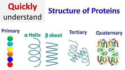
This paragraph introduces the concept of protein secondary structure, which is the pattern that arises from hydrogen bonds between atoms in the amino acid backbone. It explains that these interactions are localized and not covalent bonds. The two most common types of secondary structures are the alpha helix and beta pleated sheets, with a brief mention of the triple helix found in structural proteins like collagen. The alpha helix is characterized by hydrogen bonds between the carbonyl oxygen and the hydrogen of the amide bond, forming a spiral staircase shape. The paragraph also discusses the representation of these structures using ribbon diagrams and the significance of hydrogen bonds in forming the alpha helix.

05:00

πŸ“œ Beta Pleated Sheets and Hydrophobic Interactions

This paragraph delves into the secondary structure of beta pleated sheets, highlighting the formation of hydrogen bonds between carbonyl oxygen atoms and hydrogen atoms in the amide group of different amino acids, resulting in a sheet-like structure. It contrasts the beta pleated sheets with alpha helices, noting the presence of hydrophobic amino acids in the former and hydrophilic or polar amino acids in the latter. The paragraph also describes the use of ribbon diagrams to represent the secondary structures in proteins, with arrows indicating the direction of the amino acid chain from the N-terminus to the C-terminus.

10:03

πŸ” Triple Helix and Protein Misfolding in Disease

This paragraph discusses the triple helix, another type of secondary structure found in structural proteins such as collagen. It explains that the triple helix is formed by three polypeptide chains held together by hydrogen bonds, providing strength to connective tissues. The paragraph then connects protein structure to disease, using Alzheimer's disease as an example. It describes how beta-amyloid proteins, normally in an alpha-helical form, can misfold into beta pleated sheets, leading to the formation of insoluble plaques that contribute to neuronal dysfunction and death. The paragraph emphasizes the irreversible nature of neuronal loss and the impact on memory and motor functions.

15:04

🧠 Alzheimer's Disease and Protein Aggregation

The final paragraph focuses on the role of protein misfolding in Alzheimer's disease. It describes the transformation of beta-amyloid proteins from a normal alpha-helical shape to insoluble beta-pleated sheets that aggregate into plaques, both inside and outside neurons. These plaques disrupt neuronal signaling and contribute to cell death and brain atrophy. The paragraph explains that while the official diagnosis of Alzheimer's can only be made post-mortem by examining brain tissue for neurofibrillary tangles, symptoms such as memory loss and motor function decline can be used for an unofficial diagnosis. The summary concludes by reiterating the importance of understanding protein secondary structures in the context of disease.

Mindmap

Keywords

πŸ’‘Secondary Structure

The secondary structure of a protein refers to the local folding of its peptide chains into specific conformations, primarily stabilized by hydrogen bonds. This concept is central to the video as it sets the foundation for understanding how proteins achieve their functional shapes. Examples from the script include the alpha helix and beta pleated sheets, which are the most common types of secondary structures.

πŸ’‘Alpha Helix

An alpha helix is a right-handed coiled structure formed by hydrogen bonds between the oxygen of the carbonyl group and the hydrogen of the amide bond in the next turn of the helix. It is a key secondary structure in proteins and is mentioned in the script as a twisted structure that contributes to the protein's overall shape and function.

πŸ’‘Beta Pleated Sheets

Beta pleated sheets are a type of secondary structure where hydrogen bonds form between the carbonyl oxygen atoms and the hydrogen atoms in the amide group, bending the polypeptide chain into a sheet. The script describes these as a significant component of protein structure, often containing more hydrophobic amino acids compared to alpha helices.

πŸ’‘Hydrogen Bonds

Hydrogen bonds are a type of intermolecular force that plays a crucial role in stabilizing the secondary structure of proteins. In the script, hydrogen bonds are described as the main interaction between amino acids in both alpha helices and beta pleated sheets, contributing to their respective shapes.

πŸ’‘Globular Proteins

Globular proteins are compact, roughly spherical proteins that perform specific functions. The script mentions these proteins in the context of having secondary structures like alpha helices and beta pleated sheets, which contribute to their overall three-dimensional shape and function.

πŸ’‘Tertiary Structure

The tertiary structure of a protein refers to its three-dimensional shape resulting from the folding of its peptide chain, which includes both secondary structures and additional interactions. The script discusses how the combination of alpha helices and beta pleated sheets contributes to the formation of the tertiary structure.

πŸ’‘Triple Helix

A triple helix is a type of secondary structure found in proteins like collagen, where three polypeptide chains are intertwined and held together by hydrogen bonds. The script mentions the triple helix as a unique structure that provides added strength to structural proteins.

πŸ’‘Aqueous Environment

An aqueous environment refers to a water-based medium where many biological processes, including protein folding, occur. The script discusses how the presence of water influences the distribution of hydrophobic and hydrophilic amino acids in protein structures like alpha helices and beta pleated sheets.

πŸ’‘Ribbon Diagram

A ribbon diagram is a simplified representation of protein structure where the peptide backbone is depicted as a ribbon. The script uses this term to describe how proteins are often visualized, focusing on the secondary structures like alpha helices and beta pleated sheets rather than individual atoms.

πŸ’‘Alzheimer's Disease

Alzheimer's disease is a neurodegenerative disorder characterized by memory loss and cognitive decline. The script discusses how the misfolding of beta amyloid proteins from an alpha helical shape to beta pleated sheets contributes to the formation of insoluble plaques, which are a hallmark of Alzheimer's disease.

πŸ’‘Neurofibril Tangles

Neurofibril tangles are abnormal structures found in the neurons of individuals with Alzheimer's disease. The script mentions these tangles in the context of post-mortem diagnosis, where they are observed alongside brain atrophy and the presence of beta amyloid plaques.

Highlights

The secondary structure of a protein is defined by the hydrogen bonds between atoms in the amino acid backbones, forming local structures.

Two common types of secondary structures are the alpha helix and beta pleated sheets, with the former being common in globular proteins and the latter in structural proteins.

Collagen, a structural protein, contains a triple helix, which is another type of secondary structure.

The alpha helix is characterized by hydrogen bonds between the carbonyl oxygen and the hydrogen of the amide bond in the next turn of the helix.

Hydrogen bonds in the alpha helix give it a helical, spiral staircase shape.

Ribbon diagrams are used to represent proteins, simplifying the visualization by showing secondary structures without individual atoms.

Beta pleated sheets form when hydrogen bonds occur between carbonyl oxygen atoms and hydrogen atoms in different regions of the polypeptide chain.

Hydrophobic amino acids are more common in beta pleated sheets, while hydrophilic or polar amino acids are more exposed in alpha helices.

The direction of the amino acid chain in beta pleated sheets is indicated by arrows in ribbon diagrams.

The triple helix in structural proteins like collagen is formed by three polypeptide chains held together by hydrogen bonds, providing strength.

Hydrogen bonds are the dominant force in the triple helix, similar to the alpha helix.

Protein malfunction or structural issues can lead to diseases, such as Alzheimer's, where protein secondary structures are affected.

In Alzheimer's disease, beta amyloid proteins change from alpha helices to insoluble beta pleated sheets, forming plaques that disrupt neuronal function.

Neurofibril tangles in neurons, associated with Alzheimer's, affect nerve cell transmission and contribute to the disease's progression.

The diagnosis of Alzheimer's can only be confirmed post-mortem by examining brain slices for atrophy and the presence of neurofibril tangles.

Unofficial diagnosis of Alzheimer's is based on symptoms like memory loss and loss of motor function.

The transition of beta amyloid proteins from alpha helices to beta pleated sheets in Alzheimer's leads to the formation of plaques and neuronal death.

Transcripts

play00:00

in the next section of this chapter we

play00:02

will discuss the secondary structure of

play00:05

proteins the secondary structure of a

play00:07

protein describes the structure that

play00:09

forms when amino acids form hydrogen

play00:12

bonds between the atoms in their

play00:14

backbone and the atoms on the same or

play00:17

another peptide chain so basically you

play00:20

have some interactions between amino

play00:23

acids now we're not talking about the

play00:25

covalent linkage between amino acids

play00:27

we've already established that but we're

play00:30

looking at now interactions between

play00:34

different amino acids in different

play00:36

regions but those regions are pretty

play00:38

localized two common types include the

play00:40

alpha helix and the beta pleated sheets

play00:43

and of course we're looking at the

play00:45

globular proteins or those proteins that

play00:48

carry out very particular or specific

play00:50

functions the structural proteins might

play00:53

have something called a triple helix and

play00:55

we'll see that a little bit later so in

play00:58

this lesson we're going to describe the

play01:00

two most common types of secondary

play01:02

structure for a protein the helix and

play01:06

the beta pleated sheets right and then

play01:08

we're going to describe the structure of

play01:10

collagen collagen contains the triple

play01:12

helix

play01:14

because it is a structural protein

play01:17

in the diagram to the right you will see

play01:19

an example of the alpha helix

play01:22

the baited pleated sheets and then they

play01:25

combine to create a functional protein

play01:29

this is more of a three-dimensional

play01:31

structure right our tertiary structure

play01:34

so we're going to discuss these first

play01:36

two which will interact later right to

play01:40

create the tertiary structure

play01:43

let's start by describing the

play01:45

interactions that occur between the

play01:47

amino acids when we have an alpha helix

play01:50

in our protein

play01:52

in the secondary structure of an alpha

play01:55

helix hydrogen bonds form between the

play01:58

oxygen of the carbonyl groups and the

play02:01

hydrogen of nh groups of the amide bonds

play02:04

in the next turn of the alpha helix so

play02:08

the main intermolecular force that is

play02:11

occurring between localized amino acids

play02:14

for the alpha helix and this kind of

play02:18

twisted structure

play02:19

is the hydrogen bond okay so hydrogen

play02:23

bonds are the most significant

play02:26

interaction between amino acids to form

play02:29

the alpha helix the formation of many

play02:31

hydrogen bonds along the peptide chain

play02:34

gives a helical shape of a spiral

play02:37

staircase okay so this basically this

play02:41

little twisted

play02:43

region is going to be our alpha helix

play02:46

they draw what's called the ribbon

play02:48

diagram where those amino acids are kind

play02:51

of just drawn as

play02:54

you know like solid

play02:56

ribbons and over here on the side here

play02:59

you start to see the individual atoms

play03:02

and the individual amino acids

play03:04

the reason why we draw ribbon diagrams

play03:07

is because when proteins are very large

play03:10

they actually have a ton of atoms so

play03:12

it's a little bit hard to distinguish

play03:14

between amino acids anyways but you will

play03:18

see different uh representations of a

play03:21

protein so you have your molecules right

play03:25

and then the ribbon diagram showing your

play03:28

alpha helix

play03:30

let's take a quick look at the actual

play03:33

hydrogen bond so

play03:35

here it mentions that you have an oxygen

play03:38

of the carbonyl that is going to

play03:40

hydrogen bond

play03:42

with the hydrogen of the next amino

play03:45

acids um amide bond right so here is our

play03:50

amide bond which is the carbon of the

play03:52

carbonyl

play03:54

from the previous amino acid bonded to

play03:56

the nitrogen of the ammonium region

play03:59

right and so this nitrogen from the next

play04:03

amino acid is going to interact well not

play04:06

the nitrogen itself but the hydrogen

play04:09

which is partially positive is going to

play04:11

interact with the partially negative

play04:14

oxygen of the carbonyl and of course we

play04:18

said that the interaction is via

play04:20

hydrogen bonding

play04:22

so let's take a look at the beta pleated

play04:24

sheets in the secondary structure of a

play04:27

beta pleated sheet hydrogen bonds form

play04:30

between the carbonyl oxygen atoms and

play04:32

the hydrogen atoms in the amide group

play04:35

bending the polypeptide chain into a

play04:38

sheet

play04:39

so let's take a look at that interaction

play04:41

so you have your peptide chain here and

play04:45

so of course this peptide chain is

play04:48

starting to fold right and we said that

play04:50

the fold is happening in localized

play04:53

regions so this carbonyl oxygen is going

play04:57

to hydrogen bond to

play05:00

the hydrogen of the nitrogen

play05:02

participating in the amide bond of a

play05:06

different amino acid in a different

play05:08

region not necessarily

play05:10

right next to it right like the actual

play05:12

peptide bond so remember these are only

play05:16

interactions not covalent linkages

play05:18

so we have our hydrogen bond there

play05:22

the difference between the secondary

play05:23

structure of the beta plated sheets and

play05:26

those of the alpha helices is the

play05:28

presence of hydrophobic amino acids if

play05:32

you have hydrophobic amino acids they

play05:35

probably want to be tucked away in this

play05:38

chain here right away from the water so

play05:42

for beta plated sheets you'll probably

play05:44

see more hydrophobic amino acids than in

play05:48

the alpha helices for the alpha helix

play05:51

you'll see

play05:53

a tight interaction so more polar right

play05:56

more hydrogen bonds but also you will

play06:00

see that there is a lot of amino acids

play06:03

out here that are going to be exposed

play06:06

to the water right we're in an aqueous

play06:08

environment so you'll probably see more

play06:12

hydrophilic amino acids or more polar

play06:16

amino acids in the

play06:18

alpha helices

play06:22

so as we previously mentioned we can

play06:25

represent the structure of a protein

play06:27

using the ribbon model or the ribbon

play06:30

diagram the most common

play06:33

features of the ribbon diagram are the

play06:35

secondary structures including the alpha

play06:38

helix right you don't see individual

play06:41

atoms anymore only a general structure

play06:45

right so it's like if i zoomed out and

play06:47

kind of looked at the molecule by

play06:50

zooming out you just kind of see

play06:51

everything combined together creating

play06:54

this organization so you'll see the

play06:57

ribbon model um for the alpha helix

play07:00

right and then also for the beta pleated

play07:03

sheets for the beta pleated sheets they

play07:06

show you arrows to represent the

play07:08

direction of the amino acid chain so

play07:11

this is the n-terminus and then we're

play07:13

going this way to the c terminus

play07:17

and then some of these will have many

play07:19

beta plated sheets until you get to the

play07:23

c terminus right so once you start to

play07:26

kind of put this together or the protein

play07:28

itself starts to put itself together

play07:30

you'll see many beta plated sheets and

play07:33

alpha helices

play07:35

so a protein with alpha helices and beta

play07:38

pleated sheets is found here

play07:40

you can look for the n terminal usually

play07:44

they will label it for you

play07:46

and it will be in one of these

play07:49

non

play07:51

helical or

play07:53

sheeted

play07:54

regions so they're just kind of

play07:56

represented by the strand itself and you

play07:59

will also be able to find the c terminus

play08:03

the end of the protein they're not

play08:06

labeled in this particular diagram and

play08:08

it's really hard to see the c and the n

play08:12

terminus now if you're looking at the

play08:14

three-dimensional structure you might be

play08:16

able to um use a program that shows the

play08:20

three-dimensional structure that will

play08:22

allow you to rotate the molecule to find

play08:24

the beginning and the end of the chain

play08:27

or also help you to kind of see what's

play08:30

on the back here well we know it's a

play08:31

beta pleated sheet but what's the

play08:33

direction of that strand in the back

play08:36

right that sheet so you can use some

play08:40

different programs to help you visualize

play08:43

in three dimensions and basically kind

play08:45

of turn the molecule around

play08:48

another type of secondary structure

play08:50

includes the triple helix we talked a

play08:53

little bit about how the triple helix

play08:55

was present in a lot of structural

play08:57

proteins so in a triple helix three

play09:01

polypeptide chains are woven together

play09:04

hydrogen bonds hold the chains together

play09:07

giving the polypeptide the added

play09:09

strength of typical structural proteins

play09:12

like collagen connective tissue skin

play09:16

tendons and cartilage

play09:18

so here you have kind of like the

play09:20

winding of the three polypeptide chains

play09:24

right it's starting to wind and then

play09:26

over here it's really gotten tight

play09:29

collagen fibers are triple helices of

play09:31

polypeptide chains held together by

play09:33

hydrogen bonds so of course just like in

play09:37

the alpha helix which only involves one

play09:39

strand the main type of interaction

play09:42

among amino acids

play09:45

in different regions right here you have

play09:47

three different chains interacting

play09:50

with one another through hydrogen bonds

play09:53

so the hydrogen bond is the dominant

play09:55

force here

play09:57

so let's try this learning check

play09:59

indicate the type of protein structure

play10:03

in the primary

play10:06

sequence the alpha helix the beta plated

play10:10

sheet and the triple helix

play10:13

okay so letter a polypeptide chains held

play10:17

side by side by hydrogen bonds

play10:20

sequence of amino acids in a polypeptide

play10:23

chain

play10:24

corkscrew shape with hydrogen bonds

play10:27

between amino acids and three peptide

play10:30

chains woven like a rope okay so let's

play10:33

match the

play10:35

type of structure to the description so

play10:38

polypeptide chains held side by side by

play10:41

hydrogen bonds so this is going to be

play10:44

our beta pleated sheets right

play10:47

side by side to create those um sheets

play10:51

so here we're going to say this is a

play10:55

right

play10:56

and then the sequence of amino acids in

play10:58

a polypeptide chain this is going to be

play11:01

the primary sequence

play11:05

and then the corkscrew shape with

play11:07

hydrogen bonds between amino acids

play11:10

that's the alpha helix so this is c and

play11:14

then of course the three polypeptide

play11:16

chains woven like a rope is the triple

play11:19

helix

play11:20

let's see if we were correct

play11:22

okay so the polypeptide chains held side

play11:25

by side by hydrogen bonds

play11:28

are in fact the beta pleated sheets the

play11:31

sequence of amino acids in a polypeptide

play11:33

chain is the primary structure of the

play11:36

protein a corkscrew shape with hydrogen

play11:40

bonds between amino acids is the alpha

play11:42

helix and then the three peptide chains

play11:45

woven like a rope is the triple helix

play11:49

in the beginning of our lessons on

play11:51

proteins we talked a little bit about

play11:54

how

play11:55

any time a disease state is occurring we

play11:58

will look to see if there is a protein

play12:01

involved a lot of the time you have a

play12:03

malfunction of a protein or a structural

play12:06

problem with that protein which causes

play12:09

it to malfunction

play12:11

let's take a look at an example of a

play12:14

protein secondary structure and how this

play12:17

can lead to disease

play12:19

alzheimer's disease is a form of

play12:21

dementia in which a person has

play12:23

increasing memory loss and inability to

play12:26

handle daily tasks

play12:28

so how does this come about how does

play12:31

this disease arise

play12:33

in the brain of a normal person small

play12:36

beta amyloid proteins made up of 42

play12:38

amino acids exist in the alpha helical

play12:42

form

play12:43

in the brain of a person with

play12:45

alzheimer's disease the beta amyloid

play12:48

proteins change shape from the normal

play12:50

alpha helices which are soluble

play12:53

to the sticky beta pleated sheets which

play12:57

are insoluble right they form clusters

play13:00

of insoluble protein fragments called

play13:03

plaques

play13:04

so these plaques continue to build until

play13:08

eventually the neuron can't handle it so

play13:11

one they can't pass the neuronal signal

play13:15

and two they eventually die

play13:17

neuron cells don't replenish themselves

play13:20

quickly like say skin cells right skin

play13:23

cells will regenerate quite easily and

play13:27

quite quickly

play13:28

however the neurons don't so once those

play13:31

nerve cells die they don't come back so

play13:35

a lot of the regions that are affected

play13:37

by these

play13:39

by this disease

play13:41

and the beta amyloid protein problem is

play13:44

the memory region right the hippocampus

play13:46

of the brain and other motor regions

play13:49

right those that are responsible for our

play13:53

normal functions so somebody that dies

play13:56

of alzheimer's will actually die because

play13:59

their motor functions are not working

play14:01

for example they can no longer breathe

play14:04

because the nerves aren't firing to send

play14:06

the signal to tell the body to breathe

play14:09

or to swallow or to digest and so all of

play14:13

those body functions are starting to

play14:16

shut down

play14:17

unfortunately

play14:19

the diagnosis of alzheimer's

play14:22

can only be done post-mortem because

play14:24

slices of the brain must be taken and

play14:27

studied and basically we look at the

play14:30

atrophy or the brain kind of shrinking

play14:33

or starting to die

play14:35

and then we look for the presence of

play14:37

neurofibril tangles

play14:40

in the neurons that affect the

play14:41

transmission of the nerve cells so again

play14:45

the official diagnosis can only be done

play14:48

postmortem but other

play14:51

symptoms can be used to unofficially

play14:53

diagnose someone as having alzheimer's

play14:56

disease like memory loss

play14:58

and loss of motor function

play15:01

so let's take a look at these diagrams

play15:04

here you have a normal or a healthy

play15:09

beta amyloid that is found in its alpha

play15:12

helical form

play15:14

but with somebody that has alzheimer's

play15:17

disease or that's in a disease state

play15:19

these alpha helices are not necessarily

play15:23

folded correctly and instead they are

play15:27

converted into the beta pleated sheets

play15:30

so here's our diseased

play15:34

state right the alzheimer's

play15:37

and of course we said that now that

play15:40

alpha helix has been converted into beta

play15:44

pleated sheets which create these large

play15:48

plaques the large plaques will prevent

play15:51

the nerve impulse from firing from

play15:54

neuron to neuron right so here you have

play15:56

your healthy

play15:58

neuron

play16:00

if you look closely at the diseased

play16:02

state here the cell contains not only

play16:06

the plaques inside the cell right but

play16:10

you also have a buildup of these beta

play16:13

amyloid plaques outside the cell so it

play16:16

affects transmission in two ways so it's

play16:18

blocking transmission here so receiving

play16:21

the signal from the previous cell

play16:23

but also you have way too much stuff in

play16:27

here and these large tangles that are

play16:30

found within the cell um are actually

play16:33

going to prevent the transmission as

play16:35

well and again eventually these cells

play16:38

will die because they can't handle all

play16:40

of those um beta plated sheets right

play16:43

those plaques that are formed

play16:46

those cells don't regenerate quite

play16:48

quickly and so then you start to have

play16:52

atrophy of the brain

play16:54

memory loss and of course motor

play16:56

functions are also lost

play16:59

in patients with alzheimer's disease

play17:01

beta amyloid proteins change from a

play17:04

normal alpha helical shape to beta

play17:07

pleated sheets that are insoluble

play17:10

aggregates they form plaques right they

play17:12

aggregate together they stick to one

play17:14

another

play17:16

and of course these plaques remain in

play17:18

the in the neuron and of course

play17:20

eventually the neuron will die

play17:22

so this concludes our lesson on the

play17:24

secondary structure of a protein in the

play17:28

next lesson we'll talk about the

play17:29

tertiary structure

Rate This
β˜…
β˜…
β˜…
β˜…
β˜…

5.0 / 5 (0 votes)

Related Tags
Protein StructureSecondary StructureAlpha HelixBeta Pleated SheetsHydrogen BondsGlobular ProteinsStructural ProteinsTriple HelixCollagenAlzheimer's DiseaseProtein Misfolding