Respiratory System Anatomy (v2.0)
Summary
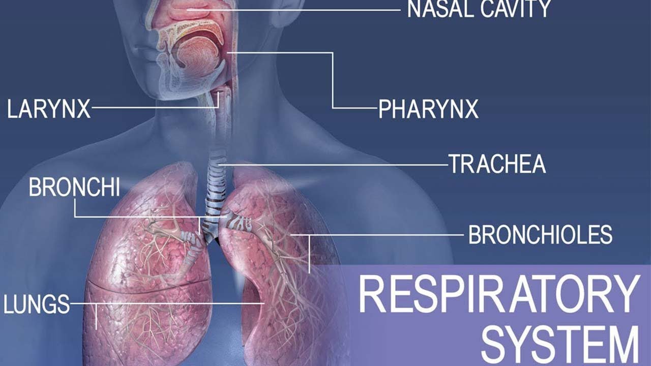
TLDRThe respiratory system is essential for oxygen intake and carbon dioxide release, divided into upper and lower tracts. The upper tract includes the nasal cavity with conchae for air processing, the pharynx with three parts, and the larynx with the epiglottis and vocal cords. The lower tract comprises the trachea, bronchi, and lungs, where the trachea splits into bronchi and further into bronchioles leading to alveoli for gas exchange. The alveoli contain type I and II pneumocytes, with the latter secreting surfactant to prevent lung collapse. The lungs are protected by pleural membranes and have fissures for structural division.
Takeaways
- π The respiratory system is divided into upper and lower tracts, responsible for taking in oxygen and releasing carbon dioxide.
- π The upper respiratory tract includes the nasal cavity, pharynx, and larynx, which are essential for filtering, warming, and humidifying the air.
- π The nasal cavity features conchae or turbinates that create turbulent airflow for efficient heating, cleaning, and humidifying of air.
- π The pharynx is divided into three parts: nasopharynx, oropharynx, and laryngopharynx, each serving a distinct role in the respiratory process.
- π£οΈ The larynx, located below the hyoid bone, contains the thyroid and cricoid cartilages, and is critical for sound production and airway protection.
- π« The epiglottis in the larynx acts as a flap to prevent food and liquid from entering the airway during swallowing.
- π€ The vocal cords, both true and false, are crucial for sound production, with the true cords producing sound and the false cords providing additional support.
- πΏ The trachea, supported by rings of hyaline cartilage, splits into primary bronchi at the carina and is lined with cells that help clean the respiratory tract.
- π¨ The bronchi further divide into smaller passages, eventually leading to bronchioles and alveoli, where gas exchange occurs.
- π«οΈ Alveoli, the functional units of the lungs, contain type I and II pneumocytes; the latter secretes surfactant to prevent lung collapse.
- π The lungs are structured into lobes, with the right lung having three lobes and the left lung having two, adapted to the space occupied by the heart.
Q & A
What are the two main sections of the respiratory system?
-The respiratory system is divided into the upper respiratory tract and the lower respiratory tract.
What are the components of the upper respiratory tract?
-The upper respiratory tract consists of the nasal cavity, pharynx, and larynx.
What is the function of the nasal conchae?
-The nasal conchae create turbulent airflow for heating, cleaning, and humidifying the air.
How many parts are there in the pharynx and what are they?
-There are three parts of the pharynx: the nasopharynx, oropharynx, and laryngopharynx.
What is the role of the epiglottis in the respiratory system?
-The epiglottis closes during swallowing to protect the airway from food and liquids.
What are the true and false vocal cords, and what is their function?
-The true vocal cords produce sound, while the false vocal cords, or vestibular folds, are membranes that extend from the arytenoid cartilages.
How does the trachea contribute to the respiratory system?
-The trachea splits at the carina into two primary bronchi and contains rings of hyaline cartilage that allow for flexibility.
What is the function of the ciliated pseudostratified columnar epithelium in the trachea?
-The cilia in the trachea work to clean the respiratory tract by moving debris toward the pharynx.
Describe the structure and function of the alveolus in the lungs.
-The alveolus is the functional unit of the lung where gas exchange occurs, containing type I pneumocytes for gas exchange and type II pneumocytes that secrete surfactant.
How many lobes does the right lung have and what are they?
-The right lung has three lobes: superior, middle, and inferior lobes.
What is the purpose of the pleural cavity and the fluid within it?
-The pleural cavity contains fluid that reduces friction and helps keep the lungs inflated by sticking to them.
Outlines
π« Overview of the Respiratory System
The respiratory system is responsible for inhaling oxygen and exhaling carbon dioxide. It is divided into the upper and lower respiratory tracts. The upper tract includes the nasal cavity, pharynx, and larynx, while the lower tract comprises the trachea, bronchi, and lungs. The nasal cavity features conchae that help in filtering, warming, and humidifying the air. The pharynx has three parts: nasopharynx, oropharynx, and laryngopharynx. The larynx, located below the hyoid bone, contains the thyroid and cricoid cartilages and features the epiglottis, which protects the airway during swallowing. The vocal cords within the larynx are responsible for sound production.
Mindmap
Keywords
π‘Respiratory System
π‘Upper Respiratory Tract
π‘Nasal Cavity
π‘Conchae
π‘Pharynx
π‘Larynx
π‘Trachea
π‘Bronchi
π‘Alveolus
π‘Pleura
π‘Surfactant
Highlights
The respiratory system is essential for taking in oxygen and releasing carbon dioxide.
It is divided into the upper and lower respiratory tracts.
The upper respiratory tract includes the nasal cavity, pharynx, and larynx.
The lower respiratory tract consists of the trachea, bronchi, and lungs.
The nasal cavity contains conchae or turbinates for air processing.
Conchae are bony processes that create turbulent airflow for heating, cleaning, and humidifying air.
The pharynx is divided into three parts: nasopharynx, oropharynx, and laryngopharynx.
The larynx contains the thyroid cartilage and the epiglottis, which protects the airway during swallowing.
The vocal cords, both true and false, are located in the larynx and are crucial for sound production.
The trachea is lined with ciliated pseudostratified columnar epithelium and mucous-secreting goblet cells.
The trachea splits into primary bronchi at the carina.
Bronchi further divide into secondary, tertiary bronchi, and bronchioles.
The alveolus is the functional unit of the lung where gas exchange occurs.
Type I and II pneumocytes are present in the alveolus for gas exchange and surfactant secretion.
Surfactant reduces surface tension to prevent lung collapse.
The right lung has three lobes, while the left lung has two.
The lungs have fissures that separate lobes and contain structures like the cardiac impression and hilum.
The pleural membranes include the visceral pleura and parietal pleura, with a pleural cavity in between.
The pleural cavity contains fluid that reduces friction and helps keep the lungs inflated.
Transcripts
Lets learn about respiratory system anatomy.
The respiratory system takes in oxygen for our cells and releases carbon dioxide.
It is divided into 2 large sections.
The upper respiratory tract consists of the nasal cavity, pharynx and larynx.
The lower respiratory tract includes the trachea, bronchi and lungs.
The nasal cavity contains bony processes called conchae or turbinates.
There are superior, middle and inferior conchae.
The superior and middle conchae are processes on the ethmoid bone.
The inferioir conchae extend from the maxillary bone.
The Nasal conchae create tubulent airflow for heating, cleaning and humidifying the
air.
Air passes from the nasal cavity to the pharynx.
There are 3 parts of the pharynx.
The nasopharynx extends to the end of the soft palate, the oropharynx extends from the
soft palate to the epiglottis and the laryngopharynx from the epiglottis to the glottis.
The larynx extends below the hyoid bone and contains the large thyroid cartilage or adams
apple.
Just below the thyroid cartilage is the cricoid cartilage.
Here is a posterior view of the larynx.
We can see a cartilage flap called the epiglottis.
The epiglottis closes when swallowing to protect the airway.
We can also see the vocal cords.
The true vocal cords produce sounds.
The false vocal cords or vestibular folds are membranes extending from the arytenoid
cartilages.
Here is a close up of the vocal cords.
We can see the true and false vocal cords along with a triangular space called the glottis
which forms when the vocal cords are relaxed.
Air moves from the larynx to the trachea.
The trachea splits at an area called the carina into 2 primary bronchi.
The trachea contains rings of hyaline cartilage.
The rings are open in the back to allow for some flexibility.
The primary bronchi split into secondary bronchi which divide into tertiary bronchi which again
divide into smaller passages called bronchioles and terminal bronchioles.
The trachea is lined with ciliated pseudostratified columnar epithelium and mucous secreting goblet
cells.
The cilia work to clean the respiratory tract by moving debris toward the pharynx.
Air from the tiny respiratory bronchioles reaches the alveolar ducts and flows into
the alveolus..the functional unit of the lung where gas exchange occurs.
The alveolus contains type I pneumocytes.
where gas exchange occurs.
It also contains type II pneumocytes that secrete surfactant.
Surfactant is a substance that reduces surface tension to keep the lungs from collapsing.
The right lung has 3 lobes.
There are superior, middle and inferior lobes.
The left lung only has 2 lobes which include a superior and inferior lobe.
The right lung also has horizontal and oblique fissures.
The left lung only has an oblique fissure.
Some other structures include the cardiac impression or notch and the hilum or root
of the lung where the vessels enter.
There are 2 pleural membranes.
The visceral pleura is a thin membrane on the surface of the lung.
The parietal pleura is a membrane lining the inside of the thoracic cavity.
Between the membranes is a space called the pleural cavity.
This space contains fluid that helps reduce friction and sticks to the lungs to help to
keep them inflated.
We hope you have learned something about respiratory system anatomy and see you next time.
Browse More Related Video
5.0 / 5 (0 votes)