IMAT Biology Lesson 6.6 | Anatomy and Physiology | Respiratory System
Summary
TLDRIn this detailed lecture on the human respiratory system, Andre explores the anatomy and physiology of both the upper and lower respiratory tracts. He explains key structures like the nasal cavity, pharynx, larynx, trachea, bronchi, bronchioles, and alveoli, emphasizing their roles in gas exchange. The lecture covers the mechanics of breathing, including muscle involvement and pressure changes, and introduces lung volumes and capacities measured by spirometry. Finally, Andre touches on how gases like oxygen and carbon dioxide are transported in the blood, laying the foundation for understanding respiratory and circulatory system diseases.
Takeaways
- π§ Introduction to the anatomy and physiology of the human respiratory system.
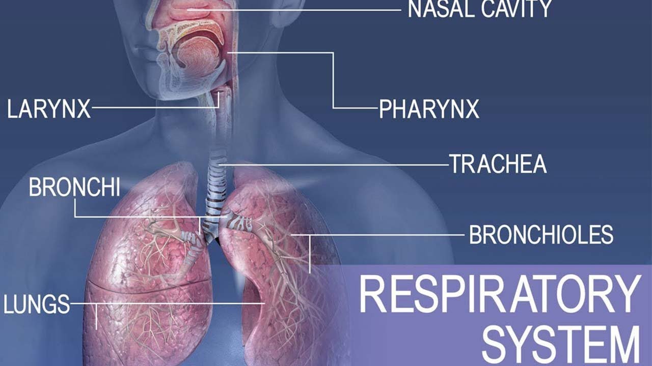
- π The upper respiratory tract includes the nasal cavity, pharynx, and larynx, which contains the vocal cords.
- π The lower respiratory tract begins with the trachea, which splits into the primary bronchi leading to the lungs.
- π¬οΈ The bronchi divide into smaller tubes called terminal bronchioles, which further divide into respiratory bronchioles and alveolar ducts ending in alveoli where gas exchange occurs.
- π« The diaphragm and intercostal muscles are crucial for lung contraction and expansion, facilitating breathing.
- π¨ Gas exchange occurs in the alveoli through diffusion, driven by differences in partial pressures of oxygen and carbon dioxide between alveoli and capillaries.
- π¦ The trachea and bronchi have cartilage and goblet cells that produce mucus, helping trap and expel pathogens.
- πͺ Smooth muscles in the trachea and bronchi can contract to adjust airflow, responding to environmental conditions or allergic reactions.
- π Gas transport in the blood: Oxygen is carried by hemoglobin, while carbon dioxide is mostly transported as hydrogen carbonate in the plasma.
- π Spirometry measures lung volumes and capacities, including tidal volume, vital capacity, and residual volume, which are critical for understanding respiratory health.
Q & A
What is the primary function of the nasal cavity in the respiratory system?
-The primary function of the nasal cavity is to filter, warm, and humidify the air we breathe in.
What are the main anatomical structures of the upper respiratory tract?
-The main anatomical structures of the upper respiratory tract include the nasal cavity, pharynx, and larynx.
What is the role of the trachea in the respiratory system?
-The trachea serves as a passage for air to travel from the upper respiratory tract to the lower respiratory tract and is supported by cartilage to keep it open.
Describe the pathway of air from the trachea to the alveoli.
-Air travels from the trachea to the primary bronchi, then to the terminal bronchioles, respiratory bronchioles, alveolar ducts, and finally reaches the alveoli where gas exchange occurs.
What is the function of the diaphragm in respiration?
-The diaphragm contracts and flattens to increase the volume of the thoracic cavity, reducing the pressure and allowing air to flow into the lungs. It relaxes to help expel air during exhalation.
How does gas exchange occur in the alveoli?
-Gas exchange occurs in the alveoli through diffusion, where oxygen moves from the alveoli into the blood in the capillaries, and carbon dioxide moves from the blood in the capillaries into the alveoli.
What are goblet cells and what is their function in the respiratory system?
-Goblet cells are cells that produce mucus to trap viruses, bacteria, and other particles, preventing them from entering the lungs and causing infection.
What is the difference between the right and left lungs in terms of lobes?
-The right lung has three lobes, while the left lung has two lobes.
Explain the concept of partial pressure in the context of gas exchange.
-Partial pressure refers to the pressure exerted by a specific gas in a mixture of gases. Gas exchange occurs because gases move from areas of higher partial pressure to areas of lower partial pressure.
What happens during anaphylactic shock in the respiratory system?
-During anaphylactic shock, the smooth muscles in the trachea and bronchus contract, narrowing the airways and making breathing difficult.
What is the function of the cilia in the respiratory system?
-Cilia are tiny hair-like projections that move mucus and trapped particles out of the respiratory tract, helping to keep the airways clear.
What are the different volumes and capacities measured in a spirogram?
-A spirogram measures tidal volume (normal breathing volume), total lung capacity, residual volume (air left in lungs after exhalation), functional residual capacity (residual volume plus expiratory reserve volume), inspiratory reserve volume, expiratory reserve volume, and vital capacity (inspiratory reserve volume plus tidal volume plus expiratory reserve volume).
How is carbon dioxide transported in the blood?
-Carbon dioxide is transported in the blood in three main ways: dissolved in plasma (5%), as hydrogen carbonate (85%) after combining with water and an enzyme, and as carbaminohemoglobin (10%) after combining with hemoglobin.
What is the significance of the pleural cavity in the respiratory system?
-The pleural cavity, the space between the parietal and visceral pleura, contains a small amount of fluid that reduces friction during breathing and helps maintain the negative pressure needed for lung expansion.
What happens to intrapleural pressure during inspiration?
-During inspiration, the intrapleural pressure decreases as the thoracic cavity volume increases, allowing air to flow into the lungs.
What is the relationship between volume and pressure in the lungs?
-Volume and pressure in the lungs are inversely related; increasing the volume of the lungs decreases the pressure, and decreasing the volume increases the pressure, facilitating the movement of air in and out of the lungs.
Outlines
π« Introduction to the Respiratory System
The video introduces the respiratory system, detailing the anatomy of the upper and lower respiratory tracts. Key structures include the nasal cavity, pharynx, larynx, trachea, bronchi, and alveoli. The importance of the diaphragm and intercostal muscles in breathing is also explained.
π¬οΈ Gas Exchange Process
This section covers the process of gas exchange in the alveoli, emphasizing the roles of oxygen and carbon dioxide partial pressures. It explains how oxygen moves from the alveoli to the capillaries and how carbon dioxide is transferred in the opposite direction.
π¨ Role of Smooth Muscle and Goblet Cells
The role of smooth muscle, goblet cells, and cilia in the respiratory system is discussed. Smooth muscles help regulate airway diameter, while goblet cells produce mucus to trap pathogens. The section also explains how these features protect against respiratory infections.
π Respiratory Physiology and Pressure Dynamics
This part details the physiology of respiration, focusing on the mechanics of inspiration and expiration. It explains how changes in volume and pressure facilitate airflow into and out of the lungs, and the roles of various pressures, including atmospheric, intrapleural, and intrapulmonary pressures.
π Lung Volumes and Capacities
The concluding section introduces the concept of lung volumes and capacities, including tidal volume, total lung capacity, residual volume, functional residual capacity, inspiratory reserve volume, expiratory reserve volume, and vital capacity. The importance of these measurements in respiratory function is highlighted.
Mindmap
Keywords
π‘Respiratory system
π‘Nasal cavity
π‘Pharynx
π‘Larynx
π‘Trachea
π‘Bronchi
π‘Alveoli
π‘Diaphragm
π‘Gas exchange
π‘Partial pressure
π‘Pleural cavity
π‘Intercostal muscles
π‘Spirogram
π‘Hemoglobin
π‘Carbonic anhydrase
π‘Tidal volume
π‘Vital capacity
Highlights
Introduction to the anatomy of the respiratory system, starting with the upper respiratory tract.
Explanation of the nasal cavity and pharynx as parts of the upper respiratory tract.
Description of the larynx and its function, including the role of the vocal cords.
Distinction between the upper and lower respiratory tracts, with the trachea marking the division.
Detailed breakdown of the lower respiratory tract, including the trachea, bronchi, and lungs.
Explanation of the bronchial tree, from primary bronchi to alveoli, and their roles in gas exchange.
Role of the diaphragm and intercostal muscles in respiration.
Identification of the pleural cavity and its components: parietal pleura, visceral pleura, and the pleural cavity space.
Mechanism of gas exchange in the alveoli, focusing on partial gas pressures of oxygen and carbon dioxide.
Differentiation of the right lung (three lobes) and left lung (two lobes).
Explanation of trachea and bronchi structures, including cartilage, goblet cells, and smooth muscle.
Function of cilia and goblet cells in protecting the respiratory system from pathogens.
Detailed look at the smooth muscle and its role in regulating airway resistance.
Carbon dioxide transportation in the blood, including the formation of hydrogen carbonate.
Overview of lung volumes and capacities, including tidal volume, residual volume, and total lung capacity.
Description of inspiratory and expiratory reserve volumes and their importance in lung function.
Definition of vital capacity and its components.
Transcripts
[Music]
hi everybody my name is andre and
welcome back to med school eu
today we are going to continue on with
this lengthy topic of anatomy physiology
of
systems
in humans and more specifically today we
are going to talk about the respiratory
system we'll begin the unit with
anatomy of the respiratory system
so
we have a couple of labels to go through
just
anatomical labels here
so beginning with the upper respiratory
tract
we got the nasal cavity up at the top
so this is the nasal
cavity
next this
little part right here at the back of
the throat that's called the pharynx
that's fairings
going a little bit lower past the
pharynx we got the larynx and in the
larynx
we've got our vocal cords so this is
where
the sound comes from that comes from the
vocal cords in the larynx now going
further down there's going to be a
divide here
between the
upper respiratory tract and the lower
respiratory tract so these all of these
anatomical structures are part of the
upper respiratory tract
and going below down here to the trachea
this is the trachea
and anything below the trachea is the
lower respiratory tract
so
splitting off from the trachea to the
right lung and the left lung this big
part right here is called the primary
bronchi
and past the the bronchi of course we've
got here the lungs and we're going to go
into further anatomy about the lungs
because there's going to be further
breakdowns and
splitting of these tubes from the
bronchi
all the way down to the alveoli and
that's what we are going to discuss in
this one so here of course we got our
trachea as you can see it's covered in
cartilage
and the the next is the bronchi which we
have labeled already as you can see
they're pretty much identical on both
sides then this moves into terminal
bronchial so that's the
the smaller division here from the
bronchus
it becomes a terminal bronchiole and
from terminal bronchial we go into
respiratory bronchiole and finally this
goes into the alveolar duct and from the
alveolar duct we're gonna have
these little grape-like structures
called alve
oli
and gas exchange occurs
in the alveoli and
sporadically in the alveolar duct
however all these other structures the
bronchioles respiratory termin terminal
and
the bronchi the bronchus and the trachea
there's going to be no gas exchange
occurring only the alveoli and
sometimes alveolar duct depending on the
thickness of it now the structure that
goes right underneath is not shown here
but the structure that goes underneath
the lungs is called the diaphragm and
the diaphragm is responsible for
contraction
of the lungs so it's going to stimulate
breathing
as well as the intercostal muscles so
the muscles that go in between here are
called inter
so intercostal muscles the one that goes
that go between between the ribs and the
diaphragm at the bottom are going to be
responsible for
inflating and deflating the lungs and
we're going to discuss their primary
functions in the next few slides as well
so there are uh three more labels here
so we got the plural cavity so there's
going to be a space between
the two plurals so the
the outer plural is called the parietal
the inner plural is called the visceral
pleura and of course the space in
between is going to be called pleural
cavity so this is just a cavity
space in between the two pleura so
that's the general respiratory system
anatomy so now we are going to talk a
little bit about gas exchange as it
occurs in the alve
alveoli so for breathing in
the gas is primarily going to be oxygen
and the gas pressure here in
the capillary
is primarily going to be carbon dioxide
so we're going to offload the carbon
dioxide and we're going to load on the
oxygen and the reason that's going to
happen is because of partial gas
pressures
so
the partial gas pressure of oxygen in
the alveoli is going to be much greater
than in the capillary therefore the net
movement of gas is going to go to the
capillary and this is just a phenomenon
called diffusion
where it goes from an area of high
concentration to an area of lower
concentration in this case we're talking
about gases therefore we're going to
refer to as partial pressure so
higher partial pressure of gas is going
to move over to
the lower partial pressure of gas now
the same thing occurs with the carbon
dioxide the
atmospheric carbon dioxide partial
pressure is extremely low compared to
the capillary carbon dioxide partial
pressure because these capillaries are
going to bring deoxygenated blood from
the body and which will have metabolites
and carbon dioxide so carbon dioxide is
going to be taken up
by the alveoli again through diffusion
the same concept
where it goes from an area of higher
partial pressure to an area of
lower partial pressure of carbon dioxide
now the exact opposite occurs once these
travel to cells so these are not alveoli
anymore these are just cells
of the body now cells of the body are
going to have lots of carbon dioxide
in high concentrations so there's going
to be high partial pressure of carbon
dioxide but they're going to have low
oxygen so
the whole purpose of the circulatory
system is to deliver nutrients and
oxygen to the cells so they can operate
and do their primary functions so once
the
blood vessels are
able to circulate from the lungs over to
the cells again they're going to go
through the heart then the entire
respiratory system they're going to end
up in various cells of the body
and they're going to offload their high
partial pressure of oxygen onto the
cells because they're going to have low
partial pressure of oxygen and they're
going to uptake the high partial
pressure of carbon dioxide again the
same concept that occurs through
diffusion now before we take a look at
this chart right here
there's another phenomenon i wanted to
mention is that
the right lung
the right lobe
of
of the lung contains
three lobes and the left side so the
left lung contains two lobes so it's
it's a little bit smaller it doesn't
split up into as many lobes
the right lung has three the left lung
has two so that's another thing uh to
just keep in mind and here with this
chart
we are going to
discuss
various structures that
are involved within each of these
anatomical positions that we discussed
earlier so the trachea number of trachea
there's only one trachea that splits off
into two bronchi
so
for each side of the lung or each
bronchus i should say is going to split
off into thousands 48 000 of terminal
bronchials
and
something very important happens here is
that the trachea and the bronchus
have cartilage these are
they're going to have cartilage in
in their structures in order to support
that volume and the air that's coming in
so they're going to have cartilage there
they're also going to have goblet cells
that
will will produce mucus and they're
going to be responsible to keep out any
viruses or any bacteria that may enter
the respiratory system they're going to
keep out the flu the cold as we are we
get in contact with the flu virus the
coronaviruses
and just the regular respiratory viruses
that we get in the winter
they are going to be caught up by these
goblet cells and the cilia
goblet cells and cilia they're
responsible for creating mucus where all
of these
respiratory illnesses and all of these
viruses are going to be caught up and
the cilia
so these are little projections
the cilia are going to be pushing up
up the wall down to the trachea and in
reverse so that you can just cough it
out or breathe it out without
the virus attaching to receptors inside
the lungs and causing
an illness now another thing to know is
the trachea the bronchus and the
terminal bronchial have smooth muscle
so this this means they could
dilate they could stretch a little bit
which involves the
elastic tissue as well
so
up to the point of the terminal
bronchiole they are going to have smooth
muscle which could control
how wide the airway is going to be
so for example if you are out in a
colder environment the smooth muscle is
typically going to contract a little bit
to create resistance for the air so the
air is not going to pass as quickly and
you are not going to get cold air
entering the alveoli because that
obviously could cause
problems for the alveolar cells
therefore in order to buffer that
temperature of the air you're going to
have smooth muscle they're going to
contract and they're going to reduce the
diameter of the trachea and the bronchus
and
something very similar happens with
anaphylactic shock or
an allergic reaction where the trachea
and the bronchus the smooth muscles
there are going to contract and they're
gonna
make a narrow airway and that's when
problems occur uh with with breathing
and anaphylactic shock so as you can see
here as i mentioned previously the site
of gas exchange so the exchange that
occurs here and here
in terms of the carbon dioxide and the
oxygen only occurs in alveoli and the
alveolar duct
primarily in the alveoli however in some
cases alveolar duct as
as well there's going to be no gas
exchange in any of the bronchioles or
the bronchus or the trachea
important thing to remember is that
trachea and bronchus have goblet cells
that produce mucus
smooth muscle and cilia
are going to be all the way down to the
terminal bronchial but not the
respiratory bronchioles
and cartilage will only reside with the
trachea and the bronchus so when we
discuss the immune response we are going
to talk about the goblet cells and the
smooth muscle cells in greater detail
next we have a quick slide about the gas
content
of the blood
so
you should be familiar with how carbon
dioxide is going to be transported
within the blood because for oxygen of
course it's going to be
loaded onto hemoglobin and then
hemoglobin is going to be
caring
for oxygen molecules per
one
molecule of hemoglobin and there's going
to be millions molecules of hemoglobin
within each erythrocyte
so
that's typically how oxygen is going to
be carried within the red blood cell
however when when we have carbon dioxide
inside circulatory system
we're going to have a bit of a different
story so first
from the respiring cells so from our
cells we're going to get carbon dioxide
that will enter
the erythrocyte five percent of carbon
dioxide will be carried in solution
meaning it's going to be carried in the
plasma now the the rest
the highlighted areas is is what's going
to be important because the carbon
dioxide is going to combine with water
in some cases
using an enzyme called carbonic
anhydrase
and and it's going to create a molecule
of hydrogen carbonate so most of carbon
dioxide is
going to be transported as hydrogen
carbonate which is this molecule right
here hco3
minus 85 percent of the carbon dioxide
will go there and 10 percent of the
carbon dioxide will be converted
to carbon amino hemoglobin which is
simply when carbon dioxide combines with
the hemoglobin within the erythrocytes
so only about 10 percent
will be
carried the same as oxygen however the
rest of the carbon dioxide most of it
will be carried in the plasma as
hydrogen carbonate so now we're going to
talk about the physiology of
respirations and we're going to begin
with inspiration which is going to be
inhaling so getting
oxygen and
air into our lungs
so we are going to first make a couple
of labels here so of course we got the
diaphragm now this i
this yellow structure right here
this is more going to be like a zoom in
of an alveoli so i'm just going to label
it as
alveolar pressure p alveolar so the
gas pressure in the alveoli
then the
gas pressure that's going to be within
the lung is called
intrapulmonary pressure
inside the pleural cavity
we are going to have intrapleural
pressure
now the pressure difference between
the
intrapleural pressure and the pressure
inside the lung is called transpulmonary
pressure so these are all the
important and also we got atmospheric so
p atm
atmospheric pressure so all of these are
going to be involved in the breathing
anatomy and the physiology
of breathing now of course we have
intercostal muscles that go around the
side of the lungs and that's going to be
the ones in between the ribs
so let's go over some of the sequences
of events that occur
so we first are going to begin with
the inspiratory muscles that are going
to contract and that's going to be the
diaphragm so the diaphragm the
intercostal muscles are going to make
the rib cage
to push outward and up so the rib
this is how we are going to create more
volume
within
the
lungs
so we must remember that volume and
pressure
are inversely related so if we increase
the volume we are going to decrease the
pressure if we increase the pressure
we're going to decrease the volume so
what this does what the sequence this
first sequence does is it increases
volume because we are going to expand
the lungs and when we increase volume we
are going to decrease
the pressure
and we're going to decrease the pressure
to the point
where
the
atmospheric pressure is going to be
above
the
pressure inside the lungs and ultimately
the the pressure inside the alveoli
therefore the air can simply flow
through the fusion
from the atmosphere inside the lung and
into the alveoli because it's going to
move from
by the definition of diffusion it's
going to move from the higher partial
pressure of gas to lower partial
pressure of gas
so this what this uh what this really
does the ultimate thing is that the
intrapro
plural pressure pip
is going to decrease
because our lung volume has increased so
now because this has decreased number
three
the p atmospheric
is going to be above
p
i p
and we have
air flows inside so that's that's
typically how it occurs because
as the gases flow into the lungs down
its pressure gradient
until that intrapulmonary pressure is
zero so then
by the end of it once we fully have
inhaled
the atmospheric pressure is going to
equal to intrapulmonary
pressure
because of course the more air we get
inside
the more pressure it's going to create
and eventually it's going to equate
itself with the atmospheric pressure and
you can no longer breathe
any further you cannot breathe in any
further you cannot take up more air now
with expiration so breathing out we're
going to have something pretty much the
opposite occur so
we're going to have the inspiratory
muscles relax so the diaphragm and this
is all due to the recoil of the coastal
cartilages so if you stretch the
cartilage of the rib cage
it's obviously going to want to recoil
back because it's got its elastic fibers
and the elastic fibro fibers will
naturally want to recoil back to its
original form so that's going to create
that
pressure that tension
that
is going to counteract
the pressure that is created by the
diaphragm rising and the intercostal
muscles contracting next we're going to
have the thoracic volume
or the thoracic cavity volume decrease
meaning that it's going to increase
the pressure and as soon as the p
i p
pressure is above
the atmospheric pressure
then obviously the air
is going to flow out out into the
atmosphere because it's going to move
from higher pressure to lower pressure
environment and again it's going to
occur until finally the pip
the intrapleural pressure is going to
equate to
atmospheric pressure and you can no
longer breathe out
any further and the final thing that i'm
going to introduce you to is called a
spirogram so this is basically just a
measurement of
lung volumes and lung capacities so what
we have here is
the general breathing rate if we are
just breathing at a resting rate you're
sitting down on your chair you're
listening to this lecture your breathing
is going to be the tidal volume it's
only going to be about
maybe 500
milliliters of
of gas that's going to come in and out
in and out and that's going to be
labeled as tidal
volume
now the entire capacity of the lungs so
how how much air we could possibly have
inside the lungs if you fully breathe in
and all the air that was already in
there
that's going to be called the total lung
capacity
now there's something called residual
volume and residual volume is something
you cannot breathe out of course if you
were to breathe out
all the air that's inside your lung
your lung is going to collapse in on
itself so there's always going to be
some sort of
a little bit of air and that's typically
you see 1200 milliliters of
of air inside the lungs that is
constantly there even if you forcefully
expire you're still gonna have that
residual volume there and you cannot get
rid of it unless you puncture it along
and it flows out and at that point
you're gonna have a collapsed lung
and of course that's gonna create a lot
of problems
so that's the residual volume it's
extremely important for our vital
functions
now the volume that you could breathe
out and in addition with the residual
volume is called functional residual
capacity so that's the that's how much
that's the uh expiratory reserve volume
plus the residual volume so both of
these together an expiratory reserve
volume is how much air you could
forcefully breathe out
so if you're regularly breathing right
now and watching this lecture try to
breathe out as much as you can to the
point where you can no longer breathe
out anything else
that's your expiratory reserve volume
from the point of breathing out normally
to the point of fully forcefully
breathing out that's the expiratory
reserve volume now if you add that
together with the residual volume that
makes up functional
residual
capacity that is labeled here next we're
going to look at the inspiratory reserve
volume so that's completely the opposite
of the expiratory inspiratory reserve
volume is when you are doing your tidal
volume you're normally breathing and you
normally breathe in however
the rest that you can breathe in up to
your full
potential of breathing in that's going
to be the inspiratory reserve
volume now if you combine the expiratory
reserve volume plus tidal volume plus
inspiratory reserve volume so if you
were to
fully breathe in and then
fully breathe out
that's your vital
capacity that's what's called vital
capacity so all of these these three
different capacities and four different
types of volumes you should know by
heart so if they are identified on a
question
you would not be struggling to to know
what the question is really talking
about so this concludes our lecture on
the respiratory system in the next video
we are going to discuss the diseases
involved with the circulatory system as
well as the respiratory system
[Music]
you
5.0 / 5 (0 votes)