Auscultation of Heart Sounds | Assessing Heart Sounds | Listening to the Heart with a Stethoscope

RegisteredNurseRN
23 Jun 201610:19

Summary

TLDRIn this educational video, Sarah from Register Nurse Orion Comm teaches viewers how to auscultate heart sounds. She demonstrates the process on a real person, explaining the significance of S1 and S2 sounds, which correspond to the closure of the heart's valves. Sarah provides anatomical landmarks for accurate auscultation and discusses additional heart sounds like S3, S4, and murmurs. She offers tips for distinguishing between sounds and the importance of patient positioning to detect subtleties in heart sounds, concluding with a guide on how to identify and grade heart murmurs.

Takeaways

  • 👂 The purpose of auscultating heart sounds is to assess the heart's rhythm, rate, and the closure of heart valves.
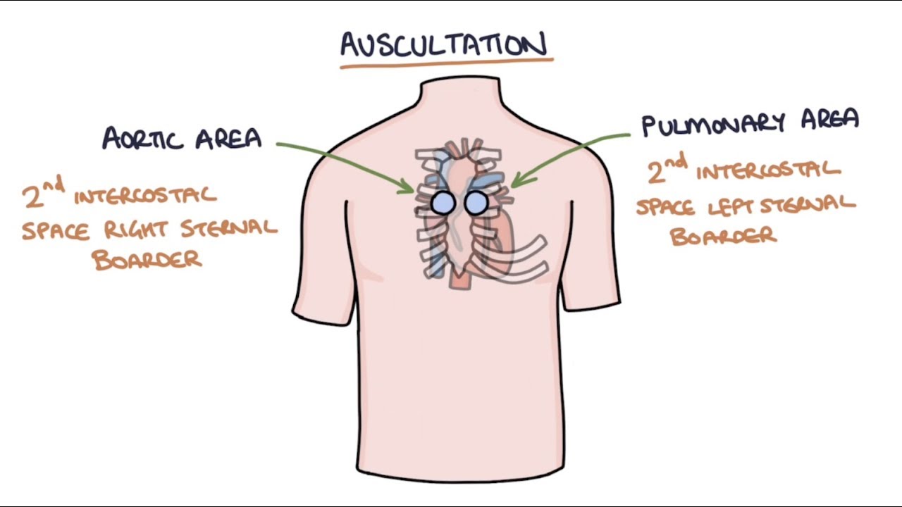
  • 📍 Heart sounds are associated with specific valves: S1 with tricuspid and mitral valves, and S2 with aortic and pulmonic valves.
  • 📌 Anatomical sites for auscultation include the aortic area, pulmonic area, the apex (heart's base), and the mitral area.
  • 🔍 Auscultation begins by finding the clavicle, angle of Louis, and the second intercostal space to locate the valves.
  • 👆 The aortic and pulmonic valves close together, producing the louder S2 sound, usually heard at the base of the heart.
  • 👇 The tricuspid and mitral valves close simultaneously to produce S1, typically louder at the apex.
  • 🎧 Using a stethoscope, start with the diaphragm to hear S1 and S2 clearly, then switch to the bell to listen for murmurs.
  • 🔊 S2 is louder at the base, and S1 is louder at the apex, which helps in distinguishing between the two sounds.
  • 🛏️ Positioning the patient differently, such as on their left side, can help in identifying additional heart sounds like S3 and S4.
  • 👂🏼 Auscultation for S3 and S4 involves listening at the apex with the bell of the stethoscope, as these sounds are low-pitched.
  • 📊 Heart murmurs are extra sounds that may indicate issues with blood flow through the heart valves and should be graded if present.

Q & A

  • What is the primary purpose of auscultating heart sounds?

    -The primary purpose of auscultating heart sounds is to assess the regularity of the heart rhythm, count the rate, and evaluate the function of the heart valves by listening to the sounds they make when closing.

  • What are the two main heart sounds, S1 and S2, associated with?

    -S1 is associated with the closing of the tricuspid and mitral valves, while S2 is associated with the closing of the aortic and pulmonic valves.

  • How can you locate the anatomical sites for auscultating heart sounds on a patient?

    -To locate the anatomical sites, find the clavicle on the patient, then go down to find the angle of Louis, where the second rib comes out. The right side of this border is the aortic valve, and the left side is the pulmonic valve. The tricuspid valve is found in the fourth intercostal space, and the mitral valve is found in the fifth intercostal space at the mid-clavicular line.

  • What is the significance of the point of maximal impulse (PMI) in auscultation?

    -The point of maximal impulse (PMI) is significant because it is where the heart's contraction is most easily felt and heard, typically located at the mitral valve area, and can help in identifying the location for auscultation.

  • What is the mnemonic 'ALL PATIENTS TAKE MEDICINE' used for in auscultation?

    -The mnemonic 'ALL PATIENTS TAKE MEDICINE' is used to remember the order of auscultation sites: Aortic, Left sternal border (mid-clavicular line), Pulmonic, Apex, and Mitral, which helps in systematically assessing the heart sounds.

  • Why is it important to differentiate between S1 and S2 when auscultating heart sounds?

    -Differentiating between S1 and S2 is important because it helps in identifying the specific valves that are closing, which is crucial for diagnosing any abnormalities in heart valve function.

  • What are some tips for distinguishing between S1 and S2 sounds?

    -S2 is typically louder at the base of the heart, while S1 is louder at the apex. Additionally, feeling the carotid pulse while listening to the apex can help identify S1, as the sound coincides with the pulse. Observing the QRS complex on a bedside monitor can also help, as S1 occurs with the spike of the QRS complex.

  • Why is the patient positioned on their left side during auscultation?

    -Positioning the patient on their left side helps because most of the heart is on the left side, and turning them to that side allows the heart to move over slightly, making it easier to hear the anatomical sites, especially the apex.

  • What are S3 and S4 heart sounds, and why are they significant?

    -S3 and S4 are extra heart sounds that can be heard in certain conditions. S3 is a sound heard just after S2 and may be normal in children and young adults but can indicate heart problems in older adults. S4 is heard just before S1 and is not typically present in a normal heart, indicating a possible issue when heard.

  • How can auscultation help in identifying heart murmurs?

    -Auscultation can help identify heart murmurs by listening for blowing or swishing noises, which may indicate turbulent blood flow due to valve issues. The patient may be asked to lean forward and exhale to enhance the sounds, and the diaphragm of the stethoscope is used to pick up these murmurs at the aortic and pulmonic areas.

  • What is the grading scale for heart murmurs, and what does it indicate?

    -The grading scale for heart murmurs ranges from one to six, with grade one being hard to hear and grade six being so loud that the stethoscope can be lifted off the chest and the murmur can still be heard. The grading indicates the intensity of the murmur, which can help in assessing the severity of the underlying condition.

Outlines

00:00

👂 Introduction to Auscultating Heart Sounds

Sarah from RegisterNurseOrion Comm introduces a tutorial on auscultating heart sounds. She plans to demonstrate how to listen to heart sounds on a real person, focusing on identifying the anatomical sites and differentiating between the sounds of the heart valves closing, such as S1 and S2. S1 is produced by the closure of the tricuspid and mitral valves, while S2 is the sound of the aortic and pulmonic valves closing. The tutorial will also cover extra heart sounds like S3, S4, and heart murmurs, and will explain the importance of assessing heart rhythm and rate.

05:05

📍 Locating Anatomical Sites for Heart Auscultation

Sarah explains the process of locating the anatomical sites for heart auscultation, starting with finding the clavicle and the angle of Louis, then identifying the intercostal spaces where the heart valves are located. She describes the positions of the aortic and pulmonic valves at the base of the heart, which are responsible for the S2 sound, and the tricuspid and mitral valves, which produce the S1 sound. She also mentions the point of maximal impulse and the distinction between the apex and the base of the heart. Sarah then demonstrates the correct placement of the stethoscope's chest piece on a patient to listen for S1 and S2, and discusses the importance of distinguishing between these sounds and looking for any splits in the sounds.

10:07

🔍 Advanced Techniques for Heart Sound Auscultation

In this part, Sarah covers advanced techniques for auscultating heart sounds, including using the bell of the stethoscope to listen for low-pitched noises like S3 and S4, and murmurs. She explains how to position the patient to better hear these sounds, such as turning them onto their left side to push the heart towards the chest wall. Sarah also provides tips for differentiating between S1 and S2 sounds, such as their relative loudness at the base and apex of the heart, and using the carotid pulse as a reference. She concludes with a demonstration of how to listen for murmurs with the patient leaning forward and exhaling, and explains the grading scale for murmurs, from barely audible to those that can be heard without a stethoscope.

📺 Conclusion and Additional Resources

Sarah concludes the tutorial by inviting viewers to watch another in-depth video about heart sounds, presumably available through a pop-up card on the screen. She thanks the viewers for watching and encourages them to subscribe to the YouTube channel for more educational content.

Mindmap

Keywords

💡Auscultation

Auscultation is a medical examination technique that involves listening to sounds within the body, typically using a stethoscope, to assess health conditions. In the context of this video, auscultation refers specifically to listening to heart sounds to evaluate the function of the heart valves and rhythm. The script describes the process of auscultating various anatomical sites on the chest to identify different heart sounds, such as S1 and S2.

💡Heart sounds

Heart sounds are the noises produced by the heart's activity, including the closing of valves and the flow of blood. The script discusses how to identify and differentiate between normal heart sounds like S1 and S2, as well as extra sounds such as S3 and S4, which can indicate certain health conditions. Heart sounds are crucial for diagnosing heart abnormalities.

💡S1 and S2

S1 and S2 refer to the first and second heart sounds, respectively. S1 is produced by the closing of the mitral and tricuspid valves, while S2 is produced by the closing of the aortic and pulmonic valves. The script emphasizes the importance of distinguishing between these sounds and describes how their loudness varies depending on the location on the chest where they are auscultated.

💡Heart valves

Heart valves are structures in the heart that regulate the flow of blood through the heart chambers, ensuring it flows in one direction. The script explains that listening to heart sounds allows healthcare providers to assess how well these valves are functioning, with S1 and S2 sounds indicating the closure of specific valves.

💡Anatomical sites

Anatomical sites refer to specific locations on the body where certain structures can be found or where certain sounds can be best auscultated. In the script, the presenter explains how to locate these sites on the chest to listen for heart sounds, such as the aortic and tricuspid valves, and how these sites relate to the heart's position and function.

💡Murmurs

A heart murmur is an abnormal sound heard during auscultation, often indicating a problem with heart function, such as a valve that is not closing properly. The script discusses how to listen for murmurs, particularly when the patient is in different positions, as they can provide important diagnostic information.

💡Stethoscope

A stethoscope is an instrument used by healthcare providers to listen to sounds from within the body. The script describes the use of a stethoscope with both its diaphragm and bell to listen to different types of heart sounds, highlighting the importance of the tool in heart auscultation.

💡Point of maximal impulse (PMI)

The point of maximal impulse (PMI) is the location on the chest where the heart's contractions are felt most strongly, typically over the left ventricle. The script mentions that the PMI is located at the mitral valve area and is an important site for auscultation.

💡Carotid artery

The carotid artery is a major blood vessel in the neck that supplies blood to the brain and head. In the script, the presenter suggests feeling the carotid artery's pulsation to help differentiate between S1 and S2 heart sounds, as the pulsation corresponds with the S1 sound.

💡QRS complex

The QRS complex is a part of the heart's electrical activity that can be seen on an electrocardiogram (ECG) and corresponds to the depolarization of the ventricles. The script mentions using the QRS complex as a visual aid to identify S1 sounds, as the sound occurs simultaneously with the spike seen on the monitor.

💡Mitral valve

The mitral valve is one of the heart's atrioventricular valves, located between the left atrium and left ventricle. The script explains that the closure of the mitral valve contributes to the S1 heart sound and that auscultation at the mitral valve area can help identify this sound.

Highlights

Sarah from Register Nurse Orion Comm demonstrates how to auscultate heart sounds on a real person.

The purpose of listening to heart sounds is to ensure the rhythm is regular, count the rate, and assess heart valve closures.

S1 and S2 represent the closing of tricuspid and mitral valves, and aortic and pulmonic valves, respectively.

Anatomical sites for auscultation include the aortic, pulmonic, tricuspid, and mitral valves, and the apex of the heart.

Finding the clavicle and the angle of Louis helps identify the correct auscultation sites on the chest.

The base of the heart is where S2 is most prominently heard, while S1 is louder at the apex.

Auscultation begins with the patient sitting or lying down, using a stethoscope diaphragm first.

Mnemonic 'all patients take medicine' helps remember the auscultation order: aortic, pulmonic, tricuspid, mitral.

Listening for S1 and S2 involves distinguishing the sounds and checking for splits in valve closures.

Differentiating S1 from S2 can be aided by their loudness at the base and apex, respectively.

Feeling the carotid pulse can help identify S1 as it coincides with the pulse.

Turning the patient to their left side enhances the audibility of the apex, important for detecting S3, S4, and murmurs.

The bell of the stethoscope is used to listen for low-pitched sounds like S3, S4, and heart murmurs.

S3 and S4 are extra heart sounds heard after S2 and before S1, respectively.

Auscultation also includes having the patient lean forward and exhale to detect aortic and pulmonic murmurs.

Heart murmurs are graded on a scale from one to six, with six being the loudest.

A thrill, a vibration on the skin, can also indicate the presence of a heart murmur.

Sarah encourages viewers to watch her in-depth video on heart sounds for further familiarization.

Transcripts

play00:00

hey everyone it's sarah with register

play00:01

nurse Orion comm and in this video I'm

play00:03

going to be going over how to auscultate

play00:06

heart sounds what I'm going to be doing

play00:08

is I'm going to show you how to listen

play00:10

to heart sounds on a real person I'm

play00:13

going to show you the anatomical site

play00:14

how to identify s1 s2 talk about those

play00:18

s3 s4 heart murmurs those extra sounds

play00:21

you may hear the first let's covered the

play00:23

basics okay why do we listen to heart

play00:27

sounds what is the purpose well one

play00:29

thing is we want to make sure the rhythm

play00:31

is regular we want to count the rate but

play00:33

we're also one of the big things is that

play00:35

we're assessing how those heart valves

play00:37

are closing because whenever you are

play00:39

hearing s1 s2 those are valves valves

play00:43

closing s1 are your tricuspid and mitral

play00:48

valves closing and s2 is the sound of

play00:52

your aortic and pulmonic valves closing

play00:56

and while you're listening to heart

play00:59

sounds you'll be trying to distinguish

play01:00

am i hearing s1 s2 and then you're going

play01:03

to be positioning the patient a little

play01:05

bit different positions and going to be

play01:06

listening for those extra heart sounds

play01:08

like s3 s4 and heart murmurs so first

play01:12

let's go over the anatomical sides here

play01:15

in a second you're going to see what it

play01:17

actually looks like whenever and you're

play01:20

looking at the anatomical sites on the

play01:22

chest but let me cover them real fast if

play01:24

you want you can write this down so you

play01:25

can remember it the key to help you

play01:28

identify these anatomical sites is to

play01:30

find the clavicle on the patient and

play01:33

then go down and find the angle of Luis

play01:37

the joint little area and the second rib

play01:40

comes out from there and right below

play01:42

that we're going to start on the right

play01:44

side is intercostal space and right

play01:48

there left I mean right of that border

play01:50

is the aortic valve and the aortic valve

play01:53

represents when it closes the sound of

play01:56

s2 that in the pulmonic valve normally

play01:58

close together so when they close

play02:00

together they're similar looner valves

play02:02

you will hear s2 then right over on the

play02:06

left side in the same space

play02:08

second intercostal space you will find

play02:10

the pulmonic valve then down in the

play02:13

third space you will find herbs point

play02:16

and this is just an area where you're

play02:20

separating the base from the apex it's

play02:22

just the midway point between those two

play02:24

areas and then you have the fourth

play02:26

intercostal space which right next to

play02:29

the border of that is the tricuspid

play02:32

valve you'll find that on the left side

play02:33

as well and the tricuspid and mitral

play02:35

valves when they close together

play02:38

simultaneously you will hear s1 and they

play02:41

are filling the base then you go down a

play02:43

little bit in the fifth intercostal

play02:45

space but mid clavicular which is the

play02:48

midway point of your clavicle and you

play02:51

will find the mitral valve also called

play02:53

bicuspid valve and these are your a do

play02:56

ventricular valves your AV valves and

play02:58

this is also where you will hear the

play03:00

point of maximal impulse and also it's

play03:04

important to know the bottom part of the

play03:06

heart down in this area is that apex the

play03:09

top part of this area is called the base

play03:12

you'll want to remember that now let's

play03:14

look and auscultate these areas and see

play03:17

what they look like on a real person

play03:19

okay first I want to start out just

play03:22

showing you on the chest what is where

play03:25

you're going to actually place your

play03:26

chest piece whenever you're listening to

play03:29

the heart what I like to do whenever I'm

play03:31

first starting out either have the

play03:32

patient set up or lie down and I like to

play03:34

start and they or tick and work my way

play03:36

down remember the mnemonic all patients

play03:39

take medicine and herbs point is in

play03:42

between the pulmonic and the tricuspid

play03:44

and whenever your semilunar valves are

play03:47

your aortic and pulmonic and when they

play03:49

close you hear s2 and so you're going to

play03:52

hear s2 the most at the base of the

play03:54

heart and then whenever you're hearing

play03:57

the tricuspid and mitral which are your

play03:59

ventricular valves which are AV

play04:01

valves you're hearing s1 whenever they

play04:04

close so let's use a chest piece and

play04:06

auscultate okay whenever I'm beginning

play04:09

auscultation of the heart what I like to

play04:11

do is remove the clothing and

play04:14

like to have the patient set up you can

play04:15

also have them lay down and I listen

play04:17

with a diaphragm of my stethoscope first

play04:20

and then I'll switch to the bell and

play04:21

redo all the anatomical sites but I like

play04:24

to listen a diaphragm because you can

play04:25

hear s2 and s1 the best with this along

play04:29

with your aortic and pulmonic

play04:31

regurgitation murmurs so I start at

play04:35

theoretic remember the mnemonic all

play04:37

patients take medicine and what I'm

play04:41

going to do is I'm listening for s1 and

play04:43

s2 I'm distinguishing them and I'm also

play04:45

listening for s1 splits or s2 splits and

play04:50

this is just where the valves are not

play04:53

closing at the same time so you may hear

play04:54

a little bit of an extra noise so we're

play04:57

going to store in the aortic over here

play04:59

and what I'm hearing is love dub lub dub

play05:05

and dub is louder because dub represents

play05:08

s2 and in the base of the heart you're

play05:11

going to hear s2 louder then how you

play05:13

would hear it down there then I'm just

play05:16

going to inch over here to the pulmonic

play05:18

and I hear the same thing I don't know

play05:21

in any splitting s1 s2 or closing at the

play05:25

same time no extra heart sounds then I'm

play05:28

going to inch down to herbs point this

play05:31

is just the halfway point between the

play05:33

base and the apex of the heart now I'm

play05:37

going to inch down to the tricuspid and

play05:41

this time I'm hearing lub dub and love

play05:45

is louder because this is signifying

play05:49

more where you're going to hear s1 and

play05:52

love is represented by s1 and I hear

play05:54

that louder in this area and then I'm

play05:56

going to go over to the mitral area mid

play06:00

clavicular and this hearing the same

play06:03

thing loved a nice good rhythm and what

play06:09

I want to do is I'm just going to switch

play06:10

over to my Bell and I'm just going to

play06:12

repeat and what I'm really paying

play06:14

attention to is I'm listening for any

play06:16

type of murmurs or those low pitched

play06:18

sounds you really can't hear s3 and s4

play06:21

that great in this position that's why I

play06:26

hear in a second we're going to get on

play06:27

our left

play06:27

and you hear that best in the apex but

play06:30

what I'm listening for is maybe any

play06:32

murmurs blowing swishing noise and I'm

play06:41

not hearing anything

play06:42

now one thing you may find hard whenever

play06:45

you are auscultate is the sink wishing

play06:47

s1 from s2 and some tips to help you

play06:51

with that

play06:51

again s2 is going to be louder here at

play06:55

the base and s1 is going to be louder

play06:57

here at the apex so that can help you

play07:00

with that or if you're still having

play07:02

trouble you don't can't really

play07:04

differentiate and you can feel on the

play07:06

carotid artery and listen at the apex of

play07:09

the heart and whenever you feel a

play07:11

pulsation and you feel you hear that

play07:14

noise you've identified s1 because the

play07:17

carotid pulse ation and the sound

play07:18

signify s1 or if you have a patient on a

play07:21

bedside monitor you can look at your QRS

play07:25

complex in the AR way the big spike

play07:27

whenever you see that spike and you hear

play07:30

the noise that is s1 so those are just

play07:32

some little tips on how you can

play07:35

differentiate between s1 and s2

play07:37

now we've assist the patient onto their

play07:40

left side and the whole purpose of doing

play07:42

this is majority of your heart is on

play07:45

your left side so whenever you turn them

play07:47

have them go there it pushes the heart

play07:49

over a little bit more just so you can

play07:51

hear those anatomical sites a little bit

play07:53

better and what we are interested in is

play07:55

the apex of the heart and we're going to

play07:57

be listening with the bell of our

play07:59

stethoscope because we're listening for

play08:00

low pitched noises and if the patient

play08:04

was going to have an s3 s4 or a module

play08:07

stenosis murmur this is where we have

play08:09

most likely hear it so what we're going

play08:13

to do is just find the midclavicular the

play08:17

fifth intercostal space we're going to

play08:19

just listen over there and we're

play08:21

listening for s3 or s4 and murmurs

play08:29

and s3 is hurt after s2 so again that's

play08:34

why you have to distinguish between s1

play08:35

and s2 and s3 is going to sound like a

play08:38

love dub to love data because it's heard

play08:40

after s2 s4 is going to be heard before

play08:46

s1 and it's going to sound like this

play08:49

Tull of de mm de Tunnel lubed up and a

play08:53

murmur of course is just that blowing

play08:56

swishing noise okay last what I like to

play09:03

do is I like to have the patient set up

play09:05

and lean forward and then have them

play09:08

exhale and I'm going to listen for what

play09:12

I'm looking for is murmurs aortic and

play09:14

pulmonic murmurs and want to be

play09:15

listening at the aortic and the pulmonic

play09:17

sides with the diaphragm because it's

play09:19

good at picking up those murmurs and

play09:20

what's happening is that the chest the

play09:22

heart behind the storm is just moving a

play09:23

little bit forward so we can hear those

play09:25

anatomical positions a little bit better

play09:28

and I'm listening for like a blowing a

play09:32

swishing noise and if one's present

play09:36

you'll want to grade that and here on

play09:38

your screen you'll see what the grading

play09:40

scale is for that one a grade one is

play09:43

hard to hear and it goes all the way up

play09:45

to six and this is the loudest you could

play09:48

literally lift your chest piece off the

play09:50

patient's chest like this and you could

play09:52

hear just the blowing and swishing noise

play09:54

you could also fill in the chest a

play09:56

thrill which is like a vibration on the

play09:58

skin okay so that is how you auscultate

play10:01

heart sounds now be sure to check out my

play10:04

other video where I go in-depth about

play10:06

these heart sounds I talk in great

play10:08

detail about them a card should be

play10:10

popping up so you can access that video

play10:11

so you can familiarize yourself with

play10:13

these heart sounds thank you so much for

play10:15

watching and please consider subscribing

play10:17

to this YouTube channel

Rate This

5.0 / 5 (0 votes)

Связанные теги
Heart AuscultationMedical TrainingAnatomical SitesHeart SoundsS1 and S2Healthcare EducationStethoscope UseValve AssessmentMurmurs DetectionClinical Skills
Вам нужно краткое изложение на английском?