Pengamatan Mitosis Pada Akar Bawang dengan Metode Squash
Summary
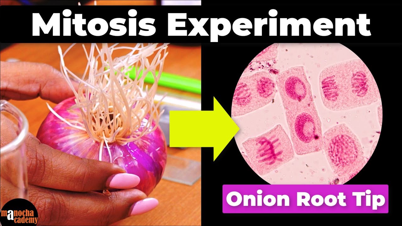
TLDRIn this educational video, viewers are guided through the process of observing mitosis in onion root tips. The tutorial begins with cultivating the roots in a damp environment or water to encourage growth. The roots are then treated with a 0.02% colchicine solution to inhibit spindle formation and enlarge chromosomes. Afterward, a Carnoy's solution is used for cell fixation. The video covers the preparation steps, including hydrolysis and staining with aceto-orcein, to reveal the chromosomes. Finally, the observer can examine the different stages of mitosis—prophase, metaphase, anaphase, and telophase—under a microscope. The video concludes with tips for preserving the samples for future observation.
Takeaways
- 🧬 The video script discusses observing mitosis in onion root tips.
- 🌱 The first step is to grow onion roots by placing them in a moist area or submerging them in water.
- 🔪 The bottom part of the onion is cleaned to ensure quick root growth.
- 💧 The root tips are then placed in a 0.02% colchicine solution for 1-2 hours to inhibit spindle formation and enlarge chromosomes.
- 🧪 After colchicine treatment, the solution is replaced with Carnoy's solution for 2-3 hours to fix and preserve the cell condition.
- 🚫 If immediate observation is not needed, the sample is stored in 70% alcohol for preservation.
- 🔬 The purpose of the treatment is to break down cell walls to facilitate the observation of mitotic division.
- 🔥 The sample is then hydrolyzed in a reaction tube at 60°C for 15 minutes, followed by rinsing and staining with 2% aceto-orcein.
- 🎨 The staining process takes 15-30 minutes or more, depending on the sample condition.
- 🔍 After staining, excess stain is washed off with 45% acetic acid, and the slide is prepared for observation under a microscope.
- 📸 The video suggests using a 100x objective lens for observation and applying immersion oil to the cover glass for clarity.
- 🖼️ The results of the mitosis observation can be preserved using a glass slide and a cover slip for permanent and repeatable viewing.
Q & A
What is the main topic of the video script?
-The main topic of the video script is learning how to observe mitosis by studying the root tips of an onion.
How should the onion root be prepared for mitosis observation?
-The onion root should be prepared by placing it in a damp area or submerging it in water to encourage root growth. The bottom part of the onion should be cleaned to facilitate quick root development.
What is the purpose of using a 0.02% colchicine solution in the process?
-The 0.02% colchicine solution is used to inhibit spindle fiber formation during cell division and to enlarge the chromosomes for easier observation.
What is the role of Carnoy's solution in the preparation process?
-Carnoy's solution is used to fix or preserve the condition of the cells after colchicine treatment, ensuring the cells are kept in a stable state for observation.
Why is it necessary to store the sample in 70% alcohol if not observed immediately?
-Storing the sample in 70% alcohol preserves it for future observation, allowing it to be kept for weeks or months without immediate analysis.
What is the purpose of the hydrolysis step in the preparation process?
-The hydrolysis step is intended to break down the intercellular bonds, making it easier to observe the mitotic divisions in the cells.
How long should the root be kept in the oven during the hydrolysis process?
-The root should be kept in the oven for 15 minutes at a temperature of 60°C during the hydrolysis process.
What is the function of aceto-orcein as a stain in the preparation process?
-Aceto-orcein is used as a stain to color the chromosomes, allowing for optimal absorption of color for clear observation under a microscope.
How long should the stained sample be left to absorb the color before observation?
-The stained sample should be left to absorb the color for 15-30 minutes, although the time may vary depending on the condition of the sample.
What should be done to clean the excess stain before observation under a microscope?
-The excess stain should be cleaned off with 45% acetic acid, followed by rinsing and blotting with a tissue to prepare the slide for clear microscopic observation.
How can the results of the mitosis observation be preserved for future reference?
-The results of the mitosis observation can be preserved by using a glass slide and cover slip, which makes the preparation permanent and allows for repeated observation.
Outlines
🌱 Observing Mitosis in Onion Root Tips
This paragraph introduces a tutorial on how to observe mitosis in onion root tips. The process begins with cultivating onion roots by placing them in a moist environment or in water to encourage growth. The roots are then treated with a 0.02% colchicine solution for 1-2 hours to inhibit spindle formation during cell division and to enlarge the chromosomes for easier observation. Afterward, the colchicine solution is replaced with Carnoy's solution for 2-3 hours to fix the cells. If immediate observation is not desired, the sample can be stored in 70% alcohol for extended periods. The purpose of these treatments is to break the cell wall bonds, facilitating the observation of mitosis in mosquito larvae. The process includes hydrolysis, cutting the root, and heating it in an oven at 60°C for 15 minutes. Afterward, the root is stained with a 2% aceto-orcein solution for 15-30 minutes to optimally absorb color. The excess stain is then washed off with 45% acetic acid, and the sample is prepared for observation under a microscope with a 100x objective lens and immersion oil.
🔬 Storing and Sharing Mitosis Observations
The second paragraph discusses the steps to store and share the results of mitosis observations. It mentions that the prepared slides can be preserved using Canada balsam or other mounting media to make them permanent and suitable for repeated observation. The paragraph also encourages viewers to support the tutorial by liking, commenting, and subscribing if they find the video helpful.
Mindmap
Keywords
💡Mitosis
💡Onion root tip
💡Colchicine
💡Chromosomes
💡Carnoy's solution
💡Alcohol 70%
💡Hydrolysis
💡Aceto-orcein
💡Cover slip
💡Phases of mitosis
💡Permanent slide
Highlights
Introduction to observing mitosis at the tip of an onion root.
Growth of onion roots by placing them in a damp area or submerging them in water.
Cleaning the bottom part of the onion to ensure rapid root growth.
Use of a 0.02% colchicine solution to inhibit spindle formation during cell division.
Purpose of colchicine treatment to enlarge chromosome size for easier observation.
Rinsing off colchicine and replacing it with Carnoy's solution for cell fixation.
Storage of samples in 70% alcohol for preservation if not observed immediately.
Explanation of the purpose of treatment to break intercellular bonds for easier observation.
Hydrolysis process using a reaction tube and oven treatment at 60°C for 15 minutes.
Careful not to dry out the root during the hydrolysis process.
Use of 2% aceto-orcein stain for optimal color absorption during observation.
Duration of staining can vary depending on the sample condition.
Rinsing off excess stain with 4-5% acetic acid to clarify the sample for observation.
Preparation of a slide by covering with a cover glass and ensuring no particles are on the sample.
Technique to prevent breaking the glass slide or cover glass during observation.
Observation of mitosis phases: prophase, metaphase, anaphase, and telophase.
Instructions on how to save the results of mitosis observation for repeated viewing.
Encouragement for viewers to like, comment, and subscribe if they find the video helpful.
Transcripts
ini memang kau selama alaikum
warahmatullahi wabarakatuh teman-teman
scorbia 21 baik kali ini kita akan
mempelajari bagaimana cara mengamati
pembelahan mitosis pada ujung akar bahwa
Hal pertama yang kita lakukan adalah
Menumbuhkan akar bawang dengan Cara
meletakkan akar bawang di tempat daerah
yang lembab atau mengecambahkan nya di
atas air jadi kita bikin seperti tusuk
sate atau seperti ini juga bisa jangan
lupa terlebih dahulu bersihkan bagian
bawah bawangnya agar akar cepat tumbuh
ujung akar bawang kemudian kita masukkan
ke dalam larutan kolkisin 0,02 persen
selama 1-2 jam Tujuannya adalah untuk
menghambat pembentukan benang spindel
pada proses pembelahan dan memperbesar
ukuran kromosom Apabila kita ingin
mengamati jumlah
Hai kromosomnya Setelah itu kita buang
larutan kolkisin tadi dan kita ganti
dengan larutan carnoy Nah karena sini
bertujuan untuk memfiksasi atau
mempertahankan kondisi selnya dan kita
lakukan sampai Slamet 2-3 jam nah ini
jika sampel tidak ingin langsung diamati
jadi kita buang dulu hartem karena esnya
dan kita ganti dengan alkohol 70 persen
dan kemudian kita simpan sampai waktu
pengamatan jadi bisa berminggu-minggu
atau berbulan-bulan tapi apabila
langsung India mati maka gak perlu
disimpan di dalam alkohol 70 persen dan
langsung diamati berikutnya adalah
masalah si tujuan dari perlakuan ini
adalah untuk memutus ikatan antar
dinding sel sehingga
I am belahan mitosis nyamuk Jadi hampir
diamati untuk model perlakuan sendiri
ini hidrolis ini cukup banyak misalnya
kita masukkan kedalam tabung reaksi
potong akar tadi kemudian kita masukkan
ke dalam oven dan dibiarkan selama 15
menit dengan suhu 60° atau seperti yang
saya lakukan dengan melalukan objek
glass diatas bunsen tetapi Ingat jangan
sampai kering dan akarnya gosong jadi
sebentar saja kemudian kita serat
sisa-sisa HCL tadi dan kita beri pewarna
yaitu aset orcein dua persen dan
dibiarkan selama 15-30 menit untuk
penyerapan warna secara optimal nah AC
resi ini fungsinya adalah untuk mewarnai
hai oh ya pewarnaan ini lamanya
tergantung pada keadaan sampel jadi bisa
15-30 menit atau bisa juga 15-45 menit
atau lebih Nah setelah 45 menit atau
lebih Kemudian sisa-sisa dari asetor
sini kita bersihkan dengan asam asetat
empat lima persen seperti ini kita Cut
kita bilas untuk memperjelas saat
pengamatan nanti
hai hai
hai hai
di tengah kemudian sisa dari asam asetat
itu kita serat dengan tisu kemudian kita
tutup dengan cover glass jangan lupa
dibawahnya diletakkan tisu dan jangan
sampai ada partikel lain diatas objek
glass atau kepergok esnya agar saat
Biskuat seperti ini kaca obyeknya atau
cover glassnya tidak pecah dan selain di
skuad seperti ini teman-teman juga bisa
memukul cover Glass itu dengan ujung
pensil yang lunak atau ada yang ada
penghapusnya Nah setelah disco seperti
ini lalu kita lakukan pengamatan nah
saya disini mengatakan nya dengan
perbesaran objektif 100 kali Nah jangan
lupa memberi minyak emersi pada
Hai cover glassnya saat menggunakan
perbesaran tersebut Nah berikut ini
adalah fase-fase pembelahan pada
pembelahan mitosis yang pertama ada
profase metafase anafase
[Musik]
hai hai
hai hai
hai lalu ada anafase i
[Musik]
hai hai
Hai kemudian ada telofase nah jika
teman-teman ingin menyimpan hasil dari
pengamatan mitosis ini kita bisa
menggunakan kutek bening untuk mengeja
WAnya atau menggunakan entelan sehingga
preparat nya menjadi berapa resmi
permanen dan dapat diamati secara
berulang-ulang dan jika teman-teman
merasa video ini bermanfaat Tolong
dukung dengan cara like comment dan
subscribe
5.0 / 5 (0 votes)