Mitosis Slide Tour (BIOL101 - Mitosis & Meiosis Lab)
Summary
TLDRThis video provides an in-depth overview of mitosis as observed in onion root tips under a microscope. The speaker highlights key cellular structures, such as the root cap and apical meristem, where mitosis occurs. Using increasing magnifications, different stages of mitosis, including prophase, metaphase, anaphase, and telophase, are explained with visual examples. The video emphasizes the appearance of chromosomes during these stages and contrasts mitosis with meiosis, noting how mitotic cells are at different stages simultaneously, unlike meiotic cells, which tend to be synchronized.
Takeaways
- 🔬 The video provides a quick overview of mitosis, focusing on plant cells in onion root tips, as seen under a microscope.
- 🌱 The root cap is clearly visible and distinct in cellularity from the root apical meristem, where cell division occurs.
- 🔍 The presenter starts with a 40x magnification (4x objective) to give an overview of the root tip structure.
- 🔎 As magnification increases to 100x and 400x, individual cells become more visible, highlighting mitosis in the meristem.
- 🧬 Cells in interphase have uniform staining, making it hard to differentiate between g1, s, and g2 phases under the microscope.
- 📊 Mitosis stages, such as prophase, metaphase, anaphase, and telophase, are visually distinguishable through chromatin condensation and chromosome alignment.
- 🧫 Prophase cells show DNA condensation, while metaphase cells have chromosomes aligned at the metaphase plate.
- 🧪 Anaphase cells display chromosomes being pulled to opposite poles, and telophase involves the reformation of nuclei and cell wall formation.
- 🔄 The video emphasizes the variety of cells seen at different stages of mitosis, unlike meiosis, where cells synchronize their stages.
- 🎥 The video aims to replicate a lab experience, allowing students to observe different mitotic stages in plant cells.
Q & A
What is being examined in the video?
-The video examines mitosis as observed in onion root tip cells under a microscope.
Why is the root cap significant in the onion root tip?
-The root cap protects the root as it pushes through the soil, and it has different cellularity and staining characteristics compared to the root apical meristem, where mitosis occurs.
Where does mitosis primarily occur in the onion root tip?
-Mitosis primarily occurs in the root apical meristem, which is located just above the root cap.
How does the appearance of cells differ between interphase and mitosis under a microscope?
-During interphase, cells have uniformly stained nuclei, while in mitosis, chromatin condenses and the nucleus becomes granular as chromosomes form.
What are the stages of mitosis mentioned in the video?
-The stages of mitosis mentioned include prophase, metaphase, anaphase, telophase, and cytokinesis.
How are the chromosomes arranged during metaphase?
-During metaphase, the chromosomes are lined up along the metaphase plate, with centromeres aligned in the middle, ready for separation.
What is the role of chromatin condensation during mitosis?
-Chromatin condensation during mitosis helps compact the DNA, making it easier to transport and divide between the two poles of the cell.
How can you differentiate between prophase and anaphase under a microscope?
-In prophase, chromatin starts to condense, making the nucleus appear granular. In anaphase, chromosomes are visibly separated and pulled toward opposite poles of the cell.
What is cytokinesis, and when does it occur?
-Cytokinesis is the final stage of cell division, where the cytoplasm divides, forming a new cell wall between the two daughter cells. It typically occurs after telophase.
How does mitosis differ from meiosis in terms of synchronization of cell stages?
-In mitosis, individual cells in a slide can be at different stages (prophase, metaphase, etc.). In meiosis, cells in a given area often progress through the same stage at the same time.
Outlines

This section is available to paid users only. Please upgrade to access this part.
Upgrade NowMindmap

This section is available to paid users only. Please upgrade to access this part.
Upgrade NowKeywords

This section is available to paid users only. Please upgrade to access this part.
Upgrade NowHighlights

This section is available to paid users only. Please upgrade to access this part.
Upgrade NowTranscripts

This section is available to paid users only. Please upgrade to access this part.
Upgrade Now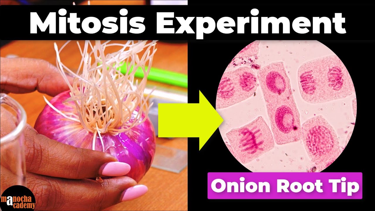


5.0 / 5 (0 votes)