Western blotting technique | principle and step by step procedure

Shomu's Biology
12 Jun 201617:59

Summary

TLDRThis video tutorial delves into Western blotting, a technique for detecting proteins in a mixture. It covers the process from protein extraction to separation via SDS-PAGE, transfer to a membrane, and detection using specific antibodies. The method's sensitivity allows for the detection of nanograms of protein and is crucial for understanding cellular processes and protein interactions. The tutorial also explains the importance of blocking to reduce background noise and the use of primary and secondary antibodies for accurate protein identification.

Takeaways

  • 🧬 Western blotting is a technique used to detect proteins in a mixture, different from Southern and Northern blotting which detect DNA and RNA respectively.
  • 🔍 It is highly sensitive, capable of detecting nanograms of proteins even when mixed with other proteins.
  • 🧴 The process begins with protein extraction from cells using detergent to break down the cell membrane, followed by centrifugation to separate the soluble protein components.
  • 🔬 Separation of proteins is achieved through SDS-PAGE (Sodium Dodecyl Sulfate Polyacrylamide Gel Electrophoresis), which separates proteins based on their mass and charge.
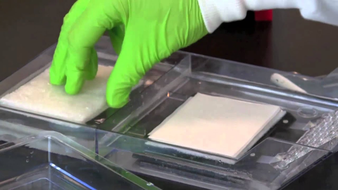
  • 🚫 The gel is fragile, so proteins are transferred onto a more robust medium like nitrocellulose or nylon membrane for further analysis.
  • 🖨 The transfer of proteins from the gel to the membrane is facilitated by applying an electric current, a process known as electroblotting.
  • 🚫 To prevent non-specific binding of antibodies, a blocking step is introduced where the membrane is treated with agents like dry milk to cover non-target areas.
  • 🔗 The primary antibody specifically binds to the target protein, but does not provide a signal for detection.
  • 🔄 After the primary antibody binding, unbound antibodies are washed away, and a secondary antibody is applied which acts as a reporter.
  • 💡 The secondary antibody is linked to an enzyme, often horseradish peroxidase (HRP), which upon reaction with a substrate, produces a detectable signal, such as light or color.
  • 📝 The final signal, whether luminescent or colored, indicates the presence and location of the target protein on the membrane, allowing for its identification and analysis.

Q & A

  • What is Western blotting?

    -Western blotting is a technique used to detect specific proteins in a sample. It involves separating proteins by size using SDS-PAGE and then transferring them onto a membrane to be detected with specific antibodies.

  • How does Western blotting differ from Southern and Northern blotting?

    -While Southern blotting is used to detect DNA and Northern blotting for RNA, Western blotting is specifically used for detecting proteins. Southern and Northern blotting techniques are quite similar, but Western blotting involves different steps, including the use of antibodies for detection.

  • What is the sensitivity of Western blotting?

    -Western blotting is a highly sensitive technique capable of detecting nanograms of protein molecules even in a mixture of other proteins.

  • What is the first step in the process of Western blotting?

    -The first step in Western blotting is the extraction of proteins from the cell. This involves treating the cell with a detergent to break down the cell membrane, followed by centrifugation to separate the soluble protein components from cellular debris.

  • What is the purpose of SDS in Western blotting?

    -SDS, or sodium dodecyl sulfate, is used in the electrophoresis step of Western blotting to denature proteins and give them a uniform negative charge, allowing them to be separated based on their mass during SDS-PAGE.

  • What is the role of the polyacrylamide gel in SDS-PAGE?

    -The polyacrylamide gel in SDS-PAGE provides a matrix with different pores that allow proteins to be separated based on their size. Smaller proteins can migrate further through the gel, while larger proteins are hindered, resulting in bands of proteins based on their mass.

  • Why is it necessary to transfer proteins from the gel to a membrane in Western blotting?

    -Transferring proteins to a membrane is necessary because the membrane is more robust and easier to handle than the gel. It allows for the application of specific antibodies for detection and is less prone to damage during the probing process.

  • What is the purpose of the blocking step in Western blotting?

    -The blocking step is used to prevent non-specific binding of antibodies to the membrane. It involves applying a blocking agent, such as dry milk, to cover areas of the membrane where the target protein is not present, reducing background noise in the final detection.

  • What are primary and secondary antibodies in the context of Western blotting?

    -Primary antibodies are specific to the target protein and bind directly to it. Secondary antibodies, which are anti-primary antibodies, bind to the primary antibodies and are often linked to an enzyme or a reporter molecule that aids in the detection of the target protein, such as through a colorimetric or luminescent reaction.

  • How is the presence of the target protein detected after the application of primary and secondary antibodies?

    -After the application of primary and secondary antibodies, a substrate is added that reacts with the enzyme linked to the secondary antibody. This reaction produces a visible color change or light emission at the location of the target protein, indicating its presence on the membrane.

  • What is the significance of the enzyme HRP in Western blotting?

    -HRP, or horseradish peroxidase, is an enzyme commonly linked to the secondary antibody in Western blotting. It catalyzes the conversion of a substrate, such as luminol, into a product that emits light, allowing for the detection of the target protein on the membrane.

Outlines

00:00

🔬 Introduction to Western Blotting Technique

The script introduces Western blotting as a technique for detecting proteins, distinguishing it from Southern and Northern blotting which are used for DNA and RNA detection, respectively. Western blotting is highlighted as a highly sensitive method capable of identifying nanograms of protein in a mixture. The script outlines the general process of blotting, emphasizing the three main stages: extraction, separation, and probing. It also mentions the use of Western blotting for detecting specific proteins, analyzing chemical reactions within cells, and understanding protein interactions.

05:02

🧬 Protein Extraction and Separation via SDS-PAGE

This paragraph delves into the first two stages of the Western blotting process: protein extraction and separation. Proteins are extracted from cells using detergents and centrifugation, resulting in a supernatant containing the protein mixture. The separation of proteins is achieved through SDS-PAGE (Sodium Dodecyl Sulfate Polyacrylamide Gel Electrophoresis), where proteins are differentiated based on their size and charge. The process is described in detail, explaining how smaller proteins migrate further in the gel due to the presence of different pore sizes in the polyacrylamide matrix.

10:03

🖨️ Transferring Proteins to a Membrane for Analysis

The script explains the transfer process of proteins from the SDS-PAGE gel to a nitrocellulose or nylon membrane, a critical step before probing. This transfer is necessary because the gel is fragile and not suitable for further probing steps. The process, known as electroblotting, involves applying an electric current that moves the proteins from the gel to the membrane. The membrane acts as a stable and durable medium for subsequent analysis, allowing for the proteins' pattern to be preserved for further detection.

15:04

🔎 Probing and Detection of Target Proteins

The final part of the Western blotting process is described in this paragraph, focusing on the probing and detection of the target protein. After transferring the proteins to the membrane, a blocking step is performed to prevent non-specific binding of antibodies. The primary antibody, specific to the target protein, is applied and binds to its antigen. Following a washing step to remove unbound antibodies, a secondary antibody linked to an enzyme is applied. This secondary antibody acts as a reporter, binding to the primary antibody and enabling the detection of the target protein through a colorimetric or luminescent reaction, depending on the substrate used.

Mindmap

Keywords

💡Western Blotting

Western blotting is a laboratory technique used to detect specific proteins in a sample of tissue homogenate or extract. It is one of the core vocabulary in the script as it is the main topic of the video. The process is described as being highly sensitive, capable of detecting nanograms of protein in a mixture, and is essential for identifying protein-protein interactions and the total proteome within a cell. The script outlines the entire process, from protein extraction to the final detection using antibodies.

💡Blotting Techniques

Blotting techniques refer to a group of methods used to transfer and detect specific molecules, such as DNA, RNA, or proteins, onto a membrane. In the script, different blotting techniques are mentioned, including Southern blotting for DNA and Northern blotting for RNA, with Western blotting being the focus. These techniques are fundamental to molecular biology and genomics research.

💡Protein Extraction

Protein extraction is the initial step in Western blotting where proteins are isolated from cells. The script describes treating cells with detergent to break down the cell membrane, followed by centrifugation to separate the soluble protein components from the cellular debris. This step is crucial as it provides the protein mixture that will be further processed in the blotting procedure.

💡SDS-PAGE

SDS-PAGE, or Sodium Dodecyl Sulfate Polyacrylamide Gel Electrophoresis, is a method described in the script for separating proteins based on their size. It is a critical step in Western blotting that allows for the visualization of proteins as distinct bands on a gel, facilitating the identification and analysis of specific proteins within a complex mixture.

💡Electrophoresis

Electrophoresis is the process used in blotting techniques to separate molecules, such as proteins, based on their charge and size. In the context of the script, electrophoresis is specifically applied to proteins in the form of SDS-PAGE, which is essential for preparing the proteins for transfer onto a membrane in Western blotting.

💡Transfer

In the script, transfer refers to the process of moving the separated protein bands from the SDS-PAGE gel onto a membrane, such as nitrocellulose or nylon. This step is unique to Western blotting and is necessary because the membrane is more stable and suitable for subsequent probing steps to detect the target protein.

💡Nitrocellulose Membrane

A nitrocellulose membrane is a type of porous membrane used in Western blotting to which proteins are transferred from the gel. The script mentions that this membrane has an affinity for proteins, allowing them to adhere to its surface after the transfer process, making it an ideal support for the detection of specific proteins.

💡Blocking

Blocking, as described in the script, is a step in Western blotting where a blocking agent, such as dry milk, is applied to the membrane to prevent non-specific binding of antibodies to the membrane. This is important to reduce background noise and ensure that the antibodies only bind to the target protein, thereby increasing the specificity of the detection.

💡Primary Antibody

The primary antibody is a specific type of antibody designed to bind to the target protein of interest. In the script, it is mentioned as the first antibody applied during the probing step of Western blotting. It is essential for the process as it directly interacts with the target protein, setting the stage for detection.

💡Secondary Antibody

The secondary antibody, as discussed in the script, is an antibody that binds to the primary antibody and is typically conjugated with an enzyme or a fluorescent tag. It acts as a reporter in Western blotting, amplifying the signal from the primary antibody's binding event, making the detection of the target protein more visible and quantifiable.

💡Horse Radish Peroxidase (HRP)

Horse Radish Peroxidase (HRP) is an enzyme used in the script's description of Western blotting as part of the detection process. When the secondary antibody is conjugated with HRP, it can catalyze a reaction with a substrate, such as luminol, to produce light. This light emission is a visual indication of the presence of the target protein, allowing for the detection and analysis of specific proteins on the membrane.

Highlights

Western blotting is a technique for detecting proteins, different from Southern and Northern blotting which detect DNA and RNA respectively.

Western blotting is highly sensitive, capable of detecting nanograms of proteins in a mixture.

The technique is used to identify specific proteins, understand chemical reactions within cells, and explore the total proteome.

Protein extraction involves treating cells with detergent to break down the cell membrane and release cytosolic components.

Centrifugation separates the supernatant containing proteins from the pellet containing cellular debris.

SDS-PAGE is used for protein separation based on size and charge, unlike DNA/RNA separation with agarose gel electrophoresis.

Proteins are negatively charged due to SDS treatment, migrating towards the positive end in the gel.

The process of transferring proteins from the gel to a membrane is called blotting, using an electric current.

Nitrocellulose or nylon membranes are used for protein transfer, as they are more robust than gels.

Blocking agents like dry milk are used to prevent non-specific antibody binding to the membrane.

Primary antibodies bind specifically to the target protein, but do not provide a detectable signal.

Secondary antibodies, linked to enzymes, act as reporters by binding to the primary antibodies.

Horseradish peroxidase (HRP) is a common enzyme used in conjunction with secondary antibodies for detection.

Luminal is a substrate that, when acted upon by HRP, produces light, indicating the presence of the target protein.

The detection of light can be captured on film, revealing the location of the target protein band.

Western blotting is crucial for identifying protein-protein interactions and their roles in cellular physiology.

The technique provides a visual confirmation of protein presence and can be used for further protein analysis.

Transcripts

play00:03

[Music]

play00:06

thank you

play00:10

welcome back friends welcome to another

play00:12

video lecture from Shamus biology and in

play00:14

this video tutorial we'll be talking

play00:16

about Western blotting we have been

play00:19

talking about different types of

play00:20

blotting techniques lately we've talked

play00:22

about southern blotting Northern

play00:24

blotting and this time we'll be talking

play00:26

about the Western blotting blotting is a

play00:28

technique of detecting macromolecules if

play00:31

you are trying to detect DNA we use

play00:33

solvent blotting if you want to detect

play00:35

RNA we use Northern blotting and if you

play00:39

want to detect proteins we use Western

play00:42

blotting now northern blotting and

play00:44

southern blotting has very very similar

play00:46

techniques which uses mostly same type

play00:48

of techniques and setup while Western

play00:50

blotting is a little different compared

play00:52

to them in Western blotting it is very

play00:54

very sensitive reaction technique that

play00:57

can even detect nanograms of protein

play01:00

molecules in the mixture of other

play01:01

proteins now there are different ways of

play01:04

detecting a Target protein from a

play01:06

mixture we can use chromatin

play01:08

immunoprecipitation we can use like

play01:10

coimino precipitation or antibody

play01:12

mediated pull down assays but in case of

play01:15

this Western blotting we detect very

play01:18

minute amount of protein that is present

play01:20

in a Cell fraction and a mixture and

play01:23

Western blotting is a widely used

play01:25

technique to not only detect a specific

play01:27

protein but also to find out different

play01:29

chemical reaction going on inside the

play01:31

cell as well as to find out the total

play01:33

proteome inside the cell as well as is

play01:36

very important to understand about the

play01:37

protein protein interactions for

play01:40

different physiological cellular

play01:41

purposes inside the cell so let us look

play01:44

at the whole process

play01:46

in the process of Western blotting again

play01:48

in any blotting processes we rely on

play01:50

three different stages the first step is

play01:53

to extraction an identification of first

play01:56

is the extraction of the molecule so in

play01:59

this case the molecule is proteins so we

play02:01

need to extract the protein content from

play02:03

a cell what we do we simply treat the

play02:05

cell with detergent that will break down

play02:08

the cell membrane so membrane pore will

play02:10

be formed and cytosolic components can

play02:12

come out then we do the spinning of that

play02:15

cell in the centrifuge that is going to

play02:17

give us a supernatant and a pellet the

play02:19

pellet contains all the cellular debris

play02:21

like cell membrane organelles and all

play02:24

the stuff while all the protein

play02:25

components are suspended it's it's

play02:27

soluble in the in the in the supernated

play02:30

so take out the supernatant and we have

play02:33

a protein mixture so start with that

play02:36

point so we have a protein

play02:39

mix

play02:41

that is isolated from cell and the

play02:44

protein mix contains so many varieties

play02:46

of protein let's see this green one

play02:49

let me draw some of the examples this

play02:51

red one

play02:54

that they have different concentrations

play02:55

as well but they have different

play02:57

varieties of

play02:59

proteins in there okay so protein mix

play03:02

then what we do the second step of any

play03:05

blotting process is separation of those

play03:08

mixture of molecules in this case we'll

play03:11

do the separation of protein the process

play03:13

of separation of protein that we use

play03:17

is

play03:19

electrophoresis

play03:21

obviously

play03:23

and the electrophoresis that we use to

play03:25

separate protein molecules from each

play03:27

other are known as

play03:29

SDS page

play03:33

sodium dodecil sulphate which is SDS

play03:37

polyacrylamide gel electrophoresis known

play03:40

as pitch so polyacrylamide jelly

play03:43

electrophoresis is a process of

play03:44

separating

play03:45

all those protein mixtures from each

play03:48

other

play03:49

now in case of Southern and West

play03:51

Northern blotting we have separated DNA

play03:54

and RNA molecules ah with the help of

play03:57

agarose gel electrophoresis but in case

play04:00

of

play04:01

in case of Western blotting we separate

play04:04

proteins with page

play04:06

it's a normal process of separating

play04:07

protein molecules in this case the the

play04:10

gel is made with polyacrylamide because

play04:13

acrylamide can attach with each other to

play04:16

make a cross linking and form a polymer

play04:18

so they are separated based on forming

play04:20

this polymers there

play04:22

okay so they are separate based on that

play04:25

so once they separate so so let me draw

play04:27

this whole thing of SDS page

play04:32

in one way we put all those known

play04:34

proteins now SDS page is a type of

play04:38

electrophoresis process which helps

play04:40

separating proteins based on their

play04:43

charge

play04:46

size

play04:49

and Mass

play04:53

mainly by their size and mainly based on

play04:55

their mass and and also shape ah mostly

play04:58

not size you can say the shape

play05:02

so its mostly the structural feature

play05:04

that helps separating proteins based on

play05:07

that charge the separation of protein

play05:09

based on the charge requires some some

play05:11

more modifications in the SDS page but

play05:13

mostly this separated by their mass now

play05:16

the idea is as this this is a

play05:19

polyacrylamide gel it carries so many

play05:22

different pores in between

play05:24

if the protein is smaller then that

play05:27

protein can migrate further but if a

play05:30

protein is large that protein will not

play05:33

migrate much that protein will halt at a

play05:37

specific point so the smaller proteins

play05:39

will migrate further so we'll get bands

play05:41

in in a in further directions while the

play05:44

larger proteins

play05:47

will be lacked behind during the

play05:49

migration and let us say the migration

play05:51

direction is this OK and again the

play05:54

directionality of this migration that we

play05:56

are talking about is also towards the

play06:00

positive end

play06:02

because the proteins that we are talking

play06:03

about here they are negatively charged

play06:05

because they treat it with SDS so the

play06:08

negatively charged so they will migrate

play06:09

towards the positive end

play06:12

okay so that is the idea

play06:15

of SDS page so with the help of this SDS

play06:18

page we separate the mixture of proteins

play06:21

based on their Mass

play06:23

and charge means or not let's talk about

play06:26

charge here we just talk about the mass

play06:28

and separation so now let's say we

play06:30

separated all the mixture of proteins

play06:32

based on their mass and then what we do

play06:35

we need to use a probe to find out our

play06:39

Target protein right

play06:42

that is the idea this is the second step

play06:44

of this blotting process every blotting

play06:46

process consists of three state the

play06:48

first step is extraction and isolation

play06:49

second step is the electrophoresis third

play06:53

step is a probing

play06:55

and before and between the

play06:56

electrophoresis and probene a new unique

play07:00

process to be done that is the transfer

play07:02

of the content that is present in the

play07:04

gel onto a membrane paper why it's

play07:07

required because you know we want to add

play07:10

probe and that probe can bind to

play07:13

different regions of this gel it should

play07:15

be properly attached to the Target

play07:17

protein only

play07:18

right and gel is really really fragile

play07:22

because the content of the gel is not

play07:24

that strong so we cannot do rest of the

play07:27

process of probing and all this

play07:29

reactions using using this gel

play07:33

so we need to transfer the components of

play07:35

the gel exactly like the same imprint

play07:37

into a nitrocellulose membrane or nylon

play07:40

membrane filter

play07:42

for this process of transfer that is the

play07:45

reason we call it as a western blotting

play07:48

that's the term blood comes in because

play07:49

it's simply blotted and simply taking

play07:51

the imprint of the gel replica onto a

play07:55

natural cellulose membrane so you take

play07:57

the nitrocellulose membrane and we put

play08:00

on top of this gel and then we apply

play08:03

current that will help flowing this

play08:06

protein molecules from the gel towards

play08:09

the membrane and those proteins will be

play08:11

attached to the membrane because this

play08:13

membrane is kind of a like sticky

play08:15

towards those macromolecules okay so

play08:18

let's see how this process is exactly

play08:20

done so let us assume SDS page is

play08:23

conducted now if you don't have any idea

play08:25

about SDS page and if you don't know

play08:26

about the details of SDS page I will

play08:28

recommend you to watch my video on SDs

play08:30

page there are plenty videos three or

play08:32

four videos available in my channel

play08:33

regarding SDS page you can watch that

play08:36

anytime but for now let us say the SDS

play08:38

page is done and we have the gel ready

play08:40

then how we transfer this component the

play08:43

way to transfer this component in case

play08:45

of Western blotting

play08:47

Electro illusion that is the process of

play08:49

transferring the component from the

play08:51

sdsgel to the Nitro cellulose membrane

play08:53

so what we do let us assume this gel as

play08:56

a as a side view so if you look at this

play08:59

gel as a side view it will look

play09:00

something like this this is the gel

play09:04

we put it in horizontal line

play09:06

and what we do now we also put

play09:09

the nitrocellulose membrane let us say

play09:12

this is the Nitro cellulose membrane

play09:16

this is the membrane

play09:18

and this is the gel

play09:22

OK and then what we do is we apply

play09:26

electrodes in both these directions okay

play09:29

so we will apply

play09:31

positively charged electrodes towards

play09:33

the membrane negatively charged

play09:35

electrodes on this towards the gel side

play09:37

that is the idea that we have

play09:40

ok now what we do once we put the

play09:42

electrodes the current will flow

play09:45

from this negative towards the positive

play09:47

electrode and as the current is Flowing

play09:49

from negative to positive the protein

play09:52

molecules that are present

play09:54

here because you know they this this

play09:56

membrane is in direct contact with the

play09:58

gel so let me do that

play10:00

it is in direct immediate contact with

play10:03

the gel

play10:05

it's an immediate contact so what is

play10:07

going on once we apply the current the

play10:09

protein molecules start flowing

play10:13

from the gel

play10:17

to the nylon filter

play10:23

that is the process and we also put

play10:26

everything in the buffer definitely we

play10:27

need to put them in buffer and

play10:28

electrolytes so that they can flow so

play10:31

once this process is done what is the

play10:33

importance of this process plotting that

play10:35

is the transfer of those components and

play10:38

proteins from the SDS page to the

play10:41

nitrocellus membrane because the

play10:43

membrane is much easier to handle it's

play10:46

more strong and we can we can use this

play10:48

membrane for other purposes so once this

play10:51

blotting is done once the transfer is

play10:53

complete then we take the membrane

play10:56

then we take this membrane here let me

play10:59

take the membrane let's draw the

play11:01

membrane

play11:02

this is the membrane and let us say in

play11:04

this membrane we find out ah some region

play11:06

of proteins different regions or bands

play11:08

that we can see for example this

play11:12

this

play11:14

is for example

play11:16

everything is present in the membrane

play11:18

now the third important stage that is a

play11:21

probing

play11:23

and hybridization in case of DNA and RNA

play11:26

blotting we simply use complementary

play11:28

strands for the hybridization assay but

play11:31

in this case hybridization won't work

play11:33

because it's protein so we need to use

play11:35

any sort of detector

play11:37

that can go and bind to the specific

play11:39

Target protein of our interest for that

play11:42

you know

play11:44

we have antibody

play11:46

antibodies can be specific it can attach

play11:49

to specific proteins so it design

play11:51

antibody against our Target protein and

play11:54

we apply that antibody to go and

play11:56

interact with our Target protein among

play11:59

the rest of the proteins in the

play12:01

nitrocellulose membrane

play12:03

but before doing that we do another step

play12:05

which is called as a blocking blocking

play12:08

is a step where we apply some blocking

play12:11

agent example one of one such blocking

play12:13

is Agent is dry milk we apply dry milk

play12:16

so that that portion covers most of the

play12:20

area because that will not bind to the

play12:22

protein or targeted protein areas but

play12:24

mostly that will bind to the rest of the

play12:27

Nitro cellulose membrane why it is

play12:29

important because if we don't do the

play12:32

blocking State and simply put the

play12:34

antibody antibody are specified antibody

play12:37

still can bind with some unspecific

play12:39

reaction with different regions antibody

play12:41

should bind with the protein but along

play12:43

with that it might also bind with some

play12:45

extra other regions of the of the

play12:47

membrane that will give us some noises

play12:50

background noises because it will be

play12:52

giving us a positive false ah positive

play12:54

signal or its a kind of a background

play12:56

noise so to reduce this noise we won't

play12:59

allow this antibody to be interacting

play13:01

with the rest of the member membranes

play13:03

region so that is the region we we kind

play13:06

of covered the rest of the membrane with

play13:08

all this blocking edges so with the help

play13:11

of buffer and blocking agents we simply

play13:13

block the rest of the portion of the

play13:16

membrane and we also use some mild

play13:18

detergent as well so it's covering rest

play13:21

of the blocking area then we apply the

play13:23

primary antibody and the antibody is

play13:26

very specific and so much specific

play13:27

towards the target protein it will not

play13:30

allow to any blocking to prevent it so

play13:32

it is going to bind for example say this

play13:34

is the target this is the target

play13:36

protein so antibody can bind to the

play13:38

Target protein and even multiple

play13:40

antibodies can bind to one protein

play13:42

content

play13:44

OK that is the first step and the

play13:46

antibody that we applied for the first

play13:48

time is known as the primary antibody is

play13:51

known as

play13:52

primary

play13:53

antibody because this is the first type

play13:55

of antibodies that we apply which will

play13:58

directly bind

play14:00

it will directly bind to the Target

play14:04

protein but primary antibody lacks in

play14:08

specific part that it will not show us

play14:11

the result because it's not a reporter

play14:16

it can only directly bind to the Target

play14:19

but it will not give us any fluorescence

play14:21

or any light or anything of that

play14:23

it's just for the proper binding so

play14:27

after binding of the primary antibody we

play14:29

we do the process of washing out so the

play14:31

rest of the Unbound antibodies are

play14:32

washed away

play14:34

then we also add a secondary antibody

play14:39

secondary antibody and now this one is

play14:42

important because secondary antibody is

play14:45

acting as a reporter

play14:49

because this secondary antibody is

play14:51

anti-primary antibody that means if this

play14:54

is the primary antibody

play14:57

one degree means primary

play14:59

secondary antibody will be anti-primary

play15:02

that means secondary antibody have the

play15:04

capability to bind with the primary

play15:06

antibody FC portions

play15:09

that will be the interaction between

play15:11

primary antibody and secondary will be

play15:14

targeted against primary so it will

play15:16

attach to the primary now secondary is

play15:18

attached with any sort of enzyme

play15:23

that is why it is called as a reporter

play15:25

primary goes and bind to our Target

play15:27

protein let us say this is the target

play15:29

protein the green color let us say this

play15:30

is the target protein primary binds to

play15:32

the Target protein we applied we do the

play15:34

washing step then you apply secondary

play15:36

antibody secondary antibody is attached

play15:38

to the enzyme it is binding to the

play15:40

primary antibody so we have a proper

play15:42

binding done

play15:43

now here the secondary antibody attached

play15:46

to the enzyme so if after this we add

play15:49

the substrate

play15:52

and the substrate will be converted into

play15:54

product by the attached enzyme that is

play15:57

present there with the secondary

play15:58

antibody FC region it will be converting

play16:00

the substrate into product

play16:02

now the product that we prepare

play16:04

sometimes give us color so if we do this

play16:07

reaction

play16:09

in in different big in different let's

play16:11

say trays and stuff we get some color

play16:13

generates in a specific region of this

play16:16

whole membrane that is going to tell us

play16:18

that that is the region where the target

play16:20

proteins present

play16:22

or sometimes the this this enzyme or the

play16:25

chemical molecule whatever present there

play16:27

upon giving a specific substrate it will

play16:29

convert it into a product and it will

play16:31

generate light as a byproduct

play16:34

and that is very common in case of the

play16:37

western blotting technique and detection

play16:38

and the enzyme that we use is known as a

play16:41

horse release peroxidase hrp horse

play16:44

reduce peroxidase once you use hrp

play16:47

remember hrp here is attached

play16:50

and then we add the luminal

play16:53

it converts the luminal and break it

play16:55

down and as a byproduct of this reaction

play16:58

it provides us light

play17:00

and we can show the light that is coming

play17:03

out and that can be easily detected with

play17:05

a extra film because that that film is

play17:08

completely uh like dark and the dark you

play17:11

will see those gray faded bands due to

play17:13

the generation of the light that's going

play17:15

to give us the idea about where exactly

play17:17

the target protein is present in that

play17:20

mixture of SDS page in the in the

play17:23

picture of the SDS page so let's say you

play17:26

want to find out that protein and cut it

play17:28

out you can still do that because if you

play17:31

see where exactly the light is coming

play17:32

from so when you develop the film you'll

play17:35

only see only one band

play17:38

and in this case let's say the band is

play17:40

kind of here

play17:42

so then you can simply look at this this

play17:45

SDS page

play17:46

and you find out the band in the page so

play17:49

if you want to take that protein out you

play17:51

can simply go there and cut that portion

play17:53

out and do the rest of the job

play17:56

so that is the process

play17:58

you see

Rate This

5.0 / 5 (0 votes)

関連タグ
Western BlottingProtein DetectionMolecular BiologyTechnique TutorialSDS-PAGEBlotting TechniquesCell FractionAntibody BindingProtein AnalysisBiotechnologyLab Methods
英語で要約が必要ですか?