Pons - External and Internal (White & Grey matter) + QUIZ | Anatomy
Summary
TLDRThis educational video script delves into the anatomy of the Pons, a critical part of the brainstem. It covers the Pons' external and internal structures, highlighting its role in cranial nerve connections and the arrangement of grey and white matter. The script also discusses the Pons' interaction with other brain regions, emphasizing its importance in motor control and sensory processing.
Takeaways
- 🧠 The central nervous system is divided into the encephalon and the spinal cord, with the encephalon further divided into the brainstem, cerebellum, diencephalon, and telencephalon.
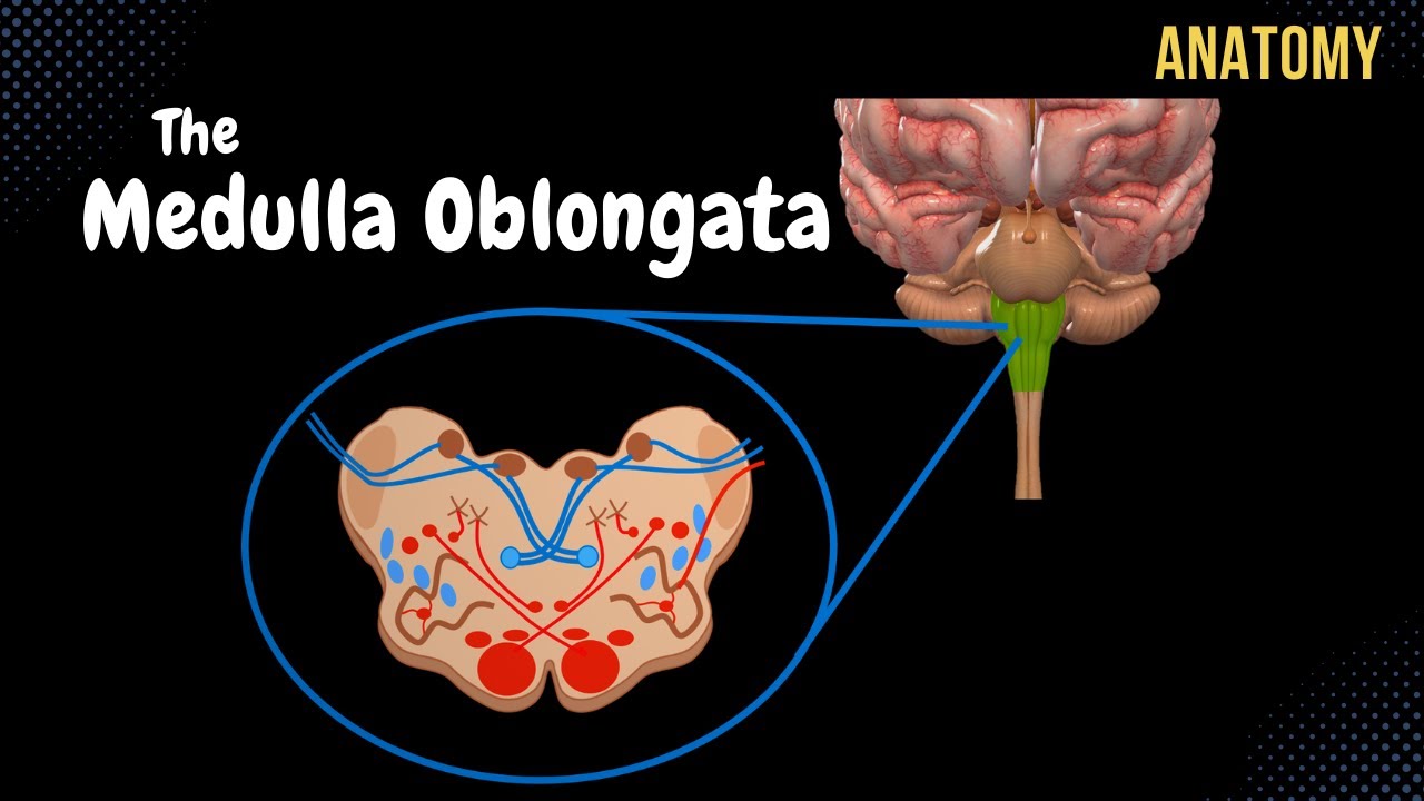
- 🌉 The brainstem includes the medulla, pons, and midbrain, and the pons is the focus of the video, located above the medulla and in front of the cerebellum.
- 👀 The pons has an anterior surface with a basilar sulcus and forms the middle cerebellar peduncles, which connect to the cerebellum.
- 🤖 Several cranial nerves emerge from the anterior part of the pons, including the 5th (trigeminal), 6th (abducens), 7th (facial), and 8th (vestibulocochlear) nerves.
- 🔍 The posterior surface of the pons is covered by the cerebellum and works with the medulla and midbrain to form structures like the rhomboid fossa.
- 📚 Grey matter in the pons contains nuclei, while white matter contains nerve fibers that form tracts.
- 🧬 The pontine nuclei in the basal part of the pons are crucial for the frontopontine tract, which synapses with the pontine nuclei and projects to the cerebellum.
- 🔄 The trapezoid body, formed by crossing fibers of the auditory nerve, divides the internal surface of the pons into the tegmentum and basal part.
- 🚀 Descending tracts in the pons include the corticopontine tracts, which are extrapyramidal and originate from various areas of the cerebral cortex.
- 🌐 Ascending tracts in the tegmentum of the pons include the medial lemniscus, spinal lemniscus, trigeminal lemniscus, and lateral lemniscus, which are involved in sensory perception.
- 🏃♂️ Descending tracts in the tegmentum of the pons include the tectospinal, rubrospinal, reticulospinal, and vestibulospinal tracts, which are involved in motor control and coordination.
Q & A
What are the two main parts of the central nervous system?
-The central nervous system consists of the encephalon and the spinal cord.
What is the brainstem composed of?
-The brainstem is composed of the Medulla, Pons, and Midbrain or mesencephalon.
What is the Pons' location in relation to other brain structures?
-The Pons is located above the medulla and in front of the Cerebellum, with the spinal cord below it.
What is the function of the basilar sulcus found on the anterior surface of the Pons?
-The basilar sulcus is a prominent groove on the anterior surface of the Pons where it turns backwards toward the Cerebellum, forming the middle cerebellar peduncles.
Which cranial nerve is associated with the lateral extraocular muscles of the eyes?
-The 6th cranial nerve, the nervus abducens, is associated with the lateral extraocular muscles of the eyes, aiding in the abduction of the eyeballs.
What is the role of the facial nerve (cranial nerve number 7)?
-The facial nerve provides motor innervation for the facial muscles, which helps with facial expressions.
How does the Pons contribute to the formation of the Rhomboid Fossa?
-The upper part of the Rhomboid Fossa is formed by the Pons, while the lower part is formed by the medulla.
What is the significance of the Trapezoid Body within the Pons?
-The Trapezoid Body is formed by fibers of the cochlear nerve that cross within the Pons to the other side, dividing the internal surface of the Pons into the Tegmentum and the Basilar Part.
What is the primary function of the Pontine Nuclei?
-The Pontine Nuclei are significant as they receive synapses from the Frontopontine Tract and then send fibers to the Cerebellum through the middle cerebellar peduncles as the Pontocerebellar tracts.
What type of tracts are found in the Basilar Part of the Pons?
-The Basilar Part of the Pons contains only descending tracts or motor tracts, including the Frontopontine tract and the Corticopontine tract.
How does the Tectospinal tract contribute to eye and neck movements?
-The Tectospinal tract, originating from the tectum of the Midbrain, is responsible for coordinated eye and neck movements, allowing the neck muscles to unconsciously follow eye movements.
Outlines
🧠 Introduction to the Pons
The script begins with an introduction to the anatomy of the Central Nervous System (CNS), focusing on the Pons, a part of the brainstem. The CNS is divided into the encephalon and the spinal cord, with the encephalon further divided into the brainstem, cerebellum, diencephalon, and telencephalon. The Pons is situated above the medulla and in front of the cerebellum. The video will cover the external surfaces of the Pons, its topography, and structures visible from anterior and posterior views. It will also explore the internal surface by slicing the Pons to observe the arrangement of grey and white matter. A quiz is mentioned at the end to aid memorization.
🔍 External Surfaces of the Pons
The script describes the external surfaces of the Pons, which include an anterior and a posterior surface. The anterior surface features the basilar sulcus in the midline and the middle cerebellar peduncles that form the structure connecting to the cerebellum. Several cranial nerves emerge from the anterior part of the Pons, including the 6th (abducens), 7th (facial), 8th (vestibulocochlear), and 5th (trigeminus) cranial nerves, each serving specific functions related to eye movement, facial expression, hearing, balance, and sensory and motor innervation of the face. The posterior surface of the Pons is covered by the cerebellum and lacks distinct structures on its own but works with the medulla and mesencephalon to form structures like the rhomboid fossa, an important landmark for tracts and nuclei.
🧬 Internal Surface and Grey Matter of the Pons
The script moves on to discuss the internal surface of the Pons, emphasizing the importance of grey matter, which contains cell bodies or nuclei, and white matter, which contains nerve fibers forming tracts. A key landmark, the Trapezoid Body, formed by fibers of the auditory nerve crossing within the Pons, divides it into dorsal (Tegmentum of Pons) and ventral (Basilar Part) regions. The Basilar part contains the Pontine Nuclei, crucial for the frontopontine tract's synapse and the subsequent pontocerebellar tracts to the cerebellum. The Tegmentum houses nuclei for cranial nerves 6-8 and the reticular formation, with small nuclei of the Trapezoid body located between the fibers of the Trapezoid body.
📚 White Matter Tracts and Summary Quiz
The script concludes with a discussion of the white matter tracts in the Pons, differentiating between descending (motor) tracts in the Basilar part, such as the frontopontine and corticopontine tracts, and ascending (sensory) tracts in the Tegmentum, including the medial lemniscus, spinal lemniscus, trigeminal lemniscus, and anterior spinocerebellar tract. It also mentions the descending tracts in the Tegmentum, like the tectospinal, rubrospinal, reticulospinal, and vestibulospinal tracts, as well as the medial longitudinal fasciculus. The script ends with a challenge for viewers to identify various structures and tracts without labels, suggesting that if they can do so, they have fully grasped the anatomy of the Pons.
Mindmap
Keywords
💡Central Nervous System
💡Pons
💡Brainstem
💡Cerebellum
💡Basilar Sulcus
💡Cranial Nerves
💡Rhomboid Fossa
💡Grey Matter
💡White Matter
💡Pontine Nuclei
💡Trapezoid Body
Highlights
The central nervous system is composed of the encephalon and the spinal cord.
The encephalon is divided into the brainstem, cerebellum, diencephalon, and telencephalon.
The brainstem includes the medulla, pons, and midbrain.
The pons is located above the medulla and in front of the cerebellum.
The external surfaces of the pons include an anterior and a posterior surface.
The anterior surface of the pons features the basilar sulcus and the middle cerebellar peduncles.
Cranial nerves VI, VII, and VIII emerge from the anterior part of the pons.
The posterior surface of the pons is covered by the cerebellum.
The rhomboid fossa is formed by the pons and the medulla.
Cerebellar peduncles are found on the posterior surface of the pons.
The internal surface of the pons is divided into the tegmentum and the basilar part.
The pontine nuclei are located in the basilar part of the pons.
The trapezoid body is formed by fibers crossing within the pons.
The tegmentum of the pons contains nuclei for cranial nerves 6-8.
The reticular formation is present in all parts of the brainstem, including the pons.
The pons contains both ascending (sensory) and descending (motor) tracts.
The corticopontine tract is an example of an extrapyramidal tract.
The medial lemniscus is an ascending tract that carries sensory information to the cerebral cortex.
The tectospinal tract is involved in coordinated eye and neck movements.
The rubrospinal tract supports and coordinates voluntary movement.
The reticulospinal tract is responsible for balance and posture.
The medial longitudinal fasciculus coordinates involuntary head and eye movements.
Transcripts
What’s up, Meditay here; let’s continue the anatomy of the Central Nervous System.
In this segment, we’ll cover the complete anatomy of Pons
So the central nervous system consists of two parts:
the encephalon and the spinal cord. The encephalon is then further divided into specific parts.
We have the brainstem, which consists of the Medulla, Pons, and the Midbrain or the
mesencephalon. We have the Cerebellum back here, then the diencephalon and the telencephalon.
Our focus in this video is going to be Pons, which is here.
So in this video, we’re first going to cover the external surfaces of the Pons. Basically,
look at its topography and what structures you’ll find from an anterior view and a posterior view.
Then we’re gonna slice up the Pons and look at the internal surface. Basically, see how the grey
matter and white matter are arranged within it. Then I’ve made a little quiz at the end which
might help you if you need to memorize. Alright, so we can start by replacing
this picture with a more realistic one. Pons is located here, lying right above
the medulla and in front of the Cerebellum. And down here, you can see the spinal cord.
Now, if we remove a part of the cerebral cortex, you’ll be able to see the rest of Pons.
As well as the mesencephalon, or the Midbrain, which is the most superior part of the brainstem.
Alright. Now. Externally, your Pons has two surfaces. It has an anterior surface and
a posterior surface. Let’s now cover the typical morphology of these two surfaces,
starting with the anterior surface first. And we’ll do that by looking at the brainstem
from this perspective; we’ll see this. So again, Pons is this prominent bulb you see here. In the
midline of the anterior surface, there s a sulcus called the basilar sulcus. Laterally on either
side, you’ll see that Pons turns backwards toward the Cerebellum. And that is because Pons forms
the middle cerebellar peduncles on either side, which continue into the Cerebellum back there.
That was the general morphology of the anterior surface of Pons,
but there are some cranial nerves that leave the brainstem at this area. We
have 12 cranial nerves in our body, and each serves its particular function in the brain,
but from the anterior part of the Pons. You’ll see a nerve that goes out between the Pyramids
of medulla oblongata and Pons. This nerve is the 6th cranial nerve, the nervus abducens,
that go to the lateral extraocular muscles of the eyes to help with the abduction of the eyeballs.
Then, between the Olives and the middle cerebellar peduncle, you’ll find cranial nerve number 7. The
facial nerve, which provides motor innervation for the facial muscles, helps with facial
expressions. And cranial nerve number 8, the vestibulocochlear nerve for hearing and balance.
The last nerve is at the surface of Pons. And that is the 5th cranial nerve, the nervus Trigeminus.
It’s a very large nerve that goes to your facial region
to provide sensory innervation, motor innervation, as well as parasympathetic innervation for glands.
So all of these nerves emerge from the anterior area of Pons.
Now let’s turn this model around and look at the posterior surface. From the posterior view, you’ll
see that the Cerebellum covers the whole posterior area of Pons. SO let’s go ahead and remove it.
So Pons is here, above the medulla and below the Midbrain, or mesencephalon.
Pons doesn’t really have any specific structure on its own. Rather it works with the medulla and the
mesencephalon in forming certain structures. One of them is the Rhomboid Fossa. The rhomboid fossa
has many nuclei for the cranial nerves and is a very important landmark for tracts and nuclei.
I’ll cover this in detail in my next video. But for now, I want you to know that the upper part
of the rhomboid fossa is formed by Pons, and the lower part is formed by the medulla. Cool.
On either side of the rhomboid fossa, you’ll see the cerebellar peduncles, which contain
fibers that run between the Cerebellum and all three parts of the brainstem. The inferior
cerebellar peduncle contains fibers that go to the medulla. The middle cerebellar peduncle contains
fibers that go to Pons. The superior cerebellar peduncle contains fibers that go to the Midbrain.
So that was all for the posterior surface of Pons. Now let’s go over to the next segment of this
video, and look at the internal surface of Pons. But before we do that, I want you to keep in mind
that grey matter always contains cell bodies, so all structures within the grey matter
are nuclei. White matter contains nerve fibers that form tracts.
Now let’s take Pons, and slice it up like this to look at the internal surface.
So this is an outline of what Pons looks like. We’ll see the Basilar Sulcus here, so this is
the anterior surface. We’ll also see the middle cerebellar peduncles and the Rhomboid Fossa back
here. I hope it makes sense at this point. Before we dig into structures on the surface of Pons,
we need to go through an important landmark that we use to separate Pons into two parts. So here we
see the internal part of the ear. The inner ear we call it with the cochlea and the vestibulum.
The cochlea, which is our primary organ of hearing, has a nerve called the cochlear
nerve. The cochlear nerve will go directly towards Pons and then ascend towards the superior temporal
gyrus as the Lateral Lemniscus. But some fibers cross to the other side and then ascend to the
superior temporal gyrus as the lateral lemniscus. Now, why am I telling you this? Because as these
fibers cross within Pons to the other side, they form the Trapezoid Body, as you see here.
And the Trapezoid body divides the internal surface of Pons into two regions. The Dorsal part
becomes the Tegmentum of Pons, and the Ventral Part becomes the Basilar Part. Let’s now go
through the Grey matter in Ventral and Dorsal parts, then do the white matter, so we’ll start
with the gray matter structures associated with the Basilar part first. The only grey
matter in the Basilar part are the Pontine Nuclei These Pontine nuclei are a very significant part
of Pons because, there’s gonna be a tract that originates outside of the primary motor cortex,
from the frontal lobe of the cortex, called Frontopontine Tract.
It will descend and synapse with the pontine nuclei, as you see here. Then once they synapse,
these fibers will go to the Cerebellum through the middle cerebellar peduncles, as
the Pontocerebellar tracts. So these fibers cross to the other side and then go to the Cerebellum.
Once they’re at the Cerebellum, the tracts will then travel towards the Red nucleus
of the Midbrain as the cerebellorubral tract and then descend as the rubrospinal tract to
support voluntary movements. So the Pontine nuclei are a very important part of Pons.
The grey matter of the Tegmentum consists of nuclei that belong to cranial nerves number
6-8 in the Rhomboid Fossa. We’ll cover the rhomboid fossa in detail in a separate video,
but for now, we’ll stick with the most important structures associated with Pons to really
understand the anatomy of it. Next, we’ll see the reticular formation. You’ll find the reticular
formation along all parts of the brainstem. Then between the fibers of the trapezoid body,
you’ll find small nuclei called the nuclei of the Trapezoid body,
which some of the fibers synapse with. So that was all the grey matter of Pons. Now
let’s go ahead and cover the white matter of the Basilar Part and the Tegmentum of Pons.
We’ll start with the basilar part. Here you’ll only find descending tracts or motor
tracts. Remember earlier when we talked about the frontopontine tract and the pontocerebellar
tracts? Where fibers from the frontal cortex go down to synapse with the pontine nuclei in Pons,
and then fibers go from Pons to the Cerebellum as pontocerebellar tracts? Pontocerebellar tracts
are descending tracts, so we’ll need to colour it as red. The other descending tracts you see
here is the frontopontine tract, you’ll see it depending on at which level you cut Pons.
But, the frontopontine tract is a part of a bigger bundle of tracts called the Corticopontine tract.
The Frontopontine tract was just an example. If the tract originates from the occipital lobe,
it’s called occipitopontine tract. If it originates from the temporal lobe,
it’s called temporopontine tract. And so on. That’s why I specifically said frontal
lobe earlier because the frontopontine tract comes from the frontal cortex.
Notice that they all originate from different areas of the cerebral cortex,
which are outside of the primary motor area of the brain. And because of that, these tracts
are considered extrapyramidal tracts. Because they don’t originate from the pyramidal cells of
the primary motor cortex. So when we use the term Corticopontine tract, you’re really talking about
the frontopontine tract, occipitopontine tract, temporopontine tract, and Parietopontine tract.
So you’ll find the corticopontine tract fibers here synapsing with the pontine nuclei. Awesome.
The other descending tract is a pyramidal tract, called the Corticospinal tract. It’s a pyramidal
tract because it comes from the pyramidal cells of the primary motor area in the cortex and descends
down to the spinal cord to innervate skeletal muscles. Alongside the corticospinal tract, you’ll
find corticonuclear tracts as well. It descends in the same areas as the corticospinal tract,
but the corticonuclear tracts are responsible for the voluntary control of muscles located in the
head and neck. So that was all the white matter of the basilar part—only descending tracts. Now let’s
do the white matter in the Tegmentum of Pons. The first one is the medial lemniscus. Now we need to
repeat a few things in order to remember this one. So here is the cross-section of the spinal cord
and the medulla. Remember that there were fibers that came from the Lower parts of the body, which
ascend as Gracile fascicle and sensory fibers that came from the upper parts of your body, which
ascend as Cuneate fascicle? I use the G in Gracile Fascicle as Genitals to remember that it comes
from the lower parts of the body. So these fibers receive conscious proprioceptive information,
as well as sensory input from mechanoreceptors, and they ascend to the gracile and cuneate nuclei
in the medulla. Then fibers there will leave as either the external arcuate fibers
or internal arcuate fibers. The internal arcuate fibers cross to the other side, and then they will
ascend as the medial lemniscus, which is what you see here in Pons. They will ascend and go
to the primary somatosensory area in the cerebral cortex so that you’re aware of sensory touch and
vibration, as well as the position of your body parts. So that is the Medial Lemniscus.
Then we have the Spinal Lemniscus. Remember when we talked about the cross-section of the medulla,
we had two spinothalamic tracts? One lateral and one anterior? They ascend together as the
spinal lemniscus, and that’s what you see here in Pons. So it ascends to the cortex,
to the primary somatosensory area as well. And they’re responsible for conscious sensory input
of Pain temperature, Pressure, and touch. So that is this one.
Next, we have the Trigemnical Lemniscus. The trigemnical lemniscus comes from the trigeminal
ganglion, which is a part of the trigeminal nerve, the 5th cranial nerve. So Pons receives sensory
input from this nerve, and then it crosses to the other side and ascent to the primary somatosensory
area. So that is the trigeminal lemniscus. Then we have the Anterior spinocerebellar tract.
Remember when we talked about the medulla, that we have posterior and anterior spinocerebellar
tracts? The posterior spinocerebellar tract will go through the inferior cerebellar peduncle
to go to the Cerebellum. But the anterior one will go through Pons and the Midbrain
and then go to the Cerebellum through the superior cerebellar peduncle. And that’s why
we see it here because it’s going up to the midbrain to eventually go to the cerebellum.
Then, I wanna repeat something I mentioned earlier in this video. About the auditory nerve called the
cochlear nerve that goes to the Pons and then ascends as the lateral lemniscus to go to the
superior temporal gyrus, which is the primary auditory area of the brain. Some fibers cross
and then ascend to the superior temporal gyrus. And the fibers that cross form the trapezoid
body. But when they ascend after crossing, they also ascend as the lateral lemniscus, which is an
ascending tract that we need to include as well. So that was all the ascending tracts in
the Tegmentum of Pons. Remember, blue represents sensory or ascending tracts, and red represents
descending or motor tracts. Now let’s do all the descending tracts in the Tegmentum of Pons.
And the first one is the Tectospinal Tract The tectospinal tract is called tectospinal
because these fibers come from the tectum of the Midbrain. So here is the Midbrain. Posteriorly,
you’ll find the tectum. So the tectospinal tract descend from here. And these are fibers
that are associated with coordinated eye and neck movements. And remember, since this tract
originates from the brainstem it is referred to as an extrapyramidal tract. Hence, it unconsciously
moves your neck muscles with your eyes. So when you look at something.
Imagine you’re looking at a hamburger, you look at it, and you keep looking at it as it passes you,
and your neck muscles unconsciously follow your eyes. That’s what this tectospinal
tract is responsible for. And that is our first descending tract of the tegmentum.
The next one is called the Rubrospinal tract. Remember we talked about the corticopontine tract,
which are fibers that originate from anywhere outside of the primary motor area that descends to
synapse with the pontine nuclei? These fibers will go to the Cerebellum as the pontocerebellar tract.
From the Cerebellum, fibers will go to the Red nuclei of the Midbrain,
as the cerebellorubral tract. And then they will descend as the rubrospinal tract. Notice that I
said these fibers do not originate from the pyramidal cells of the primary motor cortex,
that’s why they’re referred to as extrapyramidal tract. They don’t initiate movement,
but they support and coordinates the voluntary movement. So that is the rubrospinal tract.
Next, we have the reticulospinal tract, which is a part of the balance and posture system. They come
from the reticular formation inside the brainstem. The Reticular system are responsible for Sleep,
alertness, cardiovascular control, breathing and all of those vital things. But they’re also
responsible for motor control like your balance and posture through the reticulospinal tract.
So that is this one. Next, we have a vestibulospinal tract
And to understand this one, we need to involve the inner ear again, because the inner ear has
the vestibular system. The vestibular system has crystals within it that monitors your balance. And
it transmits impulses to your brain, and then down to your muscles so that you can keep your balance.
So whenever your head is tilted or you’re upside down, or you’re about to fall,
all of that is monitored and controlled by your vestibular system. So it helps with balance and
posture. Alright, let’s just clean up the labels and add a little color to differentiate them.
There is one more tract that we need to mention, which is the medial longitudinal fasciculus,
which descends and is present only in the cervical segments in the spinal cord.
This tarct coordinated involuntary movements of the head neck and eyes through synapses between
the cranial nerves 3, 4, 6, and 11. So that was all I had for the internal surface of the Pons.
I made this table for the nuclei and tracts we’ll find along the basilar part and the Tegmentum of
Pons along with a little description of them. Now, this Is where this video gets scary.
I am going to make all of these names disappear, and can you, from the beginning, tell me what is
the name of number 1, what is the name of number 4, where does number 14 go and where does number
12 go. If you can do that, then you’ve grasped the anatomy of the Pons fully. If you found this
video helpful, please put a like, comment, share, whatever you find convenient to you.
The next video is going to be about the Fourth Ventricle and the Rhomboid Fossa.
5.0 / 5 (0 votes)