Abdomen AP (KUB) View (Part - 2)

X-ray Universe
12 Apr 202408:52

Summary

TLDRThe video script discusses the process of conducting a CT scan of the kidneys, ureters, and bladder. It explains the patient preparation, the use of contrast materials, and the positioning of the patient on the scanning table. The script also details the various types of scans, such as erect, supine, and flank positions, and the importance of minimal movement during the scan. The use of radiopaque markers and the role of gas in the intestines for visualizing the kidneys and bladder are also highlighted. The video aims to educate viewers on the intricacies of CT scans for urinary system diagnostics.

Takeaways

  • 🧪 The video discusses the process of performing a CT scan of the kidneys, ureters, and bladder, providing insights into the procedure and its importance.
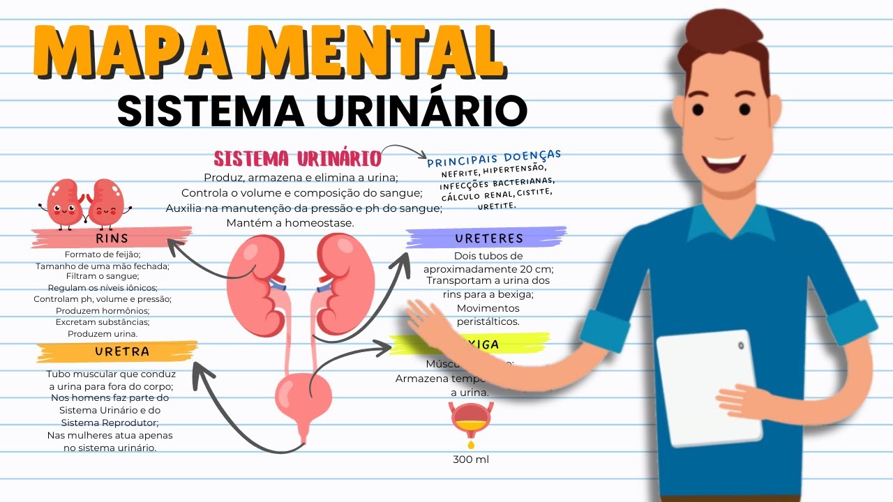
  • 📍 Kidneys are located in the retroperitoneal cavity and are bean-shaped organs responsible for filtering blood and producing urine.
  • 💧 The primary function of the kidneys is to filter blood, remove waste and excess water, and return essential minerals and water to the bloodstream.
  • 🩸 The ureters transport urine from the kidneys to the bladder, where it is stored until it is excreted through the urethra.
  • 🏥 Preparation for a CT scan involves the removal of metallic materials from the patient's west region to avoid artifacts in the images.
  • 👨‍⚕️ Patients are instructed to take laxatives and follow a clear liquid diet before the examination to ensure the digestive system is clear.
  • 🛌 Patient positioning is crucial for the CT scan, with the patient lying flat on the scanning table, centered over the table's midpoint.
  • 🔍 The use of contrast agents may be required for better visualization of the kidneys, ureters, and bladder during the scan.
  • 📏 Specific attention is given to the lower abdomen and the area 2 inches below the public symphysis to include the bladder in the scan's field of view.
  • 🎥 The radiographer must ensure that the patient remains still during the scan to prevent image blurriness or overlapping.
  • 📊 The radiographic appearance includes visualization of the entire intestine, gas shadow of the intestine, and the lower ribs to ensure proper positioning and scanning.

Q & A

  • What is the main focus of the video?

    -The main focus of the video is to explain the process of performing a CT scan of the kidneys, ureter, and bladder.

  • What is the first step in preparing a patient for a CT scan?

    -The first step in preparing a patient for a CT scan involves removing all metallic materials from the west region, which includes removing the patient's clothing and any items such as coins that may interfere with the image quality.

  • Why is it important to give the patient a low residue diet before the examination?

    -A low residue diet is preferred before the examination to minimize the bowel movements and to clear the abdomen, which helps in obtaining clear images of the abdominal organs.

  • What are the different types of scans that can be performed using the CT scan?

    -Different types of scans that can be performed using the CT scan include an acute intestinal scan, a forensic body scan, a gastro-intestinal pen, and a urological pen.

  • How is the patient positioned for the CT scan of the kidneys, ureter, and bladder?

    -The patient is positioned supine on the scanning table, with their mid-sagittal plane overlapping the center of the scanning table and the table's center line overlapping the patient's mid-sagittal plane.

  • What is the significance of the lower end of the scanning range being 2 inches below the pubic symphysis?

    -The lower end of the scanning range is placed 2 inches below the pubic symphysis to ensure that the bladder is included in the scan, as it is part of the urinary system being examined.

  • What is the role of the radiographer in the CT scan process?

    -The radiographer plays a crucial role in the CT scan process by ensuring the patient is correctly positioned, setting the scanning parameters, and capturing the images without any movement that could affect the quality of the scan.

  • What are the key components of the CT scan settings?

    -The key components of the CT scan settings include maintaining 200 mAs, a pitch of 30 to 40 mAs, a gantry speed of 0.15 to 0.2 seconds, a focus distance of 180 cm, and using a grid if necessary.

  • How does the radiographer ensure the patient remains still during the scan?

    -The radiographer instructs the patient to remain completely still during the scan to avoid any movement that could cause blurring or overlapping of the images.

  • What are the structures visualized in the radiographs shown in the video?

    -The structures visualized in the radiographs include the entire intestine, the gas shadow within the intestine, the iliac crest, the lower ribs, the pubic symphysis, the kidneys, the bladder, and the ureter.

  • Why is it important to have a clear understanding of the patient's anatomy before performing the CT scan?

    -A clear understanding of the patient's anatomy is crucial for accurately positioning the patient and setting the correct scanning parameters to ensure high-quality images that can be effectively used for diagnosis and treatment planning.

Outlines

00:00

🏥 Introduction to Kidney, Ureter, and Bladder Examination

The paragraph introduces the topic of the video, which is about how a CT scan of the kidneys, ureter, and bladder is performed. It welcomes the viewers to the X-ray University channel and explains that the kidneys, ureter, and bladder are located in the retroperitoneal cavity. The main function of the kidneys is to filter blood, removing waste and returning the filtered blood to the bloodstream. The bladder stores urine produced by the kidneys. The paragraph also discusses the preparation for an abdomen, including the removal of metallic materials from the west region, and the importance of patient positioning for the scan.

05:02

📐 Positioning and Techniques for CT Scan

This paragraph delves into the specifics of patient positioning and the technical aspects of conducting a CT scan for the kidneys, ureter, and bladder. It explains the importance of capturing the correct location and angle for the scan, including the lower and upper positions for the scan. The paragraph also discusses the use of contrast, the role of the radiographer in removing shadows, and the various pen positions used in the scan, such as the floating kidney position and the stone pen if stones are present. Additionally, it touches on the use of lead shields and the importance of minimizing patient movement during the exposure to ensure clear images.

Mindmap

Keywords

💡Kidney

The kidney is an essential organ in the human body responsible for filtering waste products from the blood and producing urine. In the context of the video, the focus is on the function and examination of the kidney, particularly in relation to the urinary tract and bladder.

💡Ureter

The ureter is a tube that connects the kidney to the bladder and is responsible for transporting urine from the kidney to the bladder for storage and eventual elimination. In the video, the location and function of the ureter are discussed as part of the urinary system's examination.

💡Bladder

The bladder is a muscular sac that stores urine produced by the kidneys until it can be excreted out of the body through the urethra. The video discusses the examination of the bladder, including its position and how it relates to other urinary system components.

💡X-ray

X-ray is a medical imaging technique that uses ionizing radiation to create images of the inside of the body. In the video, X-ray is used to visualize the urinary system, including the kidneys, ureters, and bladder, for diagnostic purposes.

💡Abdominal Preparation

Abdominal preparation involves clearing the gastrointestinal tract to minimize interference with imaging and to improve the visibility of the urinary organs during the examination. This process is crucial for accurate X-ray imaging and is mentioned in the script as part of the patient's preparation for the examination.

💡Patient Positioning

Patient positioning refers to the process of placing the patient in a specific position to facilitate the examination or procedure. In the context of the video, proper positioning is essential for obtaining clear X-ray images of the urinary system, including the kidneys, ureters, and bladder.

💡Contrast

Contrast agents are substances used in medical imaging to enhance the visibility of internal structures. In the video, contrast is likely used to improve the clarity of the X-ray images, particularly for the urinary system's examination.

💡Excretory Urography

Excretory urography is a diagnostic imaging procedure that uses X-rays and a contrast medium to visualize the urinary tract, including the kidneys, ureters, and bladder. This technique is essential for detecting abnormalities, stones, or blockages within the urinary system.

💡Floating Kidney

A floating kidney, also known as a wandering kidney, is a congenital condition where one or both kidneys are not fixed in their normal position and can move within the abdominal cavity. The video discusses the examination of kidney position, including the detection of a floating kidney.

💡Stone

Stones, or calculi, are hard deposits that can form in the kidneys and other parts of the urinary system. The presence of stones can cause pain and other complications, and their detection is a key focus of the urinary system examination.

💡Radiograph

A radiograph is an image produced using X-rays, typically used in medical and dental fields to visualize internal structures. In the video, the radiograph is the resulting image from the X-ray examination of the urinary system.

Highlights

The video discusses the process of conducting a CT scan of the kidneys, ureter, and bladder.

The kidneys are located in the retroperitoneal cavity, with the right kidney positioned slightly lower than the left.

The primary function of the kidneys is to filter blood, removing waste and excess materials to form urine.

The bladder stores urine until it is excreted, and its position is noted in the pelvic region.

Preparation for the abdomen includes removal of metallic materials from the west region to avoid interference in the image.

The patient is instructed to take a laxative diet and take clear fluids the night before the examination.

During the patient's preparation, all metal materials are removed to prevent image capture issues.

The patient is positioned on the X-ray table with the center of the table aligned with the patient's midsagittal plane.

The arms are placed below the head, and the hands are placed under the scalene muscles to ensure proper positioning.

The lower end of the X-ray table is adjusted to include the pubic symphysis and the lower 2 inches of the sacrum.

The patient is instructed to lie still to avoid exposure during the procedure and to ensure clear imaging.

The CT scan is performed with 200 mAs, a range of 30 to 40 mAs, and a 0.15 to 0.2 second duration.

The focus is set to film distance, with an 180 cm grid applied automatically due to the posterior position.

The tube angle is not used, and the center will be at L3 or 2 inches above the midline of the sacrum.

The size of the X-ray used is 14x14 inches or 14x17 inches, depending on the patient's size.

In radiography, the diaphragm visualizes the entire intestine, and the gas shadow within the intestine is visualized.

The iliac crest is visualized in the lower part of the scan, and the lower end position is adjusted accordingly.

The radiography appearance includes the visualization of the head, pubic symphysis, hip joint, and sacroiliac joint.

The video encourages viewers to like, share, and subscribe for more radiography-related content and videos.

Transcripts

play00:07

केयूप केयूवी इज द फुल फॉर्म ऑफ किडनी

play00:10

यूरेटर एंड

play00:12

ब्लेडर केयूप का एक्सरे कैसे करते हैं हम

play00:15

आज के वीडियो में जानेंगे नमस्कार दोस्तों

play00:17

स्वागत है आपका एक्सरे यूनिवर्स चैनल में

play00:20

केयू बी इज किडनी यूरेटर ब्लैडर किडनी

play00:24

कहां पर लोकेटेड होती है रेट्रो

play00:26

पेरिटोनियल कैविटी में किडनी जो होती है

play00:29

वह आपकी बीन शेप की होती है ऑलमोस्ट लाइक

play00:32

अ फिस्ट साइज जो आपकी मुट्ठी होती है उसकी

play00:35

साइज की होती है किडनी का मेन फंक्शन क्या

play00:38

होता है ब्लड को आपके विटामिन मिनरल्स इन

play00:42

सब से फिल्टर करके ब्लड को वापस रिटर्न कर

play00:45

देना आपके ब्लड स्ट्रीम में और जो भी आपका

play00:48

वेस्ट मटेरियल है उसको आपको एक्सक्रीट कर

play00:51

देना बॉडी से तो जो भी आपका

play00:55

एक्सक्रीट मटेरियल होता है वो किडनी से

play00:58

ब्लैडर में पहुंचता है यू ब के थ्रू और

play01:01

ब्लेडर आपके यूरिन को स्टोर करके रखता है

play01:09

अंट्स आ जाए तो उसके एक्सरे करने के लिए

play01:11

हम करते हैं एब्डोमेन

play01:14

सुपाइम प्रिपरेशन क्या होगी पेशेंट

play01:17

प्रिपरेशन ऑल मेटलिक मटेरियल शुड बी

play01:20

रिमूव्ड फ्रॉम वेस्ट रीजन वेस्ट रीजन से

play01:23

हम सारे मेटल के मटेरियल को रिमूव करेंगे

play01:26

पेशेंट पुट ऑन हॉस्पिटल गाउन पेशेंट को हम

play01:29

हॉस्पिटल गाउन पहनने को देंगे वेस्ट

play01:31

मटेरियल से हम सारे जो मेटल के मटेरियल

play01:34

होते हैं वो इसलिए एक्सक इसलिए रिमूव

play01:36

करवाते हैं ताकि आपके वो पिक्चर में

play01:39

कैप्चर ना होए जैसा कि आप इस पिक्चर में

play01:42

देख रहे हैं कि जो आपके सेंटर रो होती है

play01:44

वो अमलास पे यहां पे दे रखी है इस जगह पे

play01:48

तो वेस्ट पे अराउंड आपके बेल्ट वगैरह यूज

play01:51

होता है अगर आपके पॉकेट्स में कॉइंस वगैरह

play01:54

है तो उसकी शैडो आपके पिक्चर में ना आए

play01:56

फिल्म प ना बने इसलिए हम मेटल के मटेरियल

play01:59

सबको मूव करवाते हैं पेशेंट इज

play02:02

इंस्ट्रक्टेड टू टेक अ लो रेसिड डाइट एंड

play02:05

टेक्स लेटिव द नाइट बिफोर द एग्जामिनेशन

play02:08

टू टेक क्लियर इमेज ऑफ एब्डोमेन जैसा कि

play02:11

मैंने पिछले वीडियो में भी बताया था हम

play02:13

पेशेंट की प्रिपरेशन के टाइम पे लेगस टव

play02:15

देते हैं और लो रेसिड डाइट प्रेफर करते

play02:17

हैं लेगिट क्या काम आते हैं पिछले वीडियो

play02:20

में डिस्क्राइब कर रखा है अगर आपने नहीं

play02:22

देखा है तो जरूर जाके देखिएगा तो लेक्ट्स

play02:25

हमारे गैसियस जो शैडो होती है उसको रिमूव

play02:28

करने के लिए हम उसको देते हैं

play02:30

और लेटिव आपके फील मटेरियल को रिमूव

play02:32

करवाती है गैस अब्जॉर्बेंट कैप्सूल भी

play02:34

देते हैं और लेक्ट्स भी देते हैं और

play02:36

पेशेंट को हम लो रेसिड्यू डाइट प्रेफर

play02:38

करते हैं एग्जामिनेशन से पहले तो यह थी

play02:41

पेशेंट की प्रिपरेशन एब्डोमेन

play02:45

सुपाइम क्या रहेगा केयूवी का देखते हैं

play02:49

एक्यूट इंटेस्ट एक्यूट पेन एक्यूट पेन के

play02:52

केसेस में हम केयूवी करते हैं इंटेस्टाइनल

play02:55

ऑब्स्ट्रक्शन अगर इंटेस्टाइन के अंदर कोई

play02:57

ऑब्स्ट्रक्शन आ रहा है उसके केसेस में हम

play02:59

एब्डोमेन एपी करते हैं फॉरेन बॉडी फॉरेन

play03:02

बॉडी अगर आपके कोई एब्डोमेन रीजन में आ गई

play03:04

है उसको देखने के लिए भी केयू बी या

play03:06

एब्डोमेन एपी प्रेफर करते हैं डिफरेंस

play03:08

बिटवीन गैस्ट्रोइंटेस्टाइनल पेन एंड

play03:10

यूरोलॉजिकल पेन गैस्ट्रोइंटेस्टाइनल और

play03:13

यूरोलॉजिकल पेन में डिफरेंस देखने के लिए

play03:15

भी हम एब्डोमेन एपी करते हैं फ्लैंक पेन

play03:18

फ्लैंक पेन जो आपके सडन अगर कोई पेन जनरेट

play03:21

हो रहा है उसको देखने के लिए भी डॉक्टर

play03:24

एब्डोमेन एपी रिकमेंड करते हैं फ्लोटिंग

play03:26

किडनी किडनी की पोजीशन देखने के लिए इफ

play03:29

किडनी आपकी नॉर्मल पोजीशन में नहीं है

play03:31

उसको देखने के लिए भी एब्डोमेन एपी प्रेफर

play03:34

किया जाता है स्टोन पेन अगर किसी के पथरी

play03:37

या स्टोन हो रखा है उसका पेन अगर जनरेट हो

play03:39

रहा है तो स्टोन की लोकेशन देखने के लिए

play03:41

भी एब्डोमेन एपी किया जाता है तो यह सारी

play03:44

थी एब्डोमेन एपी की

play03:46

इंडिकेशन पेशेंट पोजीशन क्या है देखते हैं

play03:50

पेशेंट इज आस्ड टू लाइंग डाउन इन सुपाइम

play03:53

पोजीशन ओवर द सेंटर ऑफ द एक्सरे ट्यूब तो

play03:56

यह आप एक्सरे टेबल होगा एक्चुअल में यहां

play03:59

पे

play04:01

ये एक्सरे टेबल है तो पेशेंट आपकी ये

play04:05

एक्सरे टेबल है उसके ऊपर आपके पेशेंट को

play04:08

हम बोलेंगे यह यहां पर लेटा हुआ है पेशेंट

play04:11

पेशेंट का जो मिड सेटल प्लेन होगा वो टेबल

play04:14

के मिड सजल प्लेन के ओवरलैप होना चाहिए कि

play04:17

ये जो आपकी टेबल है टेबल का जो सेंटर ऑफ

play04:20

लाइन है टेबल पे उसके ओवरलैप उसके ऊपर ही

play04:23

पेशेंट का मिड सटल प्लेन आना चाहिए कर्वड

play04:26

शुड बी रिमूव्ड फ्रॉम द पेशेंट बॉडी कर्वड

play04:29

रिमूव कर करवाएंगे पेशेंट की बॉडी से बोथ

play04:32

हैंड्स शुड बी अंडर द स्कल दोनों हाथ आपके

play04:35

स्कल के नीचे जो आपका सर है उसके नीचे

play04:37

रखवा जाएंगे एमएसपी ऑफ द पेशेंट शुड बी

play04:40

ओवर द सेंटर ऑफ द एक्सरे टेबल जो मैंने

play04:43

आपको बताया था जो पेशेंट का जो मिड सटल

play04:46

प्लेन होना चाहिए वो एक्सरे टेबल के सेंटर

play04:48

पे होना चाहिए कैसेट इज प्लेस इन लेंथ

play04:51

वाइज पोजीशन इन द बकी बकी क्या होता है

play04:54

आपकी जो एक्स्ट्रेट टेबल होती है उसके

play04:56

नीचे एक बकी पोशन होता है बकी के अंदर आप

play04:59

कैसेट को रखते हैं एक स्ट्रक्चर होता है

play05:02

उसके नीचे तो आप उसको इजली सेट कर सकते

play05:04

हैं जिस लोकेशन का आपको जिस पार्ट का आपको

play05:06

कैप्चर करना होता है वो बक्की जो होती है

play05:09

आपके एडजस्ट पेबल होती है अप डाउन आप इजली

play05:12

कर सकते हैं उसको तो बक्की के अंदर कैसेट

play05:14

को हम रखते हैं और लेंथ वाइज पोजीशन में

play05:17

हम कैसेट को उसमें रखी जाती है लोअर एंड

play05:21

ऑफ द एक्सरे कैसेट शुड बी 2 इंच बिलो द

play05:23

प्यूबिक सिंफा इसिस प्यूबिक सिंफा इसिस जो

play05:26

पोर्शन होता है आपके उससे 2 इंच नीचे रखते

play05:29

हैं हम क्यों रखते हैं क्योंकि इसमें हमें

play05:31

ब्लैडर को भी इंक्लूड करना होता है

play05:33

क्योंकि इसका नाम भी यही था किडनी यूरेटर

play05:35

ब्लैडर ब्लैडर तक पोशन देखने के लिए हमें

play05:37

यह एक्सरे किया जाता है तो आपके जो

play05:39

प्यूबिक सिंफा इसिस आ रहा है उससे 2 इंच

play05:42

नीचे हम लोअर एंड रखेंगे कैसेट

play05:46

का अपर एंड ऑफ़ द एक्सटिक एसिड शुड रिमन 2

play05:50

टू 3 इंच अबोव द सफाइड प्रोसेस जो अपर एंड

play05:53

रखेंगे हम वह ज फइड प्रोसेस से 2 से 3 इंच

play05:56

ऊपर रखेंगे सेंटर शुड बी पास्ड थ्रू अलाइक

play05:59

और थ्रू l3 और 2 इंच अबोव मिड ऑफ एस आईस

play06:05

अब जैसे कि फर्स्ट पिक में शो हो रहा था

play06:06

आपको सेंटर आप अमलास पे कर सकते हैं अन या

play06:10

फिर l3 पे कर सकते हैं या एसआईएस से 2 इंच

play06:13

ऊपर कर सकते हैं एसआईएस क्या होता है यह

play06:16

आपका पेल्विक एरिया है तो यहां पर आपके एस

play06:19

आईएस होता है तो इससे 2 इंच ऊपर समथिंग

play06:22

आपके वो अमलास पोर्शन ही आ जाता है तो

play06:24

वहां पर हमें सेंटर रे को पोजीशन करना

play06:27

होता है देर शुड बी नो मूमेंट ड रिंग द

play06:29

एक्सपोजर मूवमेंट हमें बिल्कुल भी नहीं

play06:32

होना चाहिए एक्सपोजर के टाइम पर अगर

play06:34

मूवमेंट हो जाएगा तो हमारी पिक्चर क्लियर

play06:36

नहीं आ पाएगी ओवरलैपिंग हो जाएगी या

play06:38

ब्लरनेस आ जाएगी इसलिए हम पेशेंट को पहले

play06:40

से रिकमेंड करते हैं कि आपको मूवमेंट नहीं

play06:42

करना है बिल्कुल भी यह थी हमारी पेशेंट

play06:45

पोजीशन केयूवी की एक्सपोजर चार्ट क्या

play06:48

रहेगा देखते हैं एम हम रखेंगे इसके अंदर

play06:51

200 एमएस रखेंगे 30 से 40 एमएस हम ने

play06:55

ज्यादा यूज किया है यहां पे केवीपी लेंगे

play06:57

65 से 70 टाइम होगा आपने 0.15 से 0.2

play07:02

सेकंड एफएफडी रहेगा फिल्म टू फोकस

play07:05

डिस्टेंस 180 सेमी ग्रिड अपन ने यहां पे

play07:07

यूज़ किया है क्यों किया है क्योंकि आपके

play07:09

जो कैसेट है वह बकी में पोजीशन की है बकी

play07:12

में रखी है तो बकी में रखने की वजह से

play07:14

ग्रिड ऑटोमेटिक यस हो जाता है ट्यूब एंगल

play07:17

अपन ने यूज़ नहीं किया है सेंटर यह होगा

play07:19

यहां पे l3 पे या फिर 2 इंच अबोव मिड ऑफ़

play07:22

ए आ कैसेट साइज़ यूजुअली अपन ना 14 * 14

play07:26

इंच या 14 * 17 इंच यह था अपना एक्सपोजर

play07:29

चार्ट रेडियोग्राफी अपीयरेंस क्या है

play07:31

देखते हैं रेडियोग्राफी अपीयरेंस में

play07:38

डायफास्फेट

play07:40

जो दिख रही है वो पूरी इंटेस्टाइन की है

play07:43

ये गैस के जो आपको शैडो है वो इंटेस्टाइन

play07:46

के अंदर की है गैस शैडो ऑफ इंटेस्टाइन ये

play07:49

आपकी जो गैस पोशन जो इंटेस्टाइन में बन

play07:52

जाता है तो वो इंटेस्टाइन में विजुलाइज

play07:54

होता है बोथ इलिक क्रस्ट तो यहां पे आपके

play07:57

इलिक क्रस्ट विजुलाइज हो रहा है ये

play08:00

लोअर रिप्स य यहां पर आपको लोअर रिप्स दिख

play08:02

रही है किडनी यह यहां का जो पोर्शन है वह

play08:07

यहां पर किडनी विजुलाइज होगी आपको ब्लैडर

play08:10

यहां पर आपको ब्लैडर विजुलाइज होगा और

play08:14

यूरेटर यूरेट भी इसके आसपास आपको दिखाई

play08:17

देता है तो यह सारा था आपका रेडियोग्राफी

play08:21

अपीयरेंस इसके अंदर आप देख सकते हैं फीमल

play08:24

ये हेड भी शो हो रहा है आपको प्यूबिक यहां

play08:27

पर हेड हो रहा है शो हि हिप जॉइंट शो हो

play08:30

रहा है एसआई जॉइंट शो हो रहा है तो यह

play08:33

सारे रेडियोग्राफी अपीयरेंस है जो आपको

play08:35

केयूवी के अंदर विजुलाइज होंगे तो यह था

play08:38

हमारा केयू बी एक्सप्लेन किया हुआ थैंक यू

play08:41

सो मच अगर आपको वीडियो अच्छा लगा तो लाइक

play08:43

शेयर और सब्सक्राइब जरूर कीजिए

play08:45

रेडियोग्राफी से रिलेटेड और वीडियोस देखने

play08:47

के लिए हमारे चैनल को सब्सक्राइब जरूर

play08:49

कीजिए थैंक यू

Rate This

5.0 / 5 (0 votes)

関連タグ
Kidney ImagingUrologyCT ScanPatient PrepMedical DiagnosticsHealthcareRadiologyProcedure GuideHealth Education
英語で要約が必要ですか?