Ayo Kenali Cara Menggunakan Mikroskop Cahaya dengan Baik dan Benar

Satria Production
21 Mar 202308:28

Summary

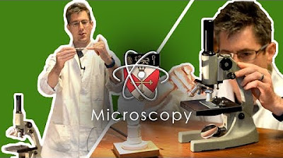
TLDRThis educational video offers a detailed tutorial on the proper use of a microscope, starting from its origin and basic parts to practical operation. It introduces the XSP 12 microscope, explaining its components like the eyepiece, objective lens, and revolver for magnification adjustment. The script guides viewers through setting up the microscope, adjusting focus and lighting, and observing a blood cell sample. It emphasizes careful handling, correct observation techniques, and post-use clean-up, concluding with an invitation to explore more educational content.

Takeaways

  • 🔬 Microscopes are essential tools for observing small or microscopic objects, derived from the Greek words 'mikros' meaning small and 'skopos' meaning to see.
  • 🌟 The XSP 12 is highlighted as a simple and affordable brand of microscope, suitable for beginners or those on a budget.
  • 👀 The main parts of a microscope include the tube, body, eyepiece with 10x magnification, objective lenses with 4x, 10x, and 40x magnifications, and a revolving nosepiece to switch between objectives.
  • 🔧 The coarse and fine adjustment knobs on the microscope body are used for adjusting the distance between the objective lens and the specimen stage for rough and fine focusing, respectively.
  • 🛠️ The stage and stage clips are used to place and secure the glass slide specimen in the center of the field of view.
  • 💡 Microscopes may have different types of mirrors for lighting conditions: flat mirrors for bright conditions and concave mirrors for less light.
  • 🌞 The diaphragm is used to regulate the amount of light entering the microscope by adjusting the size of the aperture through which light passes.
  • 🧐 Proper handling of the microscope involves holding the arm with the dominant hand and supporting the base with the other hand, being careful when moving or placing it on a table.
  • 🔍 To begin using the microscope, adjust the coarse knob to move the objective lens away from the specimen stage and align the mirror to reflect light properly for adequate illumination.
  • 🔬 When observing a specimen, place the slide on the stage, adjust the coarse knob to bring the objective lens close without touching the slide, and then use the fine adjustment to sharpen the image.
  • 🔄 After use, return the objective lens to a lower magnification, move it away from the stage, and carefully remove and store the slide before putting the microscope back in its place.

Q & A

  • What does the term 'microscope' originate from?

    -The term 'microscope' originates from the Greek words 'mikros' meaning small and 'skopos' meaning to see.

  • What are the main parts of a microscope as described in the script?

    -The main parts of a microscope described in the script are the tube, the body, the eyepiece lens, the objective lens, the revolver to change the magnification, the coarse and fine focus knobs, the stage for the specimen, the stage clips, the mirror for illumination, and the diaphragm to adjust light intensity.

  • What is the purpose of the eyepiece lens in a microscope?

    -The eyepiece lens is where the eye looks through to view the magnified image of the specimen.

  • What is the function of the objective lens in a microscope?

    -The objective lens is placed close to the specimen and is used to magnify the object being observed.

  • How can the magnification of the objective lens be changed in the described microscope?

    -The magnification of the objective lens can be changed by rotating the revolver, which allows for different lenses with varying magnifications to be used.

  • What are the two types of mirrors mentioned for illumination in the script?

    -The two types of mirrors mentioned are the flat mirror for bright light conditions and the concave mirror for less bright conditions.

  • What is the role of the diaphragm in a microscope?

    -The diaphragm is used to control the amount of light entering the microscope by adjusting the size of the aperture through which light passes.

  • How should one properly hold a microscope as per the script?

    -One should hold the microscope with the dominant hand on the arm and the other hand supporting the base, ensuring careful handling when moving or placing it on a table.

  • What is the initial step in using the microscope as described in the script?

    -The initial step is to use the coarse focus knob to move the objective lens away from the specimen stage and then adjust the mirror to reflect light into the field of view for sufficient illumination.

  • How does one adjust the focus to make the image clearer in the microscope?

    -One should slowly turn the coarse focus knob to find the object's image and then use the fine focus knob to sharpen and clarify the image.

  • What should be done after using the microscope according to the script?

    -After using the microscope, one should turn the revolver to set the objective lens to a lower magnification, move the objective lens away from the specimen stage using the coarse focus knob, remove the specimen slide from the clips, and return the microscope to its place or hand it over to the laboratory manager for further care.

Outlines

00:00

🔬 Introduction to Microscope Usage

This paragraph introduces the basics of microscope usage, starting with a greeting and moving on to explain the origin of the term 'microscope' from Greek, meaning 'small' and 'to see'. It highlights the purpose of a microscope, which is to observe small or microscopic objects. The speaker describes the specific model of microscope they are using, the XSP 12, and its components, including the eyepiece and objective lenses with different magnifications, a revolver for changing the objective lens, and the coarse and fine adjustment knobs for adjusting the distance between the lens and the specimen. The paragraph also mentions the stage, the clips for holding the slide, and the mirror for illumination, which can be adjusted depending on the lighting conditions. It concludes with a brief mention of more expensive microscopes that have additional features like adjustable stages and a condenser for focusing light.

05:02

🔍 Proper Handling and Operation of a Microscope

The second paragraph focuses on the correct way to handle and operate a microscope. It begins with the proper way to hold the microscope with the dominant hand on the arm and the other hand supporting the base. The speaker advises to be careful when moving and placing the microscope on a table. The process of using the microscope starts with adjusting the coarse knob to move the objective lens away from the specimen and aligning the mirror to reflect light properly into the field of view. The speaker then describes how to place the slide on the stage and center it in the field of view. The next steps involve using the coarse adjustment to bring the objective lens close to the specimen without touching it, followed by slowly adjusting the coarse knob to find the object's image. Once the image is found, the magnification is increased by switching to a higher power objective lens and then fine-tuning the focus with the fine adjustment knob. The paragraph concludes with instructions on how to clean and store the microscope after use, including returning the objective lens to a lower magnification, moving the lens away from the specimen, and properly storing the slide and microscope.

Mindmap

Keywords

💡Microscope

A microscope is an optical instrument designed to magnify small objects or details that are not visible to the naked eye. In the video, the term is central to the theme as it explains the use and parts of a microscope, specifically mentioning a model 'xsp 12' which is described as simple and affordable.

💡Lens

A lens is an optical device capable of refracting light to form an image. The script discusses two types of lenses in a microscope: the 'ocular lens', which is where the eye looks through, and the 'objective lens', which is close to the specimen. The lens is crucial for magnification, with the script mentioning different magnification powers such as 10x, 4x, and 40x.

💡Revolver

In the context of the video, the revolver refers to a rotating nosepiece that holds different objective lenses, allowing the user to switch between them to change the magnification power. The script describes how to use the revolver to adjust the magnification from 4x to 10x or 40x.

💡Coarse Focus Knob

The coarse focus knob is used to adjust the distance between the objective lens and the specimen stage, allowing for a rough focusing of the image. The script explains its function in setting the initial focus before fine-tuning with the fine focus knob.

💡Fine Focus Knob

The fine focus knob is used for precise focusing to make the image sharp and clear. The script illustrates its use after adjusting the coarse focus, to refine the image clarity when observing the specimen under the microscope.

💡Specimen Stage

The specimen stage is the part of the microscope where the sample or specimen is placed. The script mentions that it is used to position the 'slide' which is the glass holding the specimen, and it is central to the process of observation.

💡Slide

A slide is a thin piece of glass on which a specimen is mounted for microscopic examination. The script describes placing the slide on the specimen stage and ensuring it is centered for optimal viewing.

💡Illumination

Illumination in microscopy refers to the process of lighting the specimen to make it visible. The script discusses the use of a mirror to reflect light onto the specimen and the importance of adjusting the light intensity for proper observation.

💡Mirror

A mirror in the context of the video is a component used to direct light towards the specimen. The script mentions two types of mirrors: a flat mirror for bright conditions and a concave mirror for less light.

💡Dioptre

Dioptre refers to the unit of measurement for the strength of a lens, particularly in eyeglasses or contact lenses. Although not explicitly defined in the script, the term would relate to adjusting the microscope for the viewer's vision clarity.

💡Condenser

A condenser is a lens or system of lenses that focuses light onto the specimen to enhance contrast and resolution. The script mentions that more expensive microscopes have a condenser for better light control and image quality.

💡Procedure

The term 'procedure' in the script refers to the step-by-step method for using the microscope correctly. It is used to describe the process from setting up the microscope to observing the specimen and adjusting the focus.

Highlights

Introduction to the proper use of a microscope.

Etymology of the word 'microscope' from Greek origins.

Different brands of microscopes available today.

Description of the XSP 12 microscope, a simple and affordable model.

Explanation of the microscope's components: the tube and body.

Function of the eyepiece and objective lenses in a microscope.

The revolver mechanism for changing the magnification of the objective lens.

Different magnification powers available: 4x, 10x, and 40x.

The coarse and fine adjustment knobs for focusing.

The function of the stage and slide holder in specimen placement.

Adjustable stages in more expensive microscopes for better maneuverability.

The role of the mirror in illumination: flat for bright conditions and concave for dim conditions.

Introduction to the diaphragm for controlling light intensity.

Proper handling and transportation of the microscope.

Initial setup for using the microscope: adjusting the coarse knob and mirror for optimal light.

Observing a dry blood cell specimen preparation.

Technique for adjusting the microscope to find the specimen image.

Instructions for changing to higher magnification using the revolver.

Using the fine adjustment knob to sharpen the image.

Avoiding contact between the objective lens and the specimen during high magnification.

Post-use care: returning the objective lens to a lower magnification and adjusting the stage.

Proper storage and handling of the microscope after use.

Invitation to watch other videos by the same creator.

Transcripts

play00:02

Assalamualaikum warahmatullahi

play00:04

wabarakatuh pada video kali ini kita

play00:06

akan praktekkan Bagaimana cara

play00:08

penggunaan mikroskop dengan baik dan

play00:10

benar

play00:13

[Musik]

play00:29

mikroskop berasal dari bahasa Yunani

play00:31

mikro yang artinya kecil dan Scoopy yang

play00:35

berarti melihat intinya mikroskop adalah

play00:38

alat untuk melihat benda-benda kecil ada

play00:41

berbagai merek mikroskop di zaman

play00:42

sekarang tapi semuanya tetap sama yaitu

play00:45

untuk melihat benda-benda kecil atau

play00:47

mikroskopis

play00:54

mikroskop yang saya gunakan bermerek xsp

play00:57

12 salah satu mikroskop yang paling

play00:58

sederhana dan murah bagian-bagian

play01:01

mikroskop ini bisa kita bagi menjadi

play01:03

bagian tabung mikroskop dan badan

play01:05

mikroskop di bagian tabung mikroskop

play01:07

terdapat dua lensa pertama lensa okuler

play01:10

tempat mata kita melihat dan lensa

play01:13

objektif yang dekat dengan objek lensa

play01:15

okuler di sini memiliki perbedaan 10

play01:17

kali dan objektif 4 10 dan 40 kali di

play01:21

dekat lensa objektif terdapat revolver

play01:23

yang dapat diputar gunanya sebagai apa

play01:25

untuk mengubah perbesaran lensa objektif

play01:28

seperti yang kita tahu lensa objektif di

play01:31

sini memiliki perbesaran 4 10 dan 40

play01:33

kali dengan memutar revolver kita bisa

play01:36

mengubah ke perbesaran selanjutnya

play01:38

kemudian kita beralih ke badan mikroskop

play01:41

terdapat pom ter kasar dan pemutar halus

play01:44

fungsi dari pemutar kasar adalah untuk

play01:47

mengatur jarak antara lensa objektif

play01:49

dengan meja preparat atau meja objek

play01:52

bisa juga untuk mengatur fokus secara

play01:54

kasar

play01:57

Sedangkan untuk memutar halus berfungsi

play02:00

untuk memfokuskan bayangan supaya lebih

play02:02

tajam dan tidak buram

play02:07

selanjutnya ada meja preparat dan

play02:10

penjepit preparat fungsinya adalah meja

play02:14

preparat berfungsi sebagai tempat

play02:15

meletakkan kaca preparat sedangkan

play02:19

penjepit preparat itu bisa digunakan

play02:21

untuk menjepit preparat supaya tidak

play02:23

bergerak kemana-mana

play02:24

pada mikroskop yang lebih mahal biasanya

play02:26

memiliki preparat yang adjustable bisa

play02:29

digerakkan ke depan belakang dan ke

play02:31

samping

play02:33

bagian selanjutnya adalah cermin yang

play02:36

berperan dalam pencahayaan objek cermin

play02:38

terbagi menjadi dua yaitu cermin datar

play02:41

untuk kondisi cahaya yang terang dan

play02:43

cermin cekung untuk kondisi cahaya yang

play02:45

kurang terang

play02:46

[Musik]

play02:50

bagian selanjutnya adalah diafragma

play02:53

diafragma berfungsi untuk mengatur

play02:55

banyaknya cahaya yang masuk menggunakan

play02:57

diafragma dengan cara diputar seperti

play03:00

ini

play03:00

tujuannya adalah pada diafragma terdapat

play03:04

lubang-lubang yang memiliki ukuran yang

play03:06

berbeda sehingga kita bisa mengatur

play03:08

intensitas cahaya yang kita butuhkan

play03:13

tidak lupa juga pada kaki mikroskop dan

play03:16

leher mikroskop di samping bagian-bagian

play03:19

tadi terdapat mikroskop yang lebih mahal

play03:21

yang memiliki komponen tambahan Seperti

play03:25

contohnya pada mikroskop ini sumber

play03:27

cahaya didapat dari lampu bukan dari

play03:29

pantulan cermin pada mikroskop ini juga

play03:32

terdapat kondensor yang berfungsi untuk

play03:34

memfokuskan cahaya

play03:35

[Musik]

play03:41

cara membawa mikroskop dengan baik dan

play03:43

benar adalah seperti ini tangan dominan

play03:46

memegang leher mikroskop dan tangan

play03:48

lainnya menyanggah kaki mikroskop jika

play03:51

anda dominan lakukan seperti di video

play03:53

jika ada dominan tangan kiri lakukan

play03:55

sebaliknya biasanya mikroskop disimpan

play03:57

di rak-rak tinggi Jadi harap

play03:59

berhati-hati ketika memindahkan

play04:02

mikroskop dan meletakkannya ke meja

play04:03

lakukan dengan hati-hati

play04:06

[Musik]

play04:12

untuk memulai menggunakan mikroskop

play04:14

gunakan pemutar kasar untuk menjauhkan

play04:17

lensa objektif dari meja preparat

play04:20

karena sampai titik terjauh

play04:23

[Musik]

play04:26

kemudian Arahkan cermin hingga

play04:29

memantulkan cahaya ke bidang pandang

play04:30

sehingga bisa didapatkan suplai cahaya

play04:33

yang cukup

play04:36

amati bidang Pandang dari lensa okuler

play04:39

jika masih gelap aturlah posisi cermin

play04:41

hingga bidang pandang cukup terang

play04:51

kita di sini akan mencoba mengamati

play04:53

preparat kering dari sel darah merah

play04:56

Letakkan kaca objek di meja preparat dan

play04:59

dijepitkan pastikan objek ada di

play05:02

tengah-tengah bidang pandang

play05:04

selanjutnya dekatkan lensa objektif

play05:07

dengan meja preparat menggunakan

play05:08

komputer kasar sampai posisi paling

play05:10

dekat

play05:12

jangan sampai menyentuh ya

play05:16

tips dan trik untuk tahap ini kita bisa

play05:18

memantau dari samping mikroskop untuk

play05:21

mengiringi

play05:22

[Musik]

play05:26

selanjutnya dengan posisi mata di lensa

play05:29

okuler putarlah pemutar kasar secara

play05:31

perlahan-lahan untuk menemukan bayangan

play05:34

objek

play05:35

memutarnya pada posisi menjauhkan lensa

play05:37

objektif dari meja preparat jika

play05:39

prosedurnya benar maka akan dapat

play05:41

bayangan objek seperti ini

play05:44

selanjutnya kita tingkatkan perbesaran

play05:47

yang lebih tinggi dengan cara memutar

play05:49

revolver ke lensa objektif yang lebih

play05:51

kuat

play05:52

[Musik]

play05:56

memutar revolver sampai terdengar bunyi

play05:59

berikut

play06:07

gunakan pemutar halus untuk mempertajam

play06:09

atau memfokuskan bayangan

play06:11

[Musik]

play06:25

putar lagi revolver untuk perbesaran

play06:27

lensa objektif yang lebih kuat

play06:33

Mari kita ulangi

play06:35

lensa objektif Jadi terlihat seperti

play06:38

menyentuh preparat

play06:40

padahal sebenarnya tidak

play06:43

seperti yang kita lihat lensa objektif

play06:45

hampir menyentuh tapi tidak menyentuh

play06:47

preparat untuk menunjukkan bahwa

play06:49

prosedur yang dilakukan sudah benar Mari

play06:52

kita atur fokusnya Eits Jangan

play06:54

menggunakan pemutar kasar untuk lensa

play06:56

objektif 40 kali hanya boleh gunakan

play06:58

Ultra halus tahukah Kenapa Tulis di

play07:01

kolom komentar

play07:04

[Musik]

play07:29

setelah menggunakan mikroskop putarlah

play07:31

revolver untuk mengembalikan lensa

play07:33

objektif ke perbesaran yang lebih kecil

play07:35

kemudian menggunakan pemutar kasar

play07:37

jauhkan kembali antara lensa objektif

play07:40

dengan meja preparat

play07:44

Jangan lupa untuk mengambil kembali kaca

play07:46

preparat yang dijepitkan di penjepit

play07:48

berkarat

play07:50

lalu kembalikan mikroskop pada tempatnya

play07:53

jika ada pengurus laboratorium serahkan

play07:56

mikroskop ke pengurus untuk diurus lebih

play07:59

lanjut

play08:01

demikianlah video cara penggunaan

play08:03

mikroskop dari saya Apabila ada

play08:06

salah-salah kata saya mohon maaf semoga

play08:08

video ini dapat menjadi bermanfaat bagi

play08:11

kita semua saya akhiri wassalamualaikum

play08:14

warahmatullahi wabarakatuh

play08:17

jika anda tertarik dengan video Saya

play08:19

mungkin bisa tonton video saya yang

play08:21

lainnya

play08:21

[Musik]

Rate This

5.0 / 5 (0 votes)

Ähnliche Tags
Microscope UsageEducational VideoTutorial GuideScientific ToolsBiology LabOptics BasicsFocus TechniquesLighting AdjustmentMicroscopy 101Lab EquipmentObservation Skills
Benötigen Sie eine Zusammenfassung auf Englisch?