Light Microscopy: Function and Utility
Summary
TLDRThis video tutorial provides an in-depth introduction to the light microscope, a key tool in microbiology. It explains its function, from how light passes through lenses to magnify a specimen, to the various components like the stage, diaphragm, objective lenses, and eyepieces. The tutorial covers the correct usage, including focusing techniques, adjusting eyepieces, and using oil immersion for high magnification. The video also highlights the limitations of light microscopes in resolution, while emphasizing their accessibility, affordability, and ability to observe living specimens in real time.
Takeaways
- 😀 Light microscopes use light to magnify specimens, making them a common tool in biological studies.
- 🔬 Light microscopes can magnify images from 4x to 1,200x depending on the magnification settings.
- 🧑🔬 The microscope has several key parts: stage, stage clips, diaphragm, objectives, eyepiece, and focus knobs.
- 🔧 The coarse focus knob is used for large adjustments, while the fine focus knob is for precise focusing.
- 👁️ The eyepiece further magnifies the image, typically by a factor of 10 or 15.
- 💡 The diaphragm controls the contrast of the image by regulating the amount of light reaching the specimen.
- 🧪 The objective lenses provide different magnifications, usually between 4x and 100x, to observe specimens in detail.
- 📏 Total magnification is the product of the eyepiece magnification (10x) and the objective lens magnification.
- 🧑🔬 For 100x magnification, oil immersion is necessary to enhance the resolution and clarity of the image.
- 💡 Light microscopes are relatively inexpensive, making them common in high school and undergraduate labs for studying living specimens.
- 🔍 While useful, light microscopes have limitations in resolution and cannot observe structures as clearly as more advanced microscopes.
Q & A
What is the main function of a light microscope?
-The light microscope uses light to visualize specimens by passing light through lenses to magnify the image, allowing users to observe microorganisms and other small details.
How does a light microscope magnify an image?
-The light microscope magnifies an image by first passing light through the specimen, which is then magnified by the objective lens (4 to 100 times). The eyepiece further magnifies the image by 10 or 15 times.
What are the main components of a light microscope?
-Key components of a light microscope include the stage, stage clips, diaphragm, objective lenses, nosepiece, eyepiece, focus knobs, and light source.
What is the purpose of the diaphragm in a light microscope?
-The diaphragm controls the contrast of the image by regulating the amount of light that reaches the specimen, allowing for better visualization.
What is the difference between the coarse and fine focus knobs?
-The coarse focus knob is used for making large adjustments to the focus, while the fine focus knob is used for smaller, more precise adjustments to improve the clarity of the image.
How do you adjust the focus of a light microscope?
-To focus the microscope, first use the coarse focus knob to clear up the image, then use the fine focus knob to refine it. Additionally, adjustments may be needed for the eyepiece to accommodate different eye prescriptions.
What is the significance of the 100x objective lens in light microscopes?
-The 100x objective lens provides high magnification but requires oil immersion to improve the resolution. A drop of oil is placed on the coverslip, and the objective is rotated to immerse in the oil.
What type of specimens are commonly studied using light microscopes?
-Light microscopes are commonly used to study bacteria, tissue slices, liquid samples, and some cellular structures such as mitochondria.
How does an inverted light microscope differ from a standard light microscope?
-An inverted light microscope has the light source above and the objective below the stage, making it ideal for studying tissue cultures in liquid media. In contrast, a standard light microscope has the light source below and the objective above the specimen.
What are the advantages of using a light microscope?
-Light microscopes are easy to use, inexpensive, and capable of visualizing living specimens without significant damage, making them ideal for high school and undergraduate labs. They also provide low magnification with sufficient clarity for many basic studies.
Outlines

هذا القسم متوفر فقط للمشتركين. يرجى الترقية للوصول إلى هذه الميزة.
قم بالترقية الآنMindmap

هذا القسم متوفر فقط للمشتركين. يرجى الترقية للوصول إلى هذه الميزة.
قم بالترقية الآنKeywords

هذا القسم متوفر فقط للمشتركين. يرجى الترقية للوصول إلى هذه الميزة.
قم بالترقية الآنHighlights

هذا القسم متوفر فقط للمشتركين. يرجى الترقية للوصول إلى هذه الميزة.
قم بالترقية الآنTranscripts

هذا القسم متوفر فقط للمشتركين. يرجى الترقية للوصول إلى هذه الميزة.
قم بالترقية الآنتصفح المزيد من مقاطع الفيديو ذات الصلة

Teknik Penggunaan Mikroskop Cahaya, Gimana caranya ya??
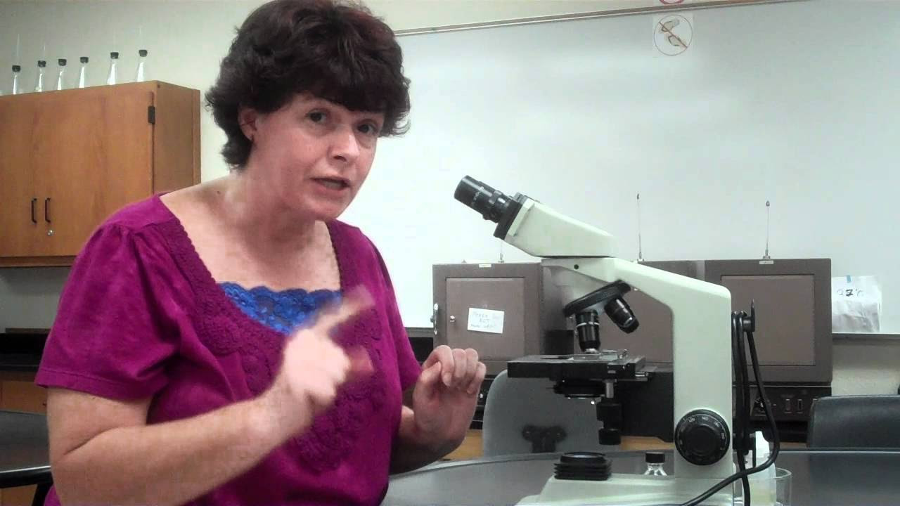
How to use a microscope and oil immersion
Ruang Lingkup dan Sejarah Mikrobiologi

Definisi dan Fungsi Mikroskop | Lomba Media Pembelajaran SIT Nurul Fikri
Introdução à MICROBIOLOGIA | Videoaula | Flavonoide #1

What Is Jira? | Jira Tool | Jira Training | Jira Tutorial For Beginners | Simplilearn
5.0 / 5 (0 votes)
