Animal Tissues
Summary
TLDRThis educational video script delves into the four primary types of animal tissues: epithelial, nervous, connective, and muscle tissues. It explains the specific functions and structures of each tissue, including the protective role of epithelial tissues, the signal-conducting function of nervous tissues, the supportive role of connective tissues, and the movement-enabling muscle tissues. The script also breaks down the different subtypes of each tissue, such as squamous, columnar, and cuboidal epithelia, sensory and motor neurons, and various connective tissues like areolar, fibrous, and cartilage. It provides a foundational understanding of the complex biological systems that support life.
Takeaways
- 🧬 Epithelial tissue is the body's protective layer, with cells often organized in a single or multi-layered structure, attached by a basement membrane.
- 🔍 Epithelial tissues can be identified by their geometric shapes, such as squamous, columnar, and cuboidal, each serving specific functions like protection, secretion, and absorption.
- 💡 Nervous tissue is responsible for conducting electrochemical signals and includes sensory neurons for sensing, motor neurons for movement, and interneurons for decision-making.
- 🧠 Nerve cells have four basic components: dendrites, cell body, axon, and axon terminals, with the position of the cell body determining the division between dendrites and axon.
- 🔗 Connective tissue supports, stabilizes, and protects the body's organs, and includes a variety of types such as areolar, fibrous, cartilage, blood, adipose, and bone tissue.
- 🩹 Areolar tissue is a basic form of connective tissue that binds epithelium and provides a loose connection between organs.
- 💪 Fibrous connective tissue is dense and strong, found in tendons and ligaments, providing flexibility without stretch.
- 🩸 Blood is the only liquid connective tissue, composed of lymphocytes, erythrocytes, and platelets, and is responsible for transporting nutrients, waste, and hormones.
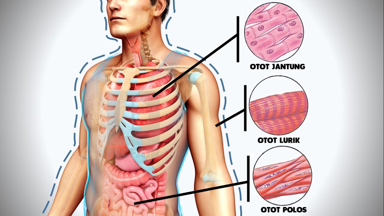
- 🏋️♂️ Muscle tissue allows for movement and is categorized into skeletal, smooth, and cardiac muscle, each with distinct characteristics and functions.
- 🦴 Skeletal muscle is striated and under voluntary control, attached to bones for movement.
- 🌀 Smooth muscle is found in the digestive system and blood vessels, responsible for involuntary movements like peristalsis and blood flow regulation.
- ❤️ Cardiac muscle is unique, found only in the heart, and moves involuntarily without instructions from the brain.
Q & A
What is the primary function of epithelial tissue?
-Epithelial tissue serves as a protective layer and lines the internal and external surfaces of the body, preventing unwanted substances from entering the body.
What is the role of the basement membrane in epithelial tissue?
-The basement membrane acts as a sticky surface that helps attach epithelial cells to the underlying tissue, ensuring they stay close together for protection.
How do squamous epithelial cells differ from columnar and cuboidal epithelial cells in terms of shape and function?
-Squamous epithelial cells are thin and irregularly shaped, often found in areas that require rapid healing like the mouth and skin. Columnar epithelial cells are elongated and often associated with secretion and absorption, found in the intestines and gallbladder. Cuboidal cells are square-like and also involved in absorption and secretion, located in sweat glands and the lining of the kidney tubes.
What are the four main types of tissues covered in the script?
-The four main types of tissues are connective, epithelial, muscle, and nervous tissue, each with specific functions and structures.
What is the primary function of nervous tissue?
-Nervous tissue is responsible for conducting electrochemical signals between the organs of the body and the brain, acting as the message pathways for the body.
How do sensory neurons differ from motor neurons and interneurons in structure and function?
-Sensory neurons are responsible for sensing and receiving information from the environment, with the cell body often off to the side. Motor neurons control movement and have a centrally located cell body. Interneurons, or relay neurons, are found in the spinal cord and brain, relaying information between sensory and motor neurons.
What is the basic structure of connective tissue?
-Connective tissue is composed of cells suspended in a fluid, matrix, or solid substance that provides support, stability, and protection to the body's organs.
What are the characteristics of areolar connective tissue?
-Areolar connective tissue is loose and binds epithelium to other tissues. It is made mostly of collagen and elastic fibers, providing a flexible yet strong connection between tissues.
How does fibrous connective tissue differ from areolar tissue?
-Fibrous connective tissue is more neatly arranged with a dense network of non-elastic collagen fibers, providing strength and flexibility without stretchiness, commonly found in tendons and ligaments.
What is the role of adipose tissue in the body?
-Adipose tissue stores excess energy in the form of fat, provides insulation for heat retention, and cushions and protects organs due to its location around them.
What are the three types of muscle tissues and their primary functions?
-Skeletal muscle is striated and responsible for voluntary movements, attached to bones. Smooth muscle is non-striated and controls involuntary movements in the digestive system and blood vessels. Cardiac muscle is also involuntary and striated, found only in the heart, responsible for its rhythmic contractions without brain instruction.
Outlines
🔬 Introduction to Animal Tissues
This paragraph introduces the topic of animal tissues, emphasizing the importance of understanding the specific functions and shapes of each tissue type. The main types of tissues discussed are connective, epithelial, muscle, and nervous tissues. The structure and function of epithelial tissue are highlighted, including its protective role and the presence of a basement membrane. The paragraph also mentions the stratified nature of epithelial cells and their geometric shapes, setting the stage for a deeper dive into the various types of epithelial tissues.
👨🏫 Exploring Epithelial Tissue Variants
This section delves into the three basic shapes of epithelial tissues: squamous, columnar, and cuboidal. Squamous epithelium is characterized by its thin, flat cells and is found in areas such as the mouth lining and skin surface. Columnar epithelium features elongated cells with oval nuclei and is associated with mucus secretion and food absorption in the digestive system. Cuboidal cells, resembling squares, are involved in absorption and secretion, commonly found in glands and the kidney. The paragraph also discusses the identification of these tissues through their cell shapes and nucleus characteristics.
💡 Nervous Tissue: The Body's Communication Network
The nervous tissue's role in conducting electrochemical signals is explored, detailing the structure and function of sensory, motor, and interneurons. Sensory neurons are responsible for receiving stimuli, motor neurons for muscle movement, and interneurons for relaying information between sensory and motor neurons. The paragraph explains the structural components of a nerve cell, including dendrites, cell body, axon, and axon terminals, and how these components contribute to the neuron's function in transmitting messages throughout the body.
🌱 Connective Tissues: Support and Stabilization
Connective tissues are introduced as a diverse group that provides support, stabilization, and protection for the body's organs. The paragraph outlines six major types of connective tissues, including areolar tissue that binds epithelium, fibrous tissue found in tendons and ligaments, cartilage that reduces friction, blood as the liquid connective tissue, adipose tissue for energy storage and insulation, and bone tissue providing the body's framework. Each type's unique structure and function are briefly described, emphasizing their role in connecting and supporting various body parts.
🏋️♂️ Muscle Tissue: The Engine of Movement
This paragraph focuses on muscle tissue, which is categorized into three types: skeletal, smooth, and cardiac muscle. Skeletal muscle is identified by its striations and multi-nucleated cells, responsible for voluntary movements. Smooth muscle, lacking striations, is involved in involuntary movements such as digestion and is found in the digestive system and blood vessels. Cardiac muscle, unique for its interconnected network of fibers, is responsible for the heart's involuntary beating. The paragraph highlights the distinct characteristics and functions of each muscle type, explaining their importance in the body's movement and regulation.
📚 Summary of Tissue Types and Functions
The final paragraph provides a comprehensive summary of the tissues covered in the script, reinforcing the key concepts and terminology. It reiterates the roles of epithelial, nerve, connective, and muscle tissues, and summarizes the functions of squamous, columnar, and cuboidal epithelia, sensory and motor neurons, and various connective tissues including areolar, fibrous, blood, bone, and adipose. The paragraph also distinguishes between the three types of muscle tissues, skeletal, smooth, and cardiac, and their respective roles in voluntary and involuntary movements. The summary serves as a recap of the essential information needed to understand the complexity and interdependence of the body's tissues.
Mindmap
Keywords
💡Epithelial Tissue
💡Connective Tissue
💡Nervous Tissue
💡Muscle Tissue
💡Squamous Epithelium
💡Columnar Epithelium
💡Cuboidal Epithelium
💡Areolar Tissue
💡Fibrous Connective Tissue
💡Chondrocytes
💡Adipose Tissue
💡Osteocytes
Highlights
Introduction to the four main types of animal tissues: connective, epithelial, muscle, and nervous tissue.
Emphasis on the link between tissue function and its shape.
Description of epithelial tissue as a simple, protective layer with few intercellular spaces.
Explanation of the basement membrane's role in holding epithelial cells together.
Identification of epithelial tissue by its geometric, square-like shape and nucleus shape variations.
Differentiation between squamous, columnar, and cuboidal epithelial tissues based on their shapes and functions.
Squamous epithelium's role in lining body surfaces and its quick healing properties.
Columnar epithelium's association with secretion of mucus and its presence in the intestines and gall bladder.
Cuboidal cells' function in absorption and secretion, found in sweat glands and kidney tubes.
Nervous tissue's function in conducting electrochemical signals and its three types of cells: sensory, motor, and interneurons.
Sensory neurons' role in sensing the environment and their unique cell body positioning.
Motor neurons' function in movement and their multi-polar structure for connecting to multiple locations.
Interneurons' function as decision-makers in the spinal cord and brain.
Connective tissues' role in supporting, stabilizing, and protecting body organs.
Areolar tissue's function in binding epithelium and its composition of collagen and elastic fibers.
Fibrous connective tissue's presence in tendons and ligaments and its non-elastic collagen structure.
Cartilage's function in preventing friction and its semi-fluid matrix with chondrocytes.
Blood as the only liquid connective tissue with its three major cell types: lymphocytes, erythrocytes, and platelets.
Adipose tissue's role in energy storage and insulation, and its composition of fat cells.
Bone tissue's structure with haversian systems and its function as the body's framework.
Muscle tissue categorization into skeletal, smooth, and cardiac muscle based on movement type and function.
Skeletal muscle's striations and multi-nucleated cells for voluntary movement.
Smooth muscle's non-striated spindle-shaped fibers for involuntary movements like breathing.
Cardiac muscle's unique branching structure, striations, and involuntary movement within the heart.
Summary of the key terminology and functions of each tissue type covered in the lesson.
Transcripts
hi everybody and welcome back today
we're going to be looking at the
different kinds of animal tissues
and going through this topic the most
important thing to keep in mind is that
every tissue has a specific function
and its function is linked to its shape
now there are four main types of tissues
that we are going to cover and that's
connective tissues
epithelial muscle and nervous tissue
now there are subdivisions of each of
these tissues we're going to go through
their specifics what they look like how
to identify
them i'm also going to look at some
micrograph pictures and some ways in
which
these can be asked in tests or exams
so to begin looking at our tissues we
need to start off with looking at
epithelial tissue one of the simplest
tissues in the body
and we need to look at its structure so
we can understand how
we use it and its function and why do we
find it in these specific locations
so our basic structure of our epithelial
tissue is very simple
the cells are often one layer thick as
we can see in this example
however they can also be multi-layered
in other words they can also be what we
call stratified
in other words you get like layers of
them on top of each other
and they're fairly simple cells but some
of the most important things we need to
remember
is that these cells are our protective
layer
and in order to hold them close together
we need something called a basement
membrane a basement membrane effectively
is a thin membrane that sort of acts as
like a sticky surface
for our cells to be attached to and
you'll notice that if you look very
carefully
there are very few intercellular spaces
in epithelial tissue
and that's because if we look at the
function you'll notice that
it is there for protection and lining
the internal and external surfaces of
the body and you don't want any openings
into the body if
that's the function you'll also notice
that there is a thin layer of connective
tissue sitting under the base
membrane and that's simply because we
need to connect
the epithelial tissue to the tissue that
is sitting underneath
this epithelial tissue which is
sometimes fat or muscle
another very defining feature of
epithelial tissues are their nuclei
now when we look at some of the examples
i'm going to tell you how to tell the
difference between
them in pictures but you want to keep an
eye out for the shape of the nucleus
some epithelial tissues they have a
circular nucleus like the one we can see
alongside
and then others have a more elongated
nucleus almost like a teardrop
shape so keep your eyes out for those
nucleus shapes and varieties the last
thing that we're going to familiarize
ourselves with
with basic basic epithelial tissues is
their overall
shape and epithelial tissues are mostly
geometric in shape
which means that if we look at these
cells these have a very square-like
shape to them
you will notice that a lot of epithelial
tissues follow a very similar pattern
they seem to have a
square or a rectangular shape to them
now let's look at the three most basic
shapes that we find epithelial tissue
in and that is a squamous collection
of cells a columnar and a cuboidal
now as you can already imagine and you
can see by the pictures
these tissues are largely based off of
their
shape and that's where they get their
name from and we're going to look at
their functions
and how best to identify them let's
start off with squamous epithelium
so what we're going to be looking for is
we want to look for a thin
irregularly shaped cell and if we have a
look at these you'll notice that
it seems as though they don't have a
very definite shape to them
sometimes they look like flattened
squares sometimes they look like a
flattened diamond shape
but essentially what you're looking for
is very flat tightly compacted cells
and you'll notice that a very defining
feature when you are looking out for
them
and how to identify them other than
their flattened appearance
is you are also looking for the
flattened nucleus
and the nucleus seems to be squashed in
this lower
area in the cell and so it's almost as
though you've taken
a circle and you sort of squashed it in
the middle so it's bulged out on either
side
where do we find the squamous epithelium
we find it in the lining of our mouth
alveoli and in the surface of our skin
and many other places
generally i like to remember squamous as
the
tissue that we often actually lose every
single day
think about all of the lining of your
mouth how it renews itself every day the
skin on the surface of your skin that
renews itself every day
so squamous tissue is something that you
are maybe very familiar with
and you'll notice that this tissue
actually heals very quickly and it needs
to
because remember epithelial tissues are
there to protect so you need to renew
that layer very quickly
now when it comes to columnar epithelium
we're looking for these
elongated cells essentially you're
looking for a very long thin rectangular
cell
and often the way we look for them and
how we identify them is we're going to
look for an oval
shaped nucleus if we have a look at the
photograph just above here
you'll notice that they're mostly
positioned pointing upwards
and they're not very circular the nuclei
so you're looking for that oval
shape now they can also be ciliated now
this columnar in this photograph is not
ciliated
but effectively what cilia look like are
these tiny hair-like structures
that grow off the surface of our
columnar epithelium and essentially all
you would call them is ciliated
columnar epithelium and basically what
they do
is all of these little cilia these
little hair-like structures they
increase the surface area of the
columnar epithelium
making a lot more surface area which
means that there's a lot more absorption
it's more efficient now generally
columnar
cells are associated with secreting
mucus
and so that's why it's important to have
a larger surface area because generally
mucus is also there for protection but
also for absorption and
the more surface area there is the
better you can do those jobs
another thing that columnar is
associated with as i mentioned is
digesting food
often this is the site of absorption
it's where
food is diet when it's digested in the
um digestive system in your
stomach and then when it moves into the
small intestines that needs to be
absorbed and it's the columnar's job to
do that
and so popular places that we find our
columnar epithelium is in your
intestines
and also in your gall bladder as well
last but not least is our cuboidal cells
and their name also gives it away they
look like
squares and so we're often looking for
these defined
square shapes um and they
also have a very very very spherical
nucleus and that is something that
really sets them apart from the other
epithelial tissues
is how circular their nuclei can be
um you'll notice that generally when we
look at cuboidal cells you'll see that
often you're given what looks like a
tube made out of them
and that's to signify generally the um
orientation the way in which they're all
they're way in which they're set inside
um
of our structures it's generally lining
tubes
and that then makes sense because if we
look at its function it's therefore
absorption and secretion
so a lot of these cuboidal cells main
job is
to absorb substances or to secrete them
and that secretion of substances can be
a variety of things it can be hormones
it can be sweat
it can be excess salts and potassium
there's a variety of things that these
cells can do
and we often find them in our sweat
glands in the thyroid gland and also
lining the tubes of the kidney
now if we move on to our next type of
tissue we're going to look at the
nervous
tissue and the nervous tissue is
responsible for
conducting electrochemical signals
between the organs of the body and the
brain
essentially these are our message
pathways it's where all the instructions
are
sent between the brain and the auger
organs of the body the effectors the
muscles
and essentially we have to get the
message from one location to the next
and we have three different kinds of
nervous cell tissues
that you'll be able to identify you must
be able to label these as well
um and it's really important to know
what they look like
because the slight differences between
them tell us
not just what message they carry but
also where and how do they connect to
other nerve cells so let's start off
with um actually i'm going to begin with
this sensory neuron over here
i'm going to start with the sensory
neuron because it is the neuron that
would be responsible for
sensing in other words the information
is coming in
to you from this cell and generally
things associated with sentry neurons
are your senses so
sight hearing smell etc and so what will
happen
is if we break down the overall
structure of a nerve cell
all nerve cells have four basic
components they have dendrites
they have a cell body they have an
axon and axon terminals and these are
the basics of what every single one of
these nerve cells
have now what's important to notice is
the difference in the structure so
our sensory neuron and how we tell what
it looks like is
its cell body which is this structure
over here
sits off to the side almost separate
it's got a little extension
and essentially what happens is this the
flow of the impulse always
enters through from the dendrites so
that means we're going to receive a
message
from your receptors maybe like your eyes
or your skin
you're going to sense the stimulus
that's going to move through the
dendrites
it's going to go past the cell body the
reason why it needs to go through it and
passed it is because the cell body will
determine where that message needs to go
next
and then it moves into the axon and then
finally out of the axon terminals
where does it go past there well i'll
get to that soon
what's really important is the naming of
these regions and so
this is the important part i want
everybody to know
the area before
your cell body is the dendrites
the whole area the area after
the cell body is known as the axon
and you're going to notice that this
label moves slightly
depending on which one we're looking at
let's look at the motor neuron
so a motor neuron is a neuron
responsible for movement
particularly with your muscles and
this is where you're going to carry out
your function so let's say for example
the sensory neuron has picked up that
you've put your hand
on a hot plate the stove is still on
now you need to move your hand away from
that hot stimulus
and so the only way to do that is for
the motor neurons to contract your
muscles and to move your hand away from
the hot object
so yet again you will notice that
this structure of the motor neuron is
slightly different you'll see that the
cell body is sitting right in the center
of our dendrites and you'll notice that
the axon label is a little sooner
and so this is what i want to clarify
with everybody because the cell body
sits inside of the dendrites
that means just this upper area
is our dendrite area whereas everything
beyond that
all this lower part here is the axon and
so
essentially wherever the cell body is
will determine where the axon is versus
where the dendrites are and so what
happens in our motor neuron
is yet again the message will come from
the dendrites
it will go down towards the axon
terminal
and then into the next neuron that's
next door
and motor neurons um are multi-polar it
means that they have lots of these
little
um dendrite branches as you can imagine
and that's because they need to be able
to send the message to multiple
locations
last but not least we have relay neurons
or we also call them interneurons um
and basically an interneuron is like the
name says like an in-between neuron
and these are the neurons that we find
mostly in the spinal cord and in the
brain
and interneurons literally relay
information between
the sensory neuron and the motor neurons
so if i were to draw in arrows
what happens is information will come in
from the sensory neuron
it will go to the relay neuron or the
interneuron a decision will be made
and then that decision will be sent out
of the interneuron
to our motor neurons to tell the motor
neurons what to do and how to
move yet again if we break down the
structure of the interneuron which is
very important
i want you to notice that the entire
area
before the cell body is the dendrite
the area after the cell body
is the axon and how do we tell that this
is an interneuron
well the cell body sits inside
of the center of our nerve cell and so
when you compare the three side by side
they have a lot of similarities but the
key difference
is where the cell body sits
moving on to connective tissues now
there are six major types of connective
tissues and essentially a connective
tissue is there to support and stabilize
and protect
your body's organs and to be a
connective tissue
you are have to have a certain structure
to be called a connective tissue
and that is you often need to be a group
of cells
surrounded by either a fluid or
a matrix and this
fluid or matrix can be a liquid but it
can also be a semi-liquid or a solid
and like the name gives away connective
tissues connect
one tissue to another some of them we're
very familiar with
and some of them are a little bit new to
us as well
so we're going to start off looking at
our
tissues um and we're going to cover
um the basics of what they all look like
how to identify them
and what are their functions now looking
at our first
type of connective tissue we are going
to look at
areolar tissue
and areolar tissue is a very basic
tissue
and what it does is it actually binds
epithelium it often sits below the
epithelial tissue
it's what we saw a little bit earlier on
in the video when we did a basic
structure of epithelium and we looked at
the cells in the basement membrane
you need to stick those epithelial cells
to the next tissue and the best way to
do that is to use areolar tissue
and in order for it to be a connective
tissue it must be cells
suspended in a matrix now this matrix is
mostly made out of collagen
and elastic fibers and we can see that
if we have a look at the diagram
the elastin fibers are the really thin
stretchy fibers
whereas the collagen fibers are much
thicker and they're not as
stretchy they give strength to this
particular connective tissue
and we often find areolar tissue in
between
our organs it actually attaches our
organs very loosely to each other so
they sort of stay in place
and it's also found like i said under
the surface of your skin
the next connective tissue is our
fibrous connective tissue
and this tissue is a little bit more
neatly arranged than its areolar chasm
and you'll notice that it's a dense
network of non-elastic collagen in other
words
we don't want this substance to be
stretchy we want it to be
strong and it needs to be flexible so it
needs to be able to
bend and twist but not stretch the
problem with stretching is that
sometimes it doesn't go back into its
original shape
now the easiest way to identify fibrous
tissue is you're going to look for these
elongated rows of fibers so if we look
up at the picture
you'll see what i mean by you're going
to look for these long
elongated fibers that are all going in
the same direction
and these little fibroblasts are the
cells
remember we're always looking for cells
that are suspended in
a matrix which in this instance the
fibroblasts are the cells
suspended in the collagen fibers now
where do we find these
well we mostly find fibrous tissue in
our tendons
and our ligaments remember that tendons
attach muscles to bones and in ligaments
you're going to join a bone to a bone
last but not least is cartilage now our
cartilage there are many different types
and i'm just going to do the very basic
idea of what cartilage is but cartilage
is
a connective tissue that provides a lot
of
prevention of friction and often yet
again it follows the same principle we
have cells suspended in a matrix in this
instance we have what we call
chondrocytes
and they are found in their sort of semi
semi-fluid
semi-solid matrix and often
cartilage has a very smooth glossy
appearance
if you have a look at the diagram you'll
see what i mean in that
each of these little purple individuals
is the cell
it's sitting in a little empty space
which we call the lacuna
or lucunai depending on how you want to
pronounce it it's this little empty
space
and these chondrocytes actually secrete
out
the fluid or the semi fluid that sits
around them that's this light blue
substance that's the actual matrix
and the purpose of cartilage is to
absorb
shock so we often find this in between
bones
and at our joints in our ear in our nose
it's important to know that there is
more than one type of cartilage
and depending on what the cartilage
needs to do
the next type of connected tissue is
blood and blood is actually the only
liquid connective tissue all the other
connective tissues are either a
semi-solid or completely solid
and when we look at blood there are
three major types of cells that we're
going to be looking at we're going to be
looking at the lymphocytes which are the
white blood cells
we're going to look at the erythrocytes
which are your red blood cells and then
you get something called platelets
platelets are technically cell
fragments they're not whole cells each
one of these cells has a different job
lymphocytes are for your immunity
and your red blood cells are for
carrying around gases
and platelets are there for blood
clotting
and so the most common functions of
blood is that we're going to transport
waste
nutrients and also hormones now blood
carries chemical messages around the
body
um often in a liquid form whereas if we
mentioned previously in the nervous
system the nervous system transports
electrical messages
and where do we find blood well we find
it in the circulatory system within all
the blood vessels the veins arteries and
capillaries remember it's important to
know that blood shouldn't be
outside of the arteries or the veins
and that might lead to internal bleeding
so you want to keep them inside the
circulatory system
next we have is the adipose tissue now
adipose tissue is largely made out of
fat cells interestingly enough you're
born with a certain amount of fat cells
and and if you gain fat the cells just
get bigger you don't actually grow
more fat cells and the purpose of
adipose tissue is to store
excess energy so this is when you don't
use all your carbohydrates up and you
need to store it for later
but it also provides insulation for heat
retention
but also to keep the body cool depending
on what needs to happen from the
external environment and where did we
find adipose tissue well we found it
around our organs it gives it a nice
insulation keeps your organs warm but
also cushioned
and it's also under your skin and it
allows your skin to insulate your heat
it allows your skin to be a bit more
supple and softer and flexible
last but not least is our bone tissue
and we'll go into bone in a more
detailed video later because it requires
a lot more explanation but the basic
idea is that bone is made up of a
network of systems
and what i mean by that is you'll notice
if you look at the diagram there seems
to be a circular structures
stacked next to each other they're
called have vision systems
and essentially what that means is these
cylindrical
or circular concentric circular
structures
give your bone their ultimate structure
and their function
and in yet again a connective tissue
style our osteocytes are these cells
that make up your bone and they secrete
the solid matrix that sits around them
so the osteocytes in this picture here
are represented by these little purple
cells
and those long sort of outgrowths are
how they secrete out your solid bone
matrix it provides a framework for your
body in other words it's
ultimately the structure of your body
because it makes your bones and it also
allows for muscle attachment
where do we find bone well we find it in
the skeletal system
finally we can look at muscle tissue and
muscle falls into three
categories we have skeletal muscle
smooth muscle and
cardiac muscle and um often the name
that
is given to them is representative of
actually what they do
and i'm going to tell you exactly how to
tell the difference between the three
starting off with skeletal muscle
so when we're looking at skeletal muscle
what we're looking for is
striations and it's one of the most
defining qualities
of skeletal muscle and striations are
these
things little stripes that run
down um the muscle fibers so you're
looking for those
striations it's one of the most
important qualities
and the reason why the striations are
there is those are the little components
that actually
contract and then relax which makes the
muscle actually move
another defining identifier for skeletal
muscle besides their stripiness
is that they have lots of nuclei so
they're multi-nucleated
and what that means is one muscle cell
will have
multiple nuclei embedded in it and you
can see that in the diagram above
all those dark little flattened circular
structures
are the multiple nuclei now skeletal
muscle is involved with voluntary
movement which means that's the movement
you choose to do that's like
running walking lifting up a glass
and those are things that are actively
happening within your brain and you you
think about doing them
where do we find the skeletal muscle
well it's attached to
our bones and it allows us to move
so when looking at smooth muscle we're
looking for muscle that is
not striated and that is really its
defining characteristic there are no
stripes and you need to look very
carefully because when you look at the
picture above
it sort of looks like those are stripes
but don't be fooled those are the muscle
fibers but if you look at the individual
fibers
you will notice that there are no stripy
appearances and what you're actually
looking at
are these thin spindle shaped fibers now
spindle
literally means if we look at the cell
shape you'll notice that it is quite
swollen in the middle and then it gets
skinnier at the end that is what spindle
means
it means it's quite big in the middle
and then it tapers off and it gets
thinner and thinner
near the edges of the cell now in terms
of its function
smooth muscle is responsible for
involuntary movement these are movements
you don't have to think about they just
happen like
blinking or coughing or breathing and so
often we find these
um muscle tissue types in the digestive
system
and in our blood vessels these are
systems that we don't have to think
about moving they just move
for us finally we're going to look at
the cardiac muscle
now the cardiac muscle is a tricky one
because we often confuse this with
skeletal muscle when we look at some of
the diagrams
and what you're going to look for is its
most defining feature which is it's that
it's a network of branch muscle fibers
and if you look at the diagram above
you'll notice what they mean
you have a muscle fiber and another
muscle
fiber and they are connected with these
branch branch-like structures that
run through them they're almost
interconnected
so you're looking for those branches to
tell you
if this is cardiac muscle or not
something else that can help you look
for
and identify cardiac muscle is it does
have striations
so that might help you tell that it's
not smooth muscle because it has
striations
but if you're going to tell the
difference between skeletal and
cardiac muscle the striations are a
little bit
fainter they're a little bit fuzzier in
this diagram
in the micrograph they're very difficult
to see so what we are looking for here
is this branchiness this almost fibrous
look to it
and that's what we're looking for that's
what's the most defining feature
in terms of its movement it's
responsible yet again for involuntary
movement
but very very specific involuntary
movement because it's only found in the
heart that's the only place it's found
hence the name
cardiac interestingly enough the heart
is
made up of a muscle the cardiac muscle
which is the only muscle in the body
that can move on its own in other words
it requires
no instructions from the brain in order
to function
in this video we have covered an
enormous amount of content with a lot of
terminology and these are the most basic
terminology words that you need to have
known from this topic
first of all we need to know that we
have four types of tissues we have the
epithelial tissue which
lines the internal instrumental surfaces
provides
protection the nerve tissues which
transport
electrochemical signals the connective
tissue which holds all the tissues
together
and our muscle tissue which allows
movement we looked at
squamous epithelium which is the
squashed squares we have the columnar
the elongated longer cells they secrete
things
and the cuboidal cells are square like
cells that line the tubes they also
secrete some substances
we looked at the sensory neuron which is
how you sense your surroundings
how you see hear touch taste motor
neurons which is how you move
it's the neurons responsible for telling
your muscles to move
and interneurons these are the neurons
that make all the decisions within the
spinal cord and the brain
we then looked at the connective tissues
areolar is loose it sits just under the
surface of the skin
and basically attaches epithelium to
other tissues
fibrous which is what we see in our
tendons and ligaments it's not very
stretchy
we've got blood the only liquid
connective tissue
bone our solid connective tissue which
provides the framework for our body
adipose which is where we have our fat
cells for insulation and also
for cushioning and then we looked at
muscle we have skeletal muscle which is
striated it can move on its
um when given instructions which means
it's voluntary and it's attached to our
bones
smooth muscle is involuntary it moves
without us telling it
it's responsible things like breathing
for digestion we find it in the
digestive system
we find it in your esophagus
and lastly we looked at your cardiac
muscle this is the muscle that the heart
is made out of it's also involuntary
muscle it moves
on its own and it's very special because
it's the only muscle in the body that
can move without being told to move by
the brain
i hope you've enjoyed this lesson and
that has been very useful to you and i
will see you all again soon
bye
浏览更多相关视频
what are tissues in human body, what are tissues made of, what are tissues class 9, Human tissues,
BIOLOGI Kelas 11 - Jaringan Hewan (Part 1) | GIA Academy
Types of Tissue Part 1: Epithelial Tissue
Animal Tissues
Tissues of Human Body | Animation | Simple Explanation
Jaringan hewan part 1 - Biologi kelas 11 SMA
5.0 / 5 (0 votes)