Type IV Hypersensitivity |T- Cell mediated Hypersensitivity |Mechanism | Examples

ilovepathology
9 Apr 202412:46

Summary

TLDRThis tutorial delves into type four hypersensitivity reactions, focusing on cell-mediated immunity. It explains how CD4+ T cells produce cytokines leading to chronic inflammation. The video outlines the pathogenesis of delayed-type hypersensitivity, highlighting T helper 1 and T helper 17 responses. It also covers clinical examples like tuberculin reactions and contact dermatitis, and discusses T cell-mediated cytotoxicity in diseases like type 1 diabetes and rheumatoid arthritis. The tutorial encourages active learning through practice tests on Visia, an engaging platform for medical knowledge.

Takeaways

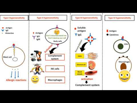
  • 🧬 Type Four Hypersensitivity, also known as cell-mediated hypersensitivity, is primarily caused by inflammation resulting from cytotoxic T cells.
  • 🔬 CD4 positive T cells produce cytokines that lead to chronic inflammation, often in response to environmental or self-antigens.
  • 🌟 Delayed Type Hypersensitivity (DTH) is a prototype of T-cell mediated inflammation, detectable within 24 to 48 hours post-antigen exposure.
  • 🔍 T helper 1 (Th1) and T helper 17 (Th17) cells are key contributors to DTH, with Th1 promoting macrophage activation and Th17 recruiting neutrophils.
  • 🛡 The pathogenesis of DTH involves the activation of CD4 positive T cells, differentiation into effector T cells, and resulting inflammation and tissue injury.
  • 🔄 Upon re-exposure to an antigen, memory T cells, which are long-lived, respond quickly to prevent further inflammation.
  • 💉 Clinical examples of CD4 positive T cell-mediated inflammation include tuberculin reactions and contact dermatitis.
  • 🛑 In persistent antigen scenarios, such as with Mycobacterium tuberculosis, T helper one cells can lead to granuloma formation, a hallmark of granulomatous inflammation.
  • 🚨 CD8 positive cytotoxic T lymphocytes kill antigen-expressing target cells through apoptosis, a key mechanism in cell-mediated immunity.
  • 🏥 Diseases mediated by T cell cytotoxicity include type 1 diabetes, graft rejection, and responses against viruses and tumor cells.
  • 📚 The script concludes with a summary of type four hypersensitivity, its mechanisms, and examples, encouraging active learning through practice tests and feedback on the Visia platform.

Q & A

  • What is type four hypersensitivity?

    -Type four hypersensitivity, also known as cell-mediated hypersensitivity, is primarily caused by inflammation resulting from cytotoxic T cells, which are produced by CD4 positive T cells.

  • What are the main cells involved in type four hypersensitivity reactions?

    -The main cells involved in type four hypersensitivity reactions are CD4 positive T cells, which produce cytokines leading to inflammation.

  • What type of inflammation is typically associated with type four hypersensitivity?

    -Type four hypersensitivity is most often associated with chronic inflammation.

  • Which T helper cells contribute to delayed type hypersensitivity?

    -T helper 1 (Th1) and T helper 17 (Th17) cells contribute to delayed type hypersensitivity.

  • What is the role of interferon gamma in T helper 1 cells?

    -Interferon gamma, produced by T helper 1 cells, promotes further helper cell development and activates other immune cells, such as macrophages.

  • How do T helper 17 cells respond to extracellular pathogens?

    -T helper 17 cells produce interleukin-17 and other cytokines and chemokines, which recruit more neutrophils and monocytes to promote inflammation against extracellular pathogens.

  • What happens to effector T cells after the inflammation is cleared?

    -After the inflammation is cleared, effector T cells transform into memory T cells, which are long-lived and respond quickly to the same antigen if encountered again.

  • What are the clinical examples of CD4 positive T cell-mediated inflammatory reactions?

    -Clinical examples of CD4 positive T cell-mediated inflammatory reactions include tuberculin reaction and contact dermatitis.

  • What is the difference between T helper 1 and T helper 17 in terms of the pathogens they target?

    -T helper 1 cells target intracellular pathogens, such as some bacteria and viruses, while T helper 17 cells handle extracellular bacteria and fungi.

  • How do activated CD8 positive cytotoxic T lymphocytes kill target cells?

    -Activated CD8 positive cytotoxic T lymphocytes kill target cells by inducing apoptosis, either through the Fas ligand and Fas receptor interaction or by forming a perforin and granzyme complex.

  • What are some diseases associated with T cell-mediated cytotoxicity?

    -Diseases associated with T cell-mediated cytotoxicity include type 1 diabetes, graft rejection, reactions against various viruses, and destruction of some tumor cells.

Outlines

00:00

🧬 Immune System Diseases: Type Four Hypersensitivity

This paragraph introduces the seventh part of a pathology tutorial series focusing on the immune system, specifically type four hypersensitivity reactions. It explains that type four hypersensitivity, also known as cell-mediated hypersensitivity, is primarily caused by inflammation due to cytotoxic T cells, which are produced by CD4 positive T cells. The paragraph outlines the process of T cell activation, differentiation into T helper 1 and T helper 17 cells, and their roles in combating intracellular and extracellular pathogens, respectively. It also describes the pathogenesis of delayed type hypersensitivity, the transformation of effector T cells into memory T cells, and the quick response upon re-exposure to the same antigen.

05:01

🛡️ Clinical Manifestations of Type Four Hypersensitivity

The second paragraph delves into the clinical examples of CD4 positive T cell-mediated inflammatory reactions, such as tuberculin reaction and contact dermatitis. It describes the process of a tuberculin reaction, including the time frame of the reaction and the histopathological features observed. The paragraph also explains granuloma formation in response to persistent or non-degradable antigens, such as Mycobacterium tuberculosis. Contact dermatitis is highlighted as a tissue injury resulting from delayed hypersensitivity, with examples like poison ivy exposure leading to rash and blister formation. Other conditions related to CD4 positive T cell-mediated inflammation, such as rheumatoid arthritis, multiple sclerosis, inflammatory bowel disease, and psoriasis, are briefly mentioned.

10:01

🗡️ CD8 Positive T Cell-Mediated Cytotoxicity

The final paragraph discusses CD8 positive cytotoxic T lymphocytes and their role in killing antigen-expressing target cells through apoptosis. It outlines the mechanisms of cell death, including Fas ligand and Fas receptor interaction, as well as the perforin and granzyme complex. The paragraph provides examples of diseases where CD8 positive T cell-mediated cytotoxicity is observed, such as type 1 diabetes, graft rejection, reactions against various viruses, and destruction of tumor cells. It also revisits rheumatoid arthritis, multiple sclerosis, inflammatory bowel disease, and psoriasis, but from the perspective of cytotoxic T cell activity. The paragraph concludes with a call to action for viewers to engage with the content through multiple choice questions on the Visia platform and encourages feedback and subscription for further educational content.

Mindmap

Keywords

💡Hypersensitivity Reactions

Hypersensitivity reactions refer to the immune system's exaggerated response to a substance that is typically harmless to others. In the video, the focus is on type four hypersensitivity, which is a cell-mediated response involving T-cells and is characterized by inflammation. This concept is central to understanding the video's theme of immune system responses to antigens.

💡Type Four Hypersensitivity

Type Four Hypersensitivity, also known as cell-mediated hypersensitivity, is the main subject of the video. It is caused by T-cells and results in chronic inflammation due to the release of cytokines. The script discusses how this type of hypersensitivity is distinct from the other types and is associated with conditions like contact dermatitis and tuberculin reactions.

💡Cytokines

Cytokines are proteins that regulate the immune response and are crucial in the context of type four hypersensitivity. The script mentions that CD4 positive T-cells produce cytokines, which lead to inflammation. Examples from the script include interferon-gamma, which is produced by T helper 1 cells and contributes to the pathogenesis of delayed type hypersensitivity.

💡CD4 Positive T Cells

CD4 positive T cells, also known as T helper cells, are a type of lymphocyte that plays a central role in the immune response. The video script explains that these cells produce cytokines that lead to inflammation, a key aspect of type four hypersensitivity. They differentiate into various subsets, such as T helper 1 and T helper 17, which have specific roles in the immune response.

💡Delayed Type Hypersensitivity

Delayed Type Hypersensitivity (DTH) is a specific type of cell-mediated immune response that occurs 24 to 48 hours after exposure to an antigen. The script describes DTH as the prototype of T-cell-mediated inflammation, with examples including the tuberculin reaction, which is characterized by the accumulation of mononuclear inflammatory cells at the site of antigen injection.

💡T Helper 1 and T Helper 17 Cells

T Helper 1 and T Helper 17 cells are subsets of CD4 positive T cells that have distinct roles in the immune response. The script explains that T helper 1 cells are involved in the response to intracellular pathogens and promote inflammation, while T helper 17 cells are involved in the response to extracellular bacteria and fungi. These cells are integral to the pathogenesis discussed in the video.

💡Inflammation

Inflammation is a protective response of the body to harmful stimuli and is a key outcome of type four hypersensitivity. The script describes how inflammation is often chronic and results from the action of cytokines produced by T cells. The video also discusses the role of inflammation in conditions like rheumatoid arthritis and multiple sclerosis.

💡Antigen Presentation

Antigen presentation is the process by which antigen-presenting cells (APCs) display antigens to T cells, initiating an immune response. The script mentions that dendritic cells present antigenic components to naive T cells, which is the first step in the activation of CD4 positive T cells and the subsequent development of type four hypersensitivity.

💡Memory T Cells

Memory T cells are a type of T cell that remains in the body long after an infection has been cleared. The script explains that effector T cells can transform into memory T cells, which respond quickly to the same antigen if encountered again. This concept is important for understanding the long-term immunity aspect of the immune response.

💡Cytotoxic T Cells

Cytotoxic T cells, particularly CD8 positive T cells, are a type of lymphocyte that can kill cells that are infected with viruses or are cancerous. The script describes how these cells can induce apoptosis in target cells, a process that is distinct from the inflammatory response seen in type four hypersensitivity but still part of the immune system's arsenal against threats.

💡Autoimmune Diseases

Autoimmune diseases are conditions where the immune system mistakenly attacks the body's own cells. The script provides examples of such diseases, including rheumatoid arthritis and multiple sclerosis, which are caused by the immune system's response to self-antigens. This concept is related to the video's discussion of T cell-mediated responses and their potential to cause harm.

Highlights

Introduction to the seventh part of the 'Diseases of the Immune System' series, focusing on type four hypersensitivity reactions.

Type four hypersensitivity, also known as cell-mediated hypersensitivity, is mainly caused by inflammation from cytotoxic T cells.

CD4 positive T cells produce cytokines leading to chronic inflammation in type four hypersensitivity.

Delayed type hypersensitivity is the prototype of T-cell mediated inflammation, detectable within 24 to 48 hours.

T helper 1 and T helper 17 cells contribute to delayed type hypersensitivity with distinct immune responses.

Pathogenesis of type four hypersensitivity involves activation of CD4 positive T cells and their differentiation into effector T cells.

Memory T cells, resulting from inflammation clearance, are long-lived and respond quickly to repeated antigen exposure.

Tuberculin reaction is a clinical example of type four hypersensitivity, characterized by induration and inflammation at the injection site.

Persistent non-degradable antigens like Mycobacterium tuberculosis can lead to granuloma formation in type four hypersensitivity.

Contact dermatitis is an example of tissue injury from delayed type hypersensitivity, often resulting in itchy rashes and blisters.

Examples of CD4 positive T cell mediated inflammation include rheumatoid arthritis, multiple sclerosis, inflammatory bowel disease, and psoriasis.

CD8 positive cytotoxic T lymphocytes kill antigen-expressing target cells through apoptosis in type one hypersensitivity.

Type 1 diabetes, graft rejection, and reactions against viruses are examples of diseases involving CD8 positive T cell-mediated cytotoxicity.

Rheumatoid arthritis involves antigens like collagen and results in chronic inflammation and joint deformity.

Multiple sclerosis is characterized by demyelination in the CNS and inflammation, leading to paralysis and optic neuritis.

Inflammatory bowel disease is an aggressive immune response to normal gut bacteria, resulting in chronic intestinal inflammation.

Psoriasis is a skin condition with unknown antigens that manifest as raised plaques.

The video concludes with a summary of type four hypersensitivity reactions, mechanisms, and examples.

Invitation to engage with the content through liking, commenting, subscribing, and sharing the video.

Transcripts

play00:01

hello everyone welcome back to this

play00:03

short tutorial from pathology M simple

play00:05

at IOP pathology.com and supported by

play00:08

visia an active learning platform this

play00:11

is the part seven of diseases of immune

play00:13

system in this section we will be

play00:16

completing the hyper sensitivity

play00:18

Reactions where I'll be discussing type

play00:20

four hyper sensitivity in the earlier

play00:23

sections we have completed type one type

play00:25

two and type three so let's move on to

play00:28

understand type four hyper sensitivity

play00:31

reactions so type four hyper sensitivity

play00:34

is also referred to as Hil mediated

play00:37

hypers sensitivity which is caused

play00:39

mainly by inflammation resulting from

play00:41

cyto mines and these cyto are produced

play00:44

by CD4 positive T cells and that's why

play00:47

it's referred to as C T cell mediated

play00:50

hyper sensitivity okay so it is these

play00:54

CD4 positive T cells which produces cyto

play00:57

kindes and that results in inflammation

play01:01

most often the inflammation is chronic

play01:04

and dist and the antigens uh implicated

play01:06

in type four hypersensitivity can be

play01:09

environmental antigens as well as self

play01:11

antigens to understand more about C cell

play01:14

mediated hypers sensitivity let's see

play01:16

this delayed type hypers sensitivity

play01:18

which is the Prototype of t- cell

play01:21

mediated inflammation here what happens

play01:24

you know there is a tissue reaction to

play01:26

antigens when it is given to an immune

play01:29

individual right and this tissue

play01:31

reaction is detectable within 24 to 48

play01:35

hours the cells which contribute to

play01:38

delayed type of hypers sensitivity are T

play01:41

helper 1 and T helper 17 cells T helper

play01:46

one is a one which is dominated by

play01:48

activated macres whereas T helper 17

play01:51

response is dominated by neutrophils

play01:54

unlike macrophases from tper one cells

play01:58

so what is a pathogenesis as I told the

play02:00

first step is the activation of CD4

play02:03

positive t- cells remember t- cells will

play02:06

be in Nave State they have to be

play02:08

activated so activation of CD4 positive

play02:11

t- cells and then these become effect

play02:14

our T cells and then the second step is

play02:17

the responses of these effector

play02:19

differentiated effector t- cells and

play02:22

finally resulting in inflammation and

play02:24

tissue injury so let's understand this

play02:27

in detail let us assume that this is a

play02:30

Nave t- cell and you have an antigen

play02:33

which to which these Nave T cells are

play02:35

exposed the antigen presenting cells

play02:37

that is the dritic cells presents

play02:39

antigenic component you know to the

play02:42

peptide from this antigen to the Nave T

play02:45

Cell which produces inin 2 which is an

play02:47

autocrine growth factor resulting in

play02:49

more and more of these t- cells and now

play02:52

these t- cells are antigen responsive t-

play02:56

cells right so these antigen responsive

play02:59

t cells further differentiate upon

play03:02

reexposure to antigen one and these

play03:04

antigen responsive te cells as I told

play03:06

you they differentiate into T helper one

play03:10

cells with the help of cyto interlukin

play03:13

12 and these t- helper cells as I told

play03:16

you they are the ones which act against

play03:17

intracellular pathogens like some

play03:20

bacteria viruses promoting inflammation

play03:23

and also activating other immune cells

play03:26

further these antigen responsive te

play03:29

cells differentiate into tper 17 cells

play03:33

with the help of cyto inin 1 6 and inin

play03:37

23 produced by these antigen presenting

play03:41

cells and these T helper 17 produces

play03:44

interin 17 inin 22 and various other

play03:48

chemokines and cytokines which recruit

play03:51

more and more neutrophils and monocytes

play03:54

which promote inflammation and this kind

play03:56

of response is important while defending

play04:00

extracellular bacteria and fungi right

play04:03

so the T helper one tackles the

play04:05

intracellular pathogens whereas T helper

play04:08

17 handles these extracellular bacteria

play04:11

T helper 1 also secretes lots and lots

play04:14

of interferon GMA which promotes further

play04:18

the helper cell development so this is

play04:20

the basic pathogenesis of delayed type

play04:23

hyper sensitivity once the inflammation

play04:26

is cleared what happens the effect our T

play04:28

cells they get transformed into memory T

play04:31

cells and these memory t- cells are long

play04:33

lived and they respond very quickly if

play04:35

the same antigen is encountered again

play04:37

and that's the role of these memory t-

play04:39

cells let's see what happens when there

play04:42

is repeat exposure to antigen so then

play04:45

the T one secretes lots and lots of

play04:47

interferon gamma you know which further

play04:50

activates macrofagos and these

play04:52

macrofagos are referred to as interferon

play04:54

gamma activated macrophages are so what

play04:56

special characteristics these macres

play04:58

have and these are the ones which have

play05:00

increased ability to phagocytose and

play05:03

kill various pathogens they also Express

play05:05

more and more class to Hy molecules on

play05:07

the surface and that results in increase

play05:10

in antigen presentation they secrete

play05:13

tubon necrosis Factor interlukin one and

play05:15

chemokines which further promote

play05:18

inflammation and lastly they also

play05:20

produce more incin 12 and we know inin

play05:23

12 is responsible for amplification of e

play05:27

one response okay more and more tper one

play05:30

cells are recruited so what are the

play05:32

clinical examples of CD4 positive t-

play05:35

cell mediated inflammatory reactions two

play05:37

most common examples are tucine reaction

play05:40

and contact dermatitis let's see what

play05:42

tubine reaction is after intracutaneous

play05:45

injection of purified protein derivative

play05:49

within 8 to 12 hours in the injected

play05:51

site there is rening and in duration

play05:55

which Peaks at around 24 to 72 hours

play05:58

this is what happens that's the

play06:00

enduration at the injection site of

play06:03

purified protein derivative which is

play06:06

speaking at around 24 to 72 hours it

play06:08

finally subsides so if you take a biopsy

play06:11

from this site what do you see the

play06:14

histopathological features include

play06:16

perivascular accumulation of mononuclear

play06:19

inflammatory cells these are the

play06:22

intradermal blood vessels and you find

play06:25

lots of these mononuclear cells

play06:27

lymphocytes and macro right that's

play06:30

called as perivascular cuffing of

play06:32

mononuclear inflammatory cells and you

play06:34

also see variable amount of dermal edema

play06:37

and fibrin deposition this is what you

play06:39

see in a tuberculin reaction now

play06:42

consider when there is a persistent or

play06:44

non-degradable antigen for example you

play06:47

know hubal baselite which is a

play06:49

microbacterium tuberculosis which

play06:51

colonizes lungs or other tissues so this

play06:54

has this basill act as an antigen which

play06:56

is non-degradable and persistent so

play06:59

there is a very strong T helper one Cell

play07:02

Activation right so once there is a t

play07:04

helper one Cell Activation we know that

play07:07

it recruits lots and lots of macres in

play07:10

the initial phases these macras are just

play07:13

not enough to tackle these tubic

play07:15

baselite what do they do they get

play07:17

transformed into epithelioid cells okay

play07:21

they have more cytoplasm they have

play07:22

elongated pale nucleus and these are

play07:25

epithelioid cells and they cluster

play07:27

together to form a granuloma okay so

play07:30

this granuloma is surrounded by a cuff

play07:33

of lymphocytes and this kind of

play07:35

inflammation is known as granulo matus

play07:38

inflammation I have discussed this in

play07:40

detail when I talked about me granul

play07:42

matus inflammation in my earlier videoos

play07:44

so you can just go back and then watch

play07:46

that video as well the second example

play07:48

which you need to understand is contact

play07:50

dermatitis which is an example of tissue

play07:52

injury resulting from delayed type of

play07:55

hyper sensitivity reaction which is a

play07:57

CD4 positive T C mediated inflammation

play08:01

one example you need to be aware is if

play08:03

an individual is exposed to this poison

play08:07

IV or poison o when when I say expose it

play08:10

has to be in terms of contact contact

play08:12

with poison IV or poison o these leaves

play08:14

contain you know rool which is an

play08:16

antigenic component which binds and

play08:19

modifies the self proteins self proteins

play08:23

means the proteins in our body and the

play08:27

peptides which are released from these

play08:28

modified proteins they are recognized by

play08:32

CD4 positive T cells right and they

play08:35

induce inflammatory response which is

play08:37

clinically manifested by rash and these

play08:39

rashes are often itchy and also result

play08:41

in blister formation and that's why it's

play08:43

referred to as blistering dermatitis or

play08:46

vesicular dermatitis other examples of

play08:49

CD4 positive T cell mediated

play08:51

inflammation include romatoid arthritis

play08:53

multiple sclerosis inflammatory bubble

play08:55

disease till now what did we see we saw

play08:57

CD4 positive T cell mediated

play09:00

inflammation now there is another entity

play09:02

which is called a cd8 positive P cell

play09:05

mediated cytotoxicity now what does that

play09:08

mean which means cd8 positive cytotoxic

play09:11

T lymphocytes they kill the antigen

play09:13

expressing target cells in in the

play09:16

earlier cases what we studied there was

play09:18

inflammation whereas in these cases we

play09:20

see that the antigen expressing target

play09:23

cells are killed so how are they killed

play09:25

see after identifying the target cells

play09:27

the activated cd8 positive cytotoxic T

play09:30

lymphocytes they kill the target cells

play09:32

by means of epopt Tois now how does this

play09:36

happen let us see this see these

play09:38

activated cd8 positive cells they

play09:41

express fast liand which activate fast

play09:45

receptor which is expressed on the

play09:47

target cells and we know fast receptor

play09:50

fast legant interaction is the one which

play09:52

activates the extrinsic pathway of

play09:55

apoptosis the second mechanism includes

play09:58

formation of perforin and granzyme

play10:01

complex so once this complex is formed

play10:04

this enters into the cytoplasm and

play10:07

within the cytoplasm of the target cell

play10:09

the perforin you know they help in

play10:12

release of the granzymes from the

play10:14

complex and these granzymes CLE and

play10:17

activate caspases which further result

play10:20

in proptosis because we know caspases

play10:23

are the most important proteases which

play10:27

result in apoptosis so what are the

play10:29

examples of CD positive t- cell mediated

play10:32

cytotoxicity I mean where the cells are

play10:34

killed the examples include the most

play10:37

common one being type 1 diabetes it can

play10:39

be graft Rejection it could be reaction

play10:41

against various viruses and even

play10:44

destruction of some tumor cells let us

play10:47

see more examples of T cell mediated

play10:50

diseases first one is rheumatoid

play10:52

arthritis where the antigen involved is

play10:54

the collagen or citrated self proteins

play10:57

the manifestations include ch arthritis

play11:00

and inflammation multiple sclerosis is

play11:02

another disease where the antigens

play11:04

involved are the protein antigens in

play11:06

mine example milin basic protein what

play11:09

happens here is that there is

play11:11

demyelination in CNS central nervous

play11:13

system with inflammation resulting in

play11:15

paralysis and even optic neuritis type

play11:18

one diabetes as I told you earlier where

play11:21

the antigen involved are the antigens of

play11:24

pancreatic highet beta cells okay and

play11:28

inflammation of these cells result in

play11:29

insulitis or chronic inflammation of

play11:32

eyelets where they result in destruction

play11:34

of beta cells leading to diabetes

play11:37

inflammatory disease another condition

play11:39

where the antigen involved is various

play11:42

entric bacteria okay you know they

play11:45

respond very aggressively to normal

play11:47

entric bacter resulting in chronic

play11:49

intestinal inflammation and the last but

play11:51

not the least is psorasis where we do

play11:54

not know what antigen is implicated but

play11:56

they manifest with dtive plages in the

play11:59

Skin So this completes type four hypers

play12:03

sensitivity if you watch this entire

play12:05

video I would request you to you know

play12:07

just click on the link below in the

play12:08

description as well as in the pin

play12:10

comment where you can solve various

play12:12

multiple choice questions from bolia I

play12:15

have tried visia and this is an amazing

play12:17

platform where you can learn very

play12:19

actively because you have various

play12:21

practice tests given you can even get

play12:23

feedback ultimately it's fun to learn so

play12:26

in summary we did learn about typ for

play12:29

hypers sensitivity reaction the

play12:30

mechanisms along with some examples

play12:33

thank you for watching if you like this

play12:35

video hit the like button do comment if

play12:37

you have any queries to ask do not

play12:40

forget to subscribe if you feel that

play12:42

this video is useful and please do share

play12:45

thank you

Rate This

5.0 / 5 (0 votes)

相关标签
Immune SystemHypersensitivityType IVInflammationCytokinesT-CellsPathologyEducationalHealthcareMedical LearningInflammatory Diseases
您是否需要英文摘要?