Development of the Face and Palate
Summary
TLDRThis script details the intricate process of embryonic development, focusing on the formation of the human face. Starting from the third week post-fertilization, the embryo's three germ layers give rise to all body tissues. By week four, the embryo's folding leads to a shrimp-like form with a developing neural tube and somites. The script describes the emergence of the stoma diem, buccopharyngeal membrane, and branchial arches, crucial for craniofacial development. It highlights the development of the nasal placodes and the fusion of maxillary and mandibular processes to form the upper and lower lips. The summary of the oral and nasal cavities, and palate development emphasizes the critical period from weeks 6 to 12, providing a comprehensive overview of facial formation in early embryonic stages.
Takeaways
- 🚀 The human embryo develops from a flat disc-shaped organism with three germ layers during the third week post-fertilization.
- 🧬 The germ layers include the endoderm, mesoderm, and ectoderm, which give rise to all organs and tissues in the body.
- 🦐 By the fourth week, the embryo takes on a more human-like form, but still resembles a shrimp, with significant development at the head end.
- 🧠 The neural tube expands to form the primitive forebrain, creating the frontal prominence, crucial for brain development.
- 🦴 The paraxial mesoderm segments to form somites, which are important for the development of the musculoskeletal system.
- 🌱 The stoma diem and buccopharyngeal membrane are key structures that develop into the oral cavity and foregut, respectively.
- 🔄 The branchial arches are formed from the mesoderm and are influenced by neural crest cells, contributing to craniofacial development.
- 👃 The nasal placodes develop into the nasal pits and eventually the nasal cavity, with the oral nasal membrane separating it from the oral cavity.
- 👶 The frontonasal process, maxillary process, and mandibular process are essential for facial development, forming the nose, upper lip, and lower jaw.
- 🦷 The mandibular process forms the lower jaw and contributes to the development of the teeth and lower lip.
- 🕊️ The palate develops from the primary and secondary palates, with the palatine shelves fusing to complete the oral and nasal cavities by week 12.
- 📈 The critical period for facial and palate development is between weeks 6 to 12, highlighting the importance of this stage in embryonic growth.
Q & A
What is the significance of the germ layers in embryonic development?
-The germ layers, consisting of the endoderm, mesoderm, and ectoderm, are crucial in embryonic development as they give rise to all the organs and tissues in the body.
How does the embryo's form change by the fourth week of development?
-By the fourth week, the embryo takes on a more human-like form due to folding along the rostral-caudal and lateral axes, although it still resembles a shrimp more than a baby.
What is the frontal prominence in the context of embryonic development?
-The frontal prominence is a bulge that forms in the primitive forebrain as the neural tube expands greatly at the head end of the embryo.
What is the role of the paraxial mesoderm in the formation of somites?
-The paraxial mesoderm partially segments rostrally to form somidomirs and fully segments caudally to form somites, which are important for the development of the musculoskeletal system.
What is the stoma diem and its significance in embryonic development?
-The stoma diem is a small pit that forms between the frontal prominence and the developing cardiac bulge, which will eventually become the oral cavity.
What is the buccopharyngeal membrane and its function during embryonic development?
-The buccopharyngeal membrane is a two-layered structure made up of ectoderm and endoderm that initially separates the stoma diem from the foregut, and later disintegrates to allow free access between the two.
What are the branchial or pharyngeal arches and their significance?
-The branchial arches are six bulges or thickenings of the mesoderm that sprout from the primitive pharynx, playing a key role in the formation of the face and neck structures.
What is the process of the formation of the nasal placodes and their role in facial development?
-Nasal placodes are patches of ectoderm on the frontal prominence that proliferate to form two thickenings, which later form the medial and lateral nasal processes, contributing to the development of the nose.
How do the maxillary and mandibular processes contribute to the formation of the face?
-The maxillary processes proliferate towards the center to form the upper lip, while the mandibular processes form the lower jaw with its teeth and the lower lip, and fuse with the maxillary processes to form the cheeks.
What is the significance of the development of the nasal sacs and the oronasal membrane?
-The nasal sacs form as the nasal pits burrow deeper and backwards, and the disintegration of the oronasal membrane at the base of the nasal sacs forms a primitive coena, connecting the nasal and oral cavities.
What is the role of the palatine shelves in the formation of the palate?
-The palatine shelves are shelf-like processes that grow vertically downwards on either side of the developing tongue, eventually fusing with each other and with the primary palate to form the secondary palate.
Why is the period between weeks 6 to 12 considered critical for face and palate development?
-This period is critical because it encompasses the development of the secondary palate, which usually completes by week 12, and any disruptions during this time can lead to facial and palate abnormalities.
Outlines

此内容仅限付费用户访问。 请升级后访问。
立即升级Mindmap

此内容仅限付费用户访问。 请升级后访问。
立即升级Keywords

此内容仅限付费用户访问。 请升级后访问。
立即升级Highlights

此内容仅限付费用户访问。 请升级后访问。
立即升级Transcripts

此内容仅限付费用户访问。 请升级后访问。
立即升级浏览更多相关视频

Gastrulation - Embryology
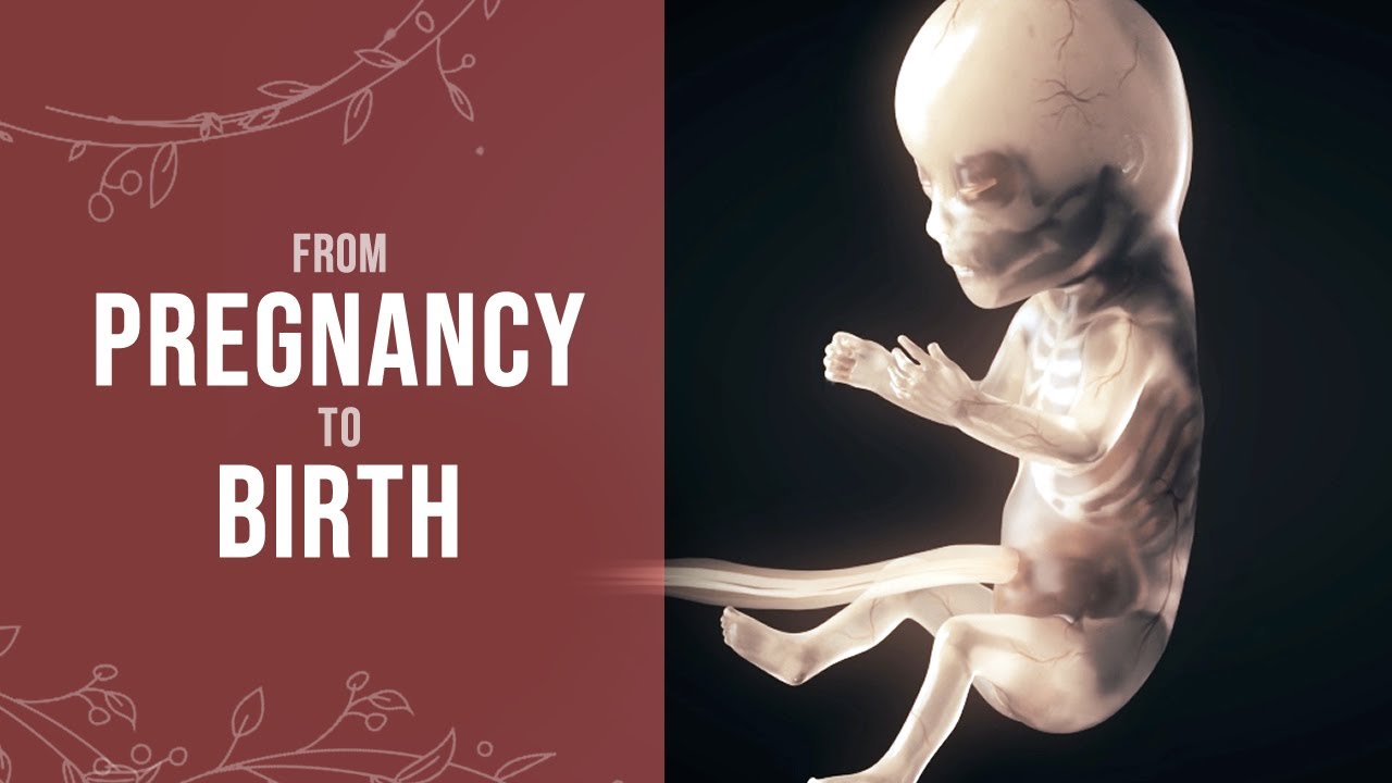
Pregnancy - How a Wonder is Born! (Animation)
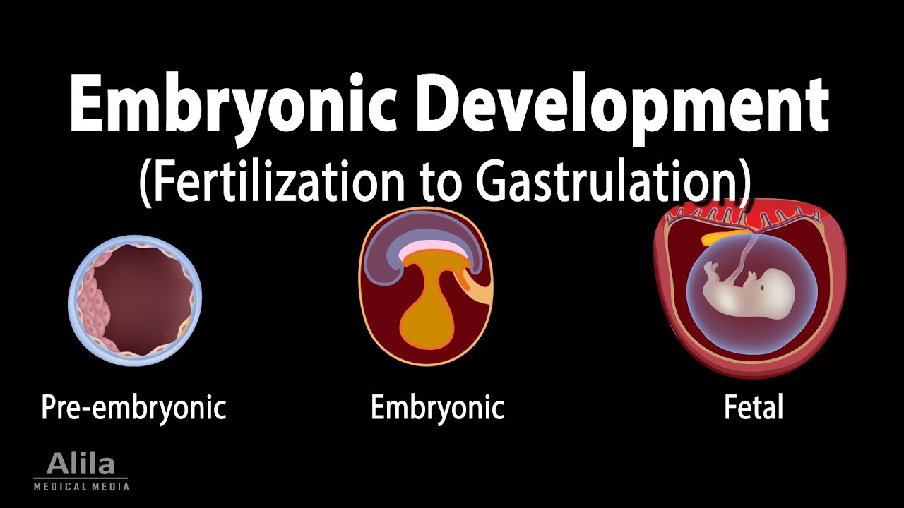
Embryology: from Fertilization to Gastrulation, Animation

EMBRIOLOGIA HUMANA | AULA 08 | TERCEIRA SEMANA DO DESENVOLVIMENTO EMBRIONÁRIO

General Embryology Review in 20 minutes
Gastrulação: Ectoderma, Mesoderma e Endoderma - Terceira Semana do Desenvolvimento (Embriologia)
5.0 / 5 (0 votes)
