Electron Microscopy (TEM and SEM)

Professor Dave Explains
3 Feb 202108:44

Summary

TLDRThe video discusses the evolution from light microscopy to electron microscopy, highlighting how electron microscopes provide over 1,000 times better resolution by using electron beams instead of light. Transmission Electron Microscopy (TEM) allows scientists to visualize small structures like proteins by recording electron energy after passing through specimens, while Scanning Electron Microscopy (SEM) captures secondary electrons emitted from specimen surfaces to study topography. Both methods offer high magnification but have limitations, such as their inability to study live specimens. They are also expensive and require extensive training.

Takeaways

  • 🔬 Light microscopy was the standard until the mid-20th century, but it still plays an important role today.
  • 👁️ Light microscopes can visualize individual cells but struggle to show structures smaller than a cell.
  • 🧪 Electron microscopy, developed in the 1930s and 40s, allowed scientists to see smaller structures with higher resolution.
  • 🔍 Electron microscopes can magnify specimens up to 1,000,000 times and achieve a resolution of 0.2 nanometers.
  • ⚡ Electron microscopy uses beams of electrons, which interact with specimens for more detailed imaging than light microscopes.
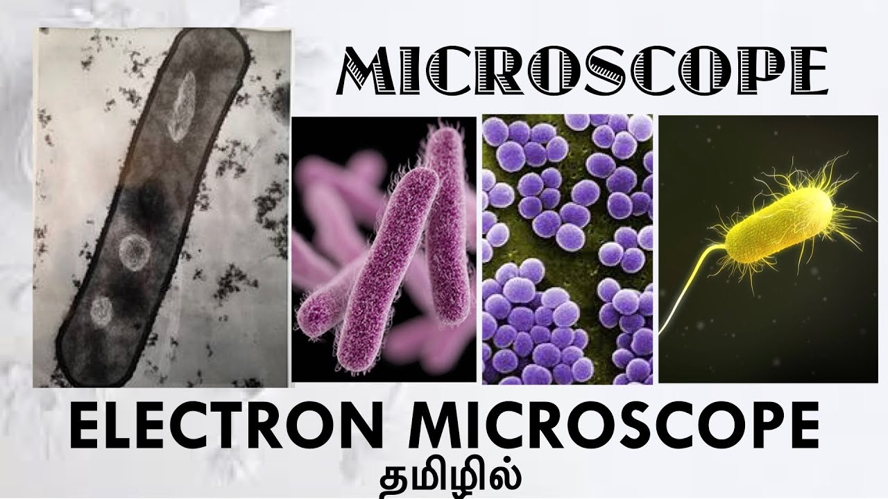
  • 🧫 Transmission Electron Microscopy (TEM) uses electron beams passing through ultra-thin, stained specimens to visualize internal structures.
  • 🛠️ TEM requires specimens to be dehydrated, embedded in resin, and sliced thinly, making it unsuitable for studying live specimens.
  • 🏞️ Scanning Electron Microscopy (SEM) creates 3D images by detecting secondary electrons emitted from the specimen’s surface.
  • 💡 SEM visualizes the topography of specimens, providing detailed surface structure analysis, unlike TEM’s flat images.
  • 💰 Both SEM and TEM are expensive and complex techniques that require extensive training, limiting their use outside specialized fields.

Q & A

  • What limitations did standard light microscopy have in studying microscopic structures?

    -Light microscopy could visualize individual cells, but it struggled to resolve structures smaller than cells, such as proteins and viruses, which limited its ability to study these smaller details.

  • How does electron microscopy achieve higher resolution than light microscopy?

    -Electron microscopy uses beams of electrons with much shorter wavelengths than visible light. This allows for much higher resolution and magnification, up to 1,000,000 times, compared to light microscopy.

  • What is Transmission Electron Microscopy (TEM), and how does it work?

    -TEM uses a beam of electrons transmitted through a specimen to produce highly detailed images. The specimen is stained with heavy metals, and electrons lose energy when passing through electron-dense regions, allowing the microscope to visualize structures like cell organelles and proteins.

  • What preparation is required for a specimen to be viewed using TEM?

    -The specimen must be dehydrated, embedded in plastic resin, and sliced into ultra-thin sections with a glass or diamond knife. These sections allow the electron beam to pass through for imaging.

  • What is Scanning Electron Microscopy (SEM), and what does it specialize in visualizing?

    -SEM specializes in studying the surface topography of samples by scanning the surface with an electron beam. The specimen is coated with vaporized gold or palladium, and the emitted secondary electrons help create a 3D image of the surface.

  • What are the main differences between TEM and SEM?

    -TEM visualizes internal structures with high resolution but produces flat images, while SEM focuses on surface topography and provides 3D images. TEM has better resolution, but SEM can visualize surface details in three dimensions.

  • What are some limitations of electron microscopy techniques like TEM and SEM?

    -Both techniques cannot be used to study living specimens, as the samples must be dehydrated and processed. Additionally, they are expensive and require extensive training to operate.

  • What role do heavy metal stains play in TEM imaging?

    -Heavy metal stains like uranyl acetate or osmium tetroxide bind to certain parts of the specimen, making them electron-dense. These regions absorb more electrons, which results in a higher contrast in the final image.

  • How are secondary electrons used in SEM imaging?

    -When the electron beam interacts with the surface of a specimen coated with gold or palladium, it causes the emission of secondary electrons. These electrons are then detected and used to visualize the surface features of the specimen.

  • Why is electron microscopy important in microbiology?

    -Electron microscopy allows microbiologists to study incredibly small structures, such as viruses, proteins, and cellular organelles, which are not visible with standard light microscopy. This has led to significant advancements in understanding the microscopic world.

Outlines

00:00

🔬 The Evolution from Light Microscopy to Electron Microscopy

This paragraph discusses the transition from light microscopy, which was dominant until the mid-20th century, to electron microscopy. As scientists became curious about structures smaller than cells, they needed more advanced tools. Electron microscopy, developed in the 1930s and 40s, offered a higher resolution, with some techniques achieving up to 0.2 nanometers, over 1,000 times better than light microscopes. This advancement allowed researchers to study finer details such as proteins and viral structures, enhancing their understanding of the microscopic world.

05:01

⚛️ How Electron Microscopy Works: Electrons Over Light

This section explains the working principle of electron microscopy, which uses beams of electrons instead of light. Electrons have a shorter wavelength than visible light, allowing for greater detail and resolution. The paragraph introduces Transmission Electron Microscopy (TEM), which involves embedding specimens in resin or staining them with heavy metals. These stained areas, called electron dense regions, lose more energy when electrons pass through, enabling high-resolution imaging. The specimen is sliced thin to allow electrons to pass through, and the image is magnified and projected using fluorescent screens and a camera.

🔧 Preparing Specimens for Transmission Electron Microscopy (TEM)

This section describes the process of preparing specimens for TEM. Specimens are dehydrated, embedded in plastic resin, and sliced into ultra-thin sections using a glass or diamond knife. These thin sections allow electrons to pass through and visualize the specimen. The electron beam is generated and accelerated through the microscope column, passing through the specimen. After passing through the electron-dense regions, the beam is magnified and projected onto a fluorescent screen, which converts electron energy into visible light for imaging.

🧪 How Transmission Electron Microscopy (TEM) Visualizes Samples

Here, the paragraph goes into more detail about the electron beam's journey. After passing through the specimen, the electrons lose energy based on the electron-dense regions, then go through objective and projector lenses that further magnify the image. Finally, the fluorescent screen translates the electron interactions into bright spots, with a camera capturing the final image. This allows TEM to visualize ultra-small structures like cell organelles, proteins, and cytoskeletal filaments. However, it is noted that TEM's limitations include its inability to image living specimens or 3D structures.

💻 Introduction to Scanning Electron Microscopy (SEM)

This paragraph introduces Scanning Electron Microscopy (SEM), which differs from TEM by visualizing surface structures. SEM specimens are coated with vaporized gold or palladium ions, and the emitted secondary electrons from the surface are detected to produce an image. Unlike TEM, SEM can visualize 3D structures but has lower resolution. Both methods require extensive training and are costly, making them tools mostly for specialized microbiology research.

🔍 How SEM Works: Scanning and Electron Detection

SEM operates by accelerating an electron beam towards the specimen, which is scanned across its surface. As electrons hit the gold- or palladium-coated surface, secondary electrons are emitted and detected. There are two detectors: the secondary electron detector and the backscatter electron detector, the latter used for crystallography and magnetic studies. However, microbiologists focus on secondary electrons to visualize surface topography, as SEM produces a 3D image by scanning the surface of the sample.

📸 The Strengths and Weaknesses of SEM

The advantages of SEM are outlined here, particularly its ability to provide 3D images and study surface topography, unlike TEM, which produces 2D images. SEM is useful for exploring depth and surface structures, but it also has limitations, including lower resolution compared to TEM and the inability to image living specimens. Both SEM and TEM require expensive equipment and advanced training, restricting their use to specialized fields.

📜 Summary of Electron Microscopy: A Powerful but Limited Tool

This final paragraph summarizes the key points covered in the tutorial. Electron microscopy, offering 1,000 times better resolution than light microscopy, uses electron beams for imaging due to their shorter wavelength. TEM is used to study internal cellular structures through electron-dense regions, while SEM is used for 3D surface imaging. Both methods are essential in microbiology but are limited by their inability to study living organisms and their high cost and complexity.

Mindmap

Keywords

💡Light Microscopy

Light microscopy is a technique that uses visible light and lenses to magnify small objects, like individual cells. It was the standard for studying microscopic organisms until the mid-20th century. Despite its limitations in resolving smaller structures, it remains highly useful today for cell visualization.

💡Electron Microscopy

Electron microscopy is a more advanced imaging technique developed in the 1930s and 1940s that uses beams of electrons instead of light to observe much smaller structures, like proteins and viruses. Its resolution is around 1,000 times better than light microscopy, making it essential for studying details at the molecular level.

💡Resolution

Resolution refers to the ability of a microscope to distinguish between two close objects. In the context of the video, electron microscopy offers a resolution of 0.2 nanometers, which is more than 1,000 times finer than light microscopy. This high resolution allows scientists to study extremely small structures like cell organelles.

💡Transmission Electron Microscopy (TEM)

TEM is a type of electron microscopy where a beam of electrons passes through an ultra-thin specimen, revealing internal structures at a high level of detail. The video explains how it is used to study proteins, viruses, and cell organelles by capturing the differing energies of electrons as they pass through electron-dense regions.

💡Scanning Electron Microscopy (SEM)

SEM is another form of electron microscopy that scans the surface of a specimen with an electron beam. Unlike TEM, SEM provides a 3D image of the sample's surface, allowing researchers to study its topography. The video mentions that SEM is useful for investigating the surface features of cells, though it has lower resolution than TEM.

💡Electron Dense Regions

Electron-dense regions are areas within a specimen that absorb more electrons due to their composition, often stained with heavy metals. In TEM, these regions lose more energy when electrons pass through them, which helps scientists visualize small structures. This concept is key in understanding how TEM provides detailed internal images.

💡De Broglie Wavelength

The de Broglie wavelength refers to the concept that particles, like electrons, exhibit wave-like properties. The video connects this to electron microscopy, explaining that the shorter wavelength of electrons compared to visible light allows for higher resolution imaging, a fundamental reason electron microscopes can see such fine details.

💡Secondary Electrons

Secondary electrons are emitted from a specimen when it is bombarded with an electron beam in SEM. These electrons are key to visualizing the sample’s surface topography. The video explains that detectors capture these secondary electrons to produce the final image, which is critical for 3D imaging in SEM.

💡Fluorescent Screen

In TEM, a fluorescent screen is used to translate electron interactions into visible images. As the electron beam passes through the sample and loses energy, the screen lights up with bright spots where the electrons hit, and this pattern is captured to create an image. This process is crucial in converting electron data into visual information.

💡Magnification

Magnification is the process of enlarging the appearance of a specimen. Electron microscopy can magnify specimens up to 1,000,000 times their original size, far beyond the limits of light microscopy. The video highlights how this extreme magnification allows scientists to study proteins, viruses, and other small structures at the molecular level.

Highlights

Light microscopy was the standard until the mid-20th century and remains useful today.

Electron microscopy, developed in the 1930s and 40s, offers much higher resolution than light microscopy.

Electron microscopes can achieve a resolution of 0.2 nanometers, 1,000 times better than light microscopy.

Electron microscopes can magnify specimens up to 1,000,000 times their original size.

Electron microscopy allows the study of smaller structures like proteins and viruses, which cannot be visualized by light microscopy.

Electron microscopes use beams of electrons with a shorter wavelength than visible light, providing higher resolution.

Transmission Electron Microscopy (TEM) visualizes fine cellular structures using heavy metal stains.

TEM requires specimens to be dehydrated, embedded in resin, and cut into ultra-thin slices.

TEM detects electron-dense regions where electrons lose energy as they pass through, creating detailed images.

Scanning Electron Microscopy (SEM) visualizes surface topography by detecting secondary electrons emitted from the specimen.

In SEM, the specimen is coated with vaporized gold or palladium to enhance electron interactions.

SEM produces 3D images of the specimen surface, unlike the flat images from TEM.

TEM is used to study internal cell structures like organelles, while SEM is better for surface topography.

Both SEM and TEM cannot visualize living specimens, as they require dehydration and coating.

Electron microscopy techniques are expensive, complicated, and primarily used by researchers in fields like microbiology.

Transcripts

play00:06

We just learned all about light microscopy, which was the standard until the middle of

play00:11

the 20th century, and remains tremendously useful today.

play00:15

But as we began to learn more about the microscopic world, using light microscopes that could

play00:20

visualize individual cells, we started to become curious about structures that are smaller

play00:25

than a cell, some details of which were difficult to visualize and study under standard light

play00:31

microscopes.

play00:33

Fortunately, electron microscopy, developed in the 1930s and 40s, proved to be much more

play00:39

powerful and allowed scientists to view these smaller structures.

play00:44

Some electron microscopy techniques employ a resolution of 0.2 nanometers, which is more

play00:50

than 1,000 times better than light microscopy.

play00:54

With this higher resolution, electron microscopes are able to magnify their specimen up to 1,000,000

play01:00

times their original size, allowing researchers to study proteins inside of cells, and the

play01:06

molecular structure of viruses, all of which led to a better understanding of the microscopic

play01:12

world.

play01:13

Of course we have to ask, how do they do this?

play01:16

Well the answer is in the name, they use beams of electrons, which interact with the sample

play01:22

as the beam is transmitted through the specimen.

play01:25

As we recall from our study of chemistry, electrons are stable, subatomic particles

play01:30

that carry a negative charge.

play01:33

Electron beams have a much shorter wavelength than the visible light that light microscopes

play01:38

use, which we refer to as the de Broglie wavelength, as we learned in the modern physics series.

play01:44

This shorter wavelength allows for a higher level of detail and resolution in imaging.

play01:51

One type of electron microscroscopy is called Transmission Electron Microscopy, or TEM.

play01:57

For TEM, specimens are fixed on a support grid and either embedded in resin or stained

play02:03

with a material like uranyl aceate or compounds of heavy metals like osmium tetroxide.

play02:11

Certain structures within the specimen will take up more of the stain than other parts

play02:16

of the specimen, and those sections are called electron dense regions.

play02:21

When electrons pass through electron dense regions they lose more energy compared to

play02:25

when they pass through sparser regions.

play02:28

This phenomenon is used by TEM to visualize the specimen at high levels of resolution

play02:34

and magnification.

play02:36

For the stained specimen to be viewed by TEM it must be dehydrated, embedded in plastic

play02:42

resin, and cut into ultra-thin slices with a glass or diamond knife.

play02:47

The sections must be ultra-thin so that the electrons can pass through them.

play02:52

When the specimen is secured in the specimen holder, an electron beam is generated and

play02:57

shot out of the electron gun.

play02:59

The electron beam is accelerated down the column of the microscope after passing through

play03:04

the anode.

play03:06

The electron beam then passes through the specimen, once again, losing energy as electrons

play03:11

pass through electron dense regions.

play03:14

The electrons then pass through the objective lens, magnifying the image, before the electrons

play03:20

pass through the projector, or ocular lens, which magnifies the image further and projects

play03:26

it onto the fluorescent screen.

play03:29

This is where we start translating the electrons into an image.

play03:33

The fluorescent screen is coated with a chemical which appears as bright spots when electrons

play03:39

come into contact with it.

play03:41

The intensity of the bright spot depends on how much energy the electrons have when they

play03:46

hit the fluorescent screen.

play03:48

The camera under the fluorescent screen then captures the image made from the bright spots

play03:53

which is displayed on a computer screen.

play03:56

Transmission electron microscopes are used to visualize structures that are too small

play04:01

to study with light microscopes such as the cell organelles, proteins, cytoskeletal filaments,

play04:08

and the components of cell membranes.

play04:10

But even with the unmatched resolution and magnification abilities of TEM, it still does

play04:17

have its limitations.

play04:19

Some of these include the fact that you cannot study living specimens or image 3D structures

play04:24

because the specimens must be dehydrated and cut into ultra-thin slices before imaging.

play04:32

The second type of electron microscopy is Scanning Electron Microscopy, or SEM.

play04:39

With SEM, specimens are coated with vaporized gold or palladium ions, because when electrons

play04:45

come into contact with this coating, they cause atoms on the surface of the specimen

play04:50

to emit electrons.

play04:52

These emitted electrons are called secondary electrons, and these are what SEMs use to

play04:58

visualize their specimen.

play05:00

After the coated specimen is secured on the stage of the SEM, the process starts out similarly

play05:06

to TEM in that an electron beam is shot out and accelerated towards the specimen.

play05:13

In the SEM, the electron beam then sweeps, or scans the surface of the specimen, which

play05:18

causes it to discharge secondary electrons.

play05:22

There are two types of detectors in the sample chamber, the secondary electron detector and

play05:28

the backscatter electron detector.

play05:31

The backscatter electron detector picks up backscatter electrons that come from deeper

play05:36

regions of the sample that are a result of elastic interactions between the beam and

play05:41

the sample.

play05:43

The results from this detector are primarily used to look at crystallography, and the magnetic

play05:48

field of the sample.

play05:49

But for the purposes of microbiology, researchers focus on the detection of secondary electrons

play05:56

which allow them to visualize the topography of the sample, or the distribution of parts

play06:01

and features on the surface of the sample.

play06:05

The detectors convert the electron signal into a light signal that is amplified by passing

play06:11

through a photomultiplier before the final image is projected on a computer screen.

play06:17

SEM imaging is primarily used to study the topography of cells and other similar structures.

play06:25

With this type of microscopy, researchers are able to investigate the depth of structures,

play06:31

which isn’t possible with TEM, as SEM produces a 3D image opposed to the flat image from

play06:37

TEM.

play06:38

Yet, just like TEM, SEM cannot visualize live specimens, and SEM has worse resolution than

play06:46

TEM.

play06:47

Both TEM and SEM are expensive and require a lot of training, so they are not likely

play06:53

to be used apart from those pursuing a microbiology career.

play06:58

We covered quite a lot in this tutorial, so let’s briefly summarize.

play07:02

Electron microscopy has around 1,000 times better resolution than light microscopy because

play07:07

electron microscopy uses electron beams instead of light, which have a shorter wavelength.

play07:14

This allows some electron microscopes to visualize specimens at 1,000,000 times their original

play07:21

size.

play07:22

Transmission Electron Microscopy, or TEM, records the differing energies that electrons

play07:27

possess after they pass through a specimen stained with heavy metals to visualize small,

play07:34

detailed structures like proteins inside of cells, and molecules associated with viral

play07:40

particles.

play07:41

Scanning Electron Microscopy, or SEM, captures the secondary electrons that are emitted from

play07:48

the surface of samples coated in vaporized gold or palladium ions to visualize the topography

play07:55

of the specimen.

play07:56

And the main drawback, as we mentioned, is that both SEM and TEM cannot be used to visualize

play08:02

living specimens, and they are also quite expensive and complicated techniques to use.

play08:09

And with that, we know a bit about electron microscopy.

Rate This

5.0 / 5 (0 votes)

相关标签
Electron MicroscopyLight MicroscopyTEMSEMMicroscopic StructuresCell BiologyProteinsVirusesHigh ResolutionScientific Imaging
您是否需要英文摘要?