Anatomia do Olho - Como Funciona o Olho Humano
Summary
TLDRIn this video, ophthalmologist Cláudio Del Claro explores the intricate anatomy of the human eye, highlighting its divine creation and complex structures. He explains how each part, from the external muscles to the internal components like the cornea, iris, and retina, plays a crucial role in vision. The video emphasizes the eye's sensitivity, its comparison to a high-performance camera, and the importance of eye care, concluding with a call to action for viewers to subscribe for more eye health content.
Takeaways
- 👁 The human eye is a divine creation and a fantastic organ, providing us with our primary sense of vision.
- 🤔 The eye, despite being small, contains highly complex structures with high metabolic cells, inspiring the invention of the camera.
- 👀 The eye's sensitivity is about 600 times greater than a camera, demonstrating its superior visual capabilities.
- 🔄 The eye's muscles allow for symmetrical and simultaneous movement, ensuring coordinated vision.
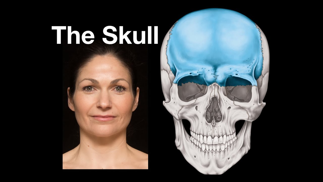
- 🛡️ The eye is protected by the orbit, a bony cavity in the skull, and surrounded by fat and orbital skin.
- 👓 Eyelids serve to cover and protect the eyeball, with a blink reflex to prevent foreign objects from entering.
- 🌞 Eyelashes protect the eyes from light and debris, enhancing protection.
- 💧 Blinking helps to spread tears across the eye surface, lubricating and cleaning the eye, which is crucial for eye health.
- 🌈 The iris controls the amount of light entering the eye by constricting or dilating the pupil, similar to a camera's diaphragm.
- 🔍 The lens of the eye adjusts focus by contracting and relaxing, similar to a camera's zoom function.
- 👁️🗨️ The retina is the eye's primary organ of vision, capturing light stimuli and transmitting them to the brain via the optic nerve.
Q & A
What makes the human eye such a remarkable organ according to the video?
-The human eye is described as a perfect creation, complex and capable of giving us our primary sense: vision. Its structures are incredibly intricate, and even though cameras have been modeled after the eye, they are still far less sensitive, with the eye being around 600 times more sensitive than a camera.
How does the musculature of the eye function to move it?
-The external muscles of the eye move it in various directions—up, down, left, right, and diagonally. These muscles work simultaneously, meaning when one eye moves in a direction, the other follows in sync, maintaining symmetrical and coordinated movements.
What role do the eyelids play in protecting the eyes?
-Eyelids protect the eyes by covering them, preventing foreign objects from entering. They also blink reflexively when anything approaches the eye, acting as a defense mechanism. Additionally, they help keep the eyes moist by spreading tears over the surface.
Why are eyelashes and eyebrows important for eye protection?
-Eyelashes help block dust, pollen, and other particles from entering the eyes, acting like a protective screen. Eyebrows help prevent sweat and rain from running into the eyes, offering further protection from external elements.
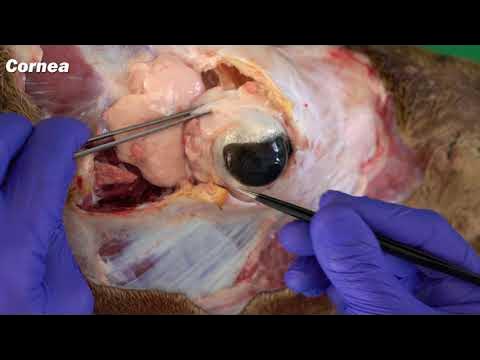
What is the function of the cornea, and why is it crucial for vision?
-The cornea is the eye's most powerful lens, playing a critical role in focusing light onto the retina. Any injury or irregularity in the cornea can severely impact vision, causing blurriness or distortion. It is also highly sensitive, filled with nerve endings that make even minor injuries very painful.
How does the iris control the amount of light entering the eye?
-The iris, the colored part of the eye, controls the size of the pupil to regulate light entry. In bright conditions, it contracts to reduce the light entering the eye, while in darker conditions, it dilates to let more light in, functioning similarly to a camera’s aperture.
What role does the crystalline lens play in vision?
-The crystalline lens focuses light onto the retina by adjusting its shape to accommodate near and far objects. It works like the zoom function on a camera, changing its shape to help the eye focus at different distances.
What is the function of the vitreous body in the eye?
-The vitreous body is a transparent gel that fills the space between the lens and the retina, helping maintain the eye’s shape. It also provides cushioning to protect the retina from potential damage in case of injury.
What happens in the retina, and why is it considered the key structure for vision?
-The retina contains photoreceptors that capture light and convert it into electrical signals, which are then sent to the brain via the optic nerve. The retina is essential for processing visual information, and any issues with it can severely affect vision.
What is the optic nerve’s role in the visual process?
-The optic nerve transmits visual information from the retina to the brain, where it is processed into recognizable images. Any damage to the optic nerve can disrupt this process, leading to vision loss, which is a major concern in conditions like glaucoma.
Outlines
👁 Anatomy of the Human Eye
The paragraph introduces the human eye as a divine creation and a fantastic organ, emphasizing its perfection and complexity. The speaker, Cláudio Del Claro, an ophthalmologist, welcomes viewers to his channel dedicated to vision care. He explains that despite the eye's small size, its structures are intricate and highly metabolically active. The eye's design has inspired the creation of the camera, yet it remains about 600 times more sensitive than a photographic camera. The paragraph outlines the external structures of the eye, including the white structure surrounded by muscles that allow eye movement, the orbital cavity, and the protective layers of fat, skin, and the eye socket.
🛡️ Protective Structures of the Eye
This section delves into the protective functions of the eye's external structures such as the eyelids and eyelashes. The eyelids have a reflex to protect the eye from foreign objects, and the eyelashes act as a first line of defense against debris. The paragraph also discusses the role of the eyelids in moistening the eye surface through blinking, which is essential for vision clarity and eye health. Additionally, the importance of eyebrows in preventing sweat and rainwater from entering the eye is highlighted.
🌈 The Iris and Its Role in Light Regulation
The paragraph focuses on the iris, the colored part of the eye, and its critical role in controlling light entry through the pupil. It explains how the iris contracts and dilates in response to light levels, protecting the eye from bright sunlight and adjusting for better vision in the dark. The text also describes the cornea as the eye's most powerful lens, affecting vision if damaged, and the high sensitivity of the cornea, which serves as an alert system for potential eye injuries.
👀 Detailed Functioning of the Eye's Internal Structures
This section provides a comprehensive overview of the eye's internal structures, including the lens (crystalline), the aqueous humor, and the ciliary body. It explains how the lens adjusts focus similar to a camera zoom, and the role of the ciliary muscles in this process. The paragraph also discusses the vitreous humor, which provides shape and protection to the eye, and the retina, the light-sensitive layer responsible for capturing visual stimuli. Special attention is given to the macula and fovea for central vision and the peripheral retina for side vision. The optic nerve's function in transmitting visual information to the brain is also highlighted, along with the choroid's role in nourishing the retina.
Mindmap
Keywords
💡Vision
💡Anatomy of the Eye
💡Cornea
💡Iris
💡Pupil
💡Lens
💡Retina
💡Optical Nerve
💡Vitreous
💡Choroidal
💡Uveitis
Highlights
The human eye is a divine creation and a fantastic organ that primarily gives us the sense of vision.
The anatomy of the eye is extremely complex, with cells having the highest metabolism compared to the rest of the body.
The human eye inspired the creation of the camera, showcasing its perfect design.
Despite technological advancements, the human eye is about 600 times more sensitive than a camera.
The eye connects us to the external world and provides us with feelings of happiness and gratitude.
External muscles around the eye enable its movement in various directions.
The eye is protected by the orbit, a bony cavity in the skull, and surrounded by fat and orbital skin.
Eyelids serve to cover and protect the eyeball, with a reflex to close when something approaches.
Eyelashes play a role in protecting the eyes from dust, pollen, and other particles.
Blinking helps to moisten and clean the eye surface, which is essential for maintaining healthy vision.
Sweat glands above the eyelids prevent sweat from entering the eyes, contributing to eye protection.
The sclera is the white part of the eye, providing support and protection against trauma.
The conjunctiva is a thin membrane that can be seen and moved over the white of the eye.
The cornea is the transparent front part of the eye and acts as the eye's most powerful optical lens.
The iris is the colored part of the eye, responsible for controlling the amount of light entering the eye.
The pupil, also known as the 'girl of the eye,' regulates light entry, similar to a camera's diaphragm.
The lens of the eye adjusts focus by contracting and relaxing, similar to a camera's zoom function.
The ciliary body produces aqueous humor and moves the lens through the ciliary muscle.
The vitreous is a transparent gel that gives the eye its shape and protects the retina.
The retina is the light-sensitive tissue at the back of the eye, responsible for capturing visual stimuli.
The optic nerve is crucial for transmitting visual information from the retina to the brain.
The choroid is a vascular tissue that nourishes the retina.
UV or uveitis is an inflammation inside the eye occurring at the junction of the iris, ciliary body, and choroid.
Transcripts
o olho humano que Criação Divina
perfeita que órgão Fantástico capaz de
nos dar o nosso principal sentido que é
a visão no vídeo de hoje eu vou detalhar
para você como este olho funciona como é
anatomia do nosso olho Bora lá
[Música]
Olá que alegria te receber aqui no meu
canal eu sou Cláudio Del Claro
oftalmologista e criei esse canal
especialmente para você para você que é
preocupado com sua visão para você que
enxergar bem é prioridade na vida pois é
aqui que você receberá as melhores
respostas então se você ainda não é
inscrito não perca tempo inscreva-se
agora mesmo clique no Sininho Porque
toda semana são três vídeos novos que
simplesmente vão te ajudar a cuidar
melhor de seus olhos o olho pode ser
muito pequeno comparado com o nosso
corpo todo mas as estruturas dentro do
olho todas as estruturas responsáveis
para nos dar a visão são muito complexas
extremamente complexas
tanto que as células de dentro do nosso
olho são as células com maior
metabolismo comparados com o corpo todo
as estruturas oculares são tão
fantásticas e como falei anteriormente
tão criadas tão perfeitamente que o
homem se inspirou nisso para criar uma
câmera fotográfica é ou não é incrível
Então hoje você vai conhecer um
pouquinho mais disso como que a visão é
formada Como que essa câmera maravilhosa
e perfeita É capaz de transmitir os
estímulos Luminosos que nós vemos aqui
na nossa frente e aí o nosso cérebro faz
o trabalho de realmente reconhecer
identificar e interpretar aquele
estímulo apesar de toda a tecnologia de
hoje apesar do homem tentar se inspirar
no olho e fazer câmeras fotográficas
cada vez melhores ainda assim está muito
a quem o nosso olho cerca de 600 vezes
mais sensibilidade de que uma câmera
fotográfica e é por isso que nós temos
Essa visão que a visão nos conecta ao
Nosso principal sentido que nos conecta
com o mundo externo e que nos faz no
sentir também que nos dá felicidade que
nos dá o sentimento de gratidão Então
bora lá conhecer como que este olho é
formado primeiro olhando aqui por fora
você vai ver que o olho ele tem essa
estrutura Branca em volta dele e que em
volta do olho existem músculos que fazem
a movimentação então é essa musculatura
externa do olho que gira o olho para um
lado para o outro para cima para baixo
na diagonal e o mais interessante é que
a musculatura é astrocular ela faz em
movimentos simultâneos então se um olho
mexe para um lado o outro acompanha
igual olha por cima o outro acompanha
igual então eles têm um funcionamento
totalmente simétrico e simultâneo e Este
globo ocular ele está protegido pela
Órbita que esta cavidade
óssea dentro do nosso crânio e envolto
por uma gordura gordura pele orbital e
ele fica aqui então
estratégicamente localizado em voto por
esta gordura e pela cavidade óssea Agora
vamos começar a analisar cada estrutura
então primeiro temos a em volta do olho
a nossas pálpebras Então as pálpebras
Tem uma função fantástica também de
cobrir e proteger o globo ocular por
isso que existe esse reflexo pau-pebral
tudo que vem se aproximando do seu olho
seja o seu dedo ou seja um objeto você
vai ter um reflexo instantâneo e a
vontade de fechar esse olho para que
nada atinja isso é fantástico Além disso
nós temos os filhos na moda da pálpebra
que também tem um papel importante na
proteção como se fosse um toldo Então a
gente vai ter uma porta que fecha e
ainda na ponta da porta a gente vai ter
esse toldo para ajudar a proteger mais
ainda então detritos poeira pólen de
flores pelos de animais eles podem ficar
aqui presos nos nossos filhos e não
entrar para dentro do olho ajuda muito e
só quem perde os cílios é que realmente
dá valor a importância deles e nota A
grande diferença que faz você ficar sem
e como outra diferença também dos filhos
é com relação a sensibilidade à luz os
cílios protegem um pouco nossos olhos da
entrada da luz então você cerrra um
pouquinho os seus olhos e aí esse todo
Ajuda também a proteger é fantástico não
é e se você já tá gostando e adorando
esse vídeo o que que você vai fazer vai
mostrar que sim que esse assunto é
importante que é legal e você vai deixar
muito mais muito like aqui e já atinge
pessoas como você amantes dos nossos
olhos pessoas interessadas em preservar
a sua visão além disso as pálpebras Elas
têm uma função importantíssima para
umidificar a nossa Cola e a superfície
ocular então a cada piscar vem uma onda
de lágrima do filme lacrimal para
lubrificar isso aí como se fosse um
para-brisa mesmo em que é jogado uma
aguinha e faz aquele movimento limpando
isso vai melhorar a sua visão e vai
proteger a sua córne esse movimento do
piscar também ela é involuntário Então
você nem sabe que tá piscando Você nem
sente que está piscando e esse piscar é
fundamental para essa lubrificação para
você manter a saúde ocular para que você
tenha uma boa visão e que para que não
tenha nenhum machucado nos olhos é
simplesmente você falar com alguma
pessoa que sofreu algum acidente ou que
teve paralisia facial que ficou com a
pálpebra aberta o estrago que faz então
esse olho começa a ficar vermelho
dolorido a começa a ver lesões na córnea
pode provocar uma infecção que a úlcera
de córnea pode provocar um embaçamento
da visão por conta da falta de
lubrificação ali então é um pepino
grande cada estrutura cada cada
estrutura aqui da anatomia do nosso olho
ela tem um papel importantíssimo e não
podemos ficar sem nenhuma delas Além
disso uma outra estrutura importante que
externa são as nossas sobrancelhas
também importantes na proteção as
sobrancelhas elas seguram o suor para
não entrar no olho água de chuva que
você pega Então realmente Também quem só
quem fica sem a sobrancelha que nota que
parece que seus olhos ficaram mais
protegidos então entrando mais realmente
no globo ocular o que que nós temos nós
temos a esclera que é toda essa parte
branca em volta do olho e a esclera um
tecido endurecido um tecido fibroso que
realmente dá essa sustentação esse
formato para o olho e protege De traumas
Lógico é um tecido muito mais resistente
em cima da esfera nós temos uma membrana
muito fina que se chama conjuntiva você
pode ver você vai conferir ali olhando
no seu espelho você pode mexer em cima
do branco do olho você vai ver que tem
uma membrana móvel essa membrana é
conjuntiva a esclera está embaixo que é
o tecido branco
agora mais aqui na frente do olho como
se fosse um vidrinho de relógio essa
parte transparentes chama córnea e a
córnea nada mais é do que a nossa lente
mais potente do olho é a nossa lente com
maior poder de óptico qualquer alteração
ali na córnea ela já prejudica a sua
visão então se você tem um machucado na
corna se você tem uma opacidade Você tem
uma irregularidade é automaticamente
isso vai impactar no resultado da sua
visão a visão vai ficar distorcida
embaçada prejudicada a córnea é
extremamente sensível e tem muitas
muitas muitas terminações nervosas por
isso se você arranha sua corna se você
machuca um pouquinho a sua córne a dor é
muito grande o incômodo é muito grande
então se você tem um cisco no seu olho
mesmo se você machucou Você sabe o
quanto que Isso incomoda e é por causa
disso por causa dessa extrema
sensibilidade extrema enervação que
existe ali e isso nada mais é também que
um sistema de defesa do nosso corpo para
te dar aquele sinal de alerta máximo que
o seu olho está em risco então machuquei
a cólera Nossa não posso machucar a
córnea que ela é importantíssima para
nossa visão o corpo já avisa para você
procurar um atendimento para você
proteger o seu olho para que aquele
machucado não prossiga Não continue
tanto que pacientes que tem algum
problema que faz com que esta inervação
não trabalha adequadamente acabam tendo
problemas mais graves porque não sentem
então eu machuquei a minha cólica minha
corda tá começando com uma infecção com
uma úlcera uma ceratite grave eu não tô
nem percebendo e aquilo ali vai piorando
piorando quando vê já tá muito mais
grave E aí nós temos logo atrás da
córnea a Íris que é a parte colorida do
olho e E aí eles existe um líquido ali
que se chama um muracoso que tem o
objetivo de manter o formato do olho que
manter essas estruturas da parte
anterior e chamamos isso de câmara
anterior e aí eles esse tecido colorido
lindo que nós ficamos tão Encantados de
ver a eles com as suas cores
maravilhosas com seus pigmentos
pintinhas que formam desenhos únicos
você sabe hoje a identificação por eles
é algo assim requintadíssimo é algo que
que é individual Seu e é utilizado por
muitos meios para você realmente ser
identificado a eles no olho ela tem um
papel fundamental de controlar a entrada
da luz então também por um reflexo que
você
inconsciente você vai ter contratura da
Íris e dilatação contratura e dilatação
de acordo com a luminosidade então se
você vai para o sol radiação Ultra
camiseta perigosa para o olho o nosso
olho automaticamente fecha a sua pupila
para que haja uma entrada menor de luz
dentro do olho em ambientes Escuros para
facilitar a sua visão essa pupila se
dilata essa bolinha de dentro ali da
Íris é chamada de pupila ou popularmente
menina do olho e a pupila nada mais é do
que o diafragma da câmera fotográfica
que o homem se inspirou Então ela regula
a entrada da luz é a pupila que dilata
ou se contrai aí logo atrás da eles nós
temos o cristalino que nada mais é do
que a lente do olho e que ela faz este
trabalho contínuo de relaxar e contrair
e relaxar e contrair para nos dar o foco
Então quando você vai focar para perto O
seu cristalino se contrai e é como se
fosse o zoom da câmera fotográfica
também ajustando as distâncias focais
ele fundamental importantíssimo também
para nossa visão em volta do cristalino
existe a musculatura ciliar e o corpo
ciliar então o corpo ciliar este corpo
ciliar ele tem Esta função de produção
do humor aquoso e de fazer essa
movimentação do cristalino através da
musculatura ciliar as fibras ondulares
ou mais conhecida por nós como zonola é
uma área de fibras musculares que
seguram a lente do olho Então o
cristalino ele é envolvido numa cápsula
se chama cápsula posterior e que liga
esta cápsula posterior ao corpo ciliar
são essas fibras ondulares então ele
fica ali seguro através das onola Então
essas estruturas anteriores aqui que vai
desde da córnea até o cristalino formam
a câmara anterior do olho já daqui para
trás que vai do cristalino Até o fundo
do olho é chamado de câmara e ele é
composto pelo vítreo pela retina e pela
coroide que é um tecido vascular que
está embaixo da retina e o vidro nada
mais é do que este gel transparente que
dá este formato para o nosso olho e
também tem uma característica de dar um
amortecimento ou caso você leve uma
pancada ele tem todo uma função
Protetora da nossa retina que é como se
fosse uma almofadinha para sua retina
não ser atingida então a retina é o
nosso principal órgão da visão que fica
logo atrás este órgão todo Amarelo
alaranjado que é a parte nervosa do
nosso olho é ali na retina através dos
fotos receptores que o estímulo luminoso
será captado então a luz ela vai entrar
dentro do olho e através de passar todas
essas estruturas ela vai sofrendo
processo de refração então a luz vai
entrando dentro do olho passando por
todas essas estruturas e a imagem deve
ser formada exatamente em cima da retina
para você ter a visão perfeita se a
imagem se forma antes da retina você tem
dificuldade se forma depois da retina
você também tem dificuldade aqui áreas
importantes da gente destacar aqui da
retina a mácula que essa área Central
que é responsável pela nossa visão
principal e um ponto ali bem em cima que
se chama fóvia que é o maior responsável
pela nossa visão em volta da macla temos
todas essas Arcadas vasculares que estão
ali nutrindo as células e aí o nervo
óptico que esta é a área mais amarela
mais clarinha
existe ainda toda a periferia da retina
porque o olho é como um globo Então
existe toda essa parte aqui periférica
que também é responsável pela nossa
visão periférica então a mácula seria
responsável pela visão central e em
volta a toda essa área ela nos dá a
visão periférica o nervo óptico é um dos
12 pares cranianos fundamental para
conduzir esse estímulo até o nosso
cérebro Então realmente é fantástico a
retina capta o estímulo luminoso e o
nervo óptico leva diante qualquer lesão
ali no nervo vai comprometer totalmente
a nossa visão É como se você tivesse um
fio de energia e cortasse tirasse algo
da tomada para de funcionar por isso que
insistimos tanto com a preservação do
nervo óptico por ela ser tão fundamental
para nossa visão e aqui lembro da doença
que é mais comum que afeta o nervo que é
o gla doença silenciosa que provoca
danos aqui na nas fibras nervosas do
nervo óptico geralmente ligada à
compressão alta e por último nós temos
esta coroide que seria esse tecido
vascular embaixo da retina que nada mais
tem como função de nutrir as células da
retina E além disso eu vou te falar de
um outro nome aqui porque talvez você vá
olhar aí vai perguntar o que que é use é
o que que é UV o que que é UV o que que
é o veit que é uma inflamação comum
dentro do olho e ela ocorre na uva a uva
nada mais é do que a junção de três
estruturas que seriam a Íris o corpo
ciliar e a coroide então depois dessa
verdadeira aula eu acho que eu consegui
falar de todas as principais estruturas
do nosso olho se você ainda ficou com
mais alguma dúvida por favor deixa aqui
nos comentários você sabe eu adoro
interagir com você e se você ficou até
aqui eu quero te dar os parabéns por ter
ficado até o final do vídeo é sinal de
que você realmente se importa com seus
olhos e se ainda não é inscrito no canal
ainda dá tempo se escreve aqui clica no
Sininho para não perder mais nenhum
vídeo Um grande beijo e eu te vejo no
nosso próximo vídeo tchau tchau
浏览更多相关视频
5.0 / 5 (0 votes)