An Approach to a Red Eye
Summary
TLDRThis video provides a comprehensive overview of the red eye, outlining the key anatomical structures of the eye and how they relate to various pathologies. It covers conditions ranging from benign issues like viral conjunctivitis and dry eyes to more severe ones such as acute angle closure glaucoma and scleritis. The video emphasizes the importance of a thorough history and ophthalmological exam in diagnosing the cause. It also highlights when urgent or emergent ophthalmological consultation is necessary, including signs like decreased visual acuity, non-responsive pupils, and severe pain.
Takeaways
- 😀 A red eye can be caused by a wide variety of conditions, ranging from benign to vision-threatening.
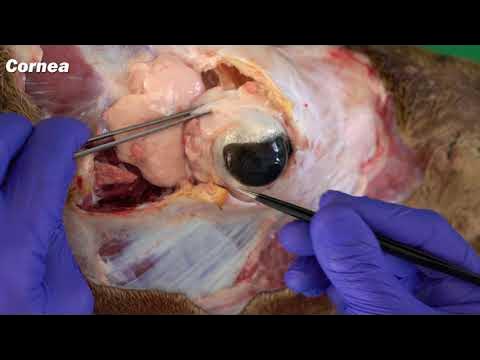
- 😀 Understanding the anatomy of the eye is crucial to diagnosing red eye conditions, as the eye has several key components such as the iris, sclera, cornea, and conjunctiva.
- 😀 Common causes of red eye include viral and allergic conjunctivitis, dry eyes, blepharitis, and a sty, which are generally benign and self-limited.
- 😀 Episcleritis and scleritis both involve inflammation of the sclera, but they differ in severity of pain, onset, and potential impact on vision.
- 😀 Anterior uveitis, inflammation of the iris and/or ciliary body, is often associated with systemic inflammatory diseases and can lead to vision impairment.
- 😀 Acute angle-closure glaucoma is a serious condition that can lead to severe pain, decreased vision, and possible eye damage, requiring urgent ophthalmological intervention.
- 😀 Redness in the eye (hyperemia) can be classified into superficial (conjunctivitis) or deep (scleritis, keratitis), with different causes and severity.
- 😀 Corneal abrasions and foreign bodies in the eye cause significant pain and require specific treatment to avoid complications.
- 😀 Hyphema (bleeding in the anterior chamber) and hypopyon (pus in the anterior chamber) are serious conditions that can result from trauma or infection and require immediate attention.
- 😀 The history and ophthalmological exam are typically enough to diagnose most red eye conditions, with further testing and referrals needed only in serious cases like glaucoma, scleritis, or severe keratitis.
Q & A
What is the main focus of the video on red eye?
-The video provides an overview of red eye, emphasizing the key causes of the condition that non-ophthalmologists need to know, including when emergent referral to an ophthalmologist is necessary.
What are the three main components of the eye visible from the front?
-The three main components visible from the front are the iris, the pupil, and the sclera. The iris is the pigmented ring, the pupil is the hole in the center of the iris, and the sclera is the white part surrounding the iris.
What is the difference between episcleritis and scleritis?
-Episcleritis refers to inflammation of the episclera, with mild pain and no vision changes. Scleritis, involving inflammation of the sclera proper, is more painful and can compromise vision. Scleritis also shows immobile vessels and is not relieved by topical anesthetics.
How can a subconjunctival hemorrhage be distinguished from other causes of red eye?
-A subconjunctival hemorrhage appears as a confluent blood pattern, typically without pain or vision impairment. It is usually spontaneous but can be triggered by sneezing, coughing, or straining. In contrast, other red eye conditions may show distinct blood vessel patterns or pain.
What is the significance of ciliary flush in diagnosing red eye conditions?
-Ciliary flush refers to a band of fine vessels surrounding the cornea and is often seen in conditions such as keratitis and uveitis. It is a key sign distinguishing these from other causes of red eye, like conjunctivitis.
What are the key characteristics of viral conjunctivitis?
-Viral conjunctivitis is typically caused by adenovirus, presents with watery discharge, and has mild or no pain. It often occurs with a coexisting upper respiratory infection and does not involve the cornea.
What are the primary causes of bacterial conjunctivitis, and how does it present?
-Bacterial conjunctivitis is commonly caused by Streptococcus pneumoniae and Haemophilus influenzae in children, and Staphylococcus aureus in adults. It presents with copious purulent discharge, mild pain, and watery discharge, with normal visual acuity and no corneal involvement.
What are the red flag findings that suggest emergent ophthalmologic referral?
-Red flag findings include decreased visual acuity, non-responsive pupil, hyphema or hypopion, corneal opacity, large or non-healing corneal abrasion, non-removable foreign body, ciliary flush, and moderate to severe pain or photophobia.
What is the role of fluorescein dye in diagnosing red eye conditions?
-Fluorescein dye is used to highlight corneal defects, such as abrasions or foreign bodies. It can also help visualize keratitis and other corneal pathologies that may not be visible with the naked eye.
How does acute angle closure glaucoma manifest and why is it urgent?
-Acute angle closure glaucoma presents with severe eye pain, decreased visual acuity, a fixed mid-dilated pupil, ciliary flush, and corneal cloudiness. It is a vision-threatening emergency due to the potential for rapid increase in intraocular pressure.
Outlines

This section is available to paid users only. Please upgrade to access this part.
Upgrade NowMindmap

This section is available to paid users only. Please upgrade to access this part.
Upgrade NowKeywords

This section is available to paid users only. Please upgrade to access this part.
Upgrade NowHighlights

This section is available to paid users only. Please upgrade to access this part.
Upgrade NowTranscripts

This section is available to paid users only. Please upgrade to access this part.
Upgrade NowBrowse More Related Video

HEINE Direct Ophthalmoscopy — How to perform Ophthalmoscopy

Cahaya dan Alat Optik: Indra Penglihatan Manusia (Bagian-Bagian Mata) - SMP Kelas 8 | Part 1

Medical Terminology Signs and Symptoms and Pathology of the Eyes
Anatomy of the Eye, Eye muscles and associated structures

Eye Models and Explanations

PARTS OF THE EYE AND THEIR FUNCTIONS
5.0 / 5 (0 votes)