Eye Models and Explanations
Summary
TLDRThis educational video explains the anatomy of the eye using a detailed model. It covers various eye structures, such as the surrounding bones, muscles, and tissues, and their functions, including the six muscles responsible for eye movement. The video explores key components like the cornea, sclera, iris, pupil, lens, retina, and optic nerve, along with their roles in vision. Additionally, it explains conditions like myopia, hypermetropia, and presbyopia. The video concludes with a discussion on how visual information is processed in the brain, providing viewers with a comprehensive understanding of the eye’s structure and function.
Takeaways
- 😀 The human eye is surrounded by facial bones, including the brow, nose, and cheekbones, which protect and shape the eye.
- 😀 There are six muscles attached to the eyeball that control its movement, including the superior rectus, superior oblique, lateral rectus, and inferior rectus muscles.
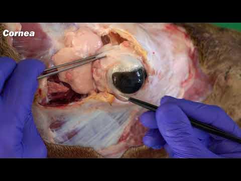
- 😀 The cornea, a transparent layer at the front of the eye, does not have blood vessels and is responsible for focusing light entering the eye.
- 😀 The sclera, the white part of the eye, protects the internal structures and helps maintain the shape of the eye.
- 😀 The iris controls the size of the pupil, which adjusts to control how much light enters the eye.
- 😀 The pupil dilates in low light and constricts in bright light to regulate light intake and maintain optimal vision.
- 😀 The lens focuses light onto the retina, and it changes shape depending on whether you are looking at something close or far away (accommodation).
- 😀 If the lens loses its ability to accommodate, vision issues like myopia (nearsightedness) or hypermetropia (farsightedness) can occur.
- 😀 The retina contains a highly sensitive area called the macula, which is responsible for sharp, detailed vision, and the fovea centralis, where light should ideally focus.
- 😀 The optic nerve transmits visual signals from the retina to the brain for processing, translating them into visual images.
- 😀 The blind spot, located at the optic disc, is the point where blood vessels enter the retina, and it lacks photoreceptor cells, causing a lack of vision in that area.
Q & A
What are the six muscles responsible for moving the eyeball?
-The six muscles responsible for moving the eyeball are: Superior Rectus, Superior Oblique, Lateral Rectus, Medial Rectus, Inferior Rectus, and Inferior Oblique.
What is the function of the iris in the eye?
-The iris regulates the amount of light entering the eye by controlling the size of the pupil, which can dilate or constrict depending on the intensity of the light.
What is the role of the cornea in the eye?
-The cornea is a transparent membrane at the front of the eye that helps focus light as it enters the eye. It is made of protein and does not contain blood vessels.
How does the lens in the eye help with vision?
-The lens focuses light onto the retina, adjusting its shape to focus on objects at different distances. This ability is called accommodation, and it helps the eye focus on both near and far objects.
What is the macula, and why is it important for vision?
-The macula is a small area in the retina responsible for sharp central vision. It contains a high concentration of photoreceptor cells that allow us to see fine details.
What is the fovea centralis, and what is its role in vision?
-The fovea centralis is a small pit in the center of the macula that provides the sharpest vision. It contains only cone cells, which are responsible for color vision and visual acuity.
What is the blind spot in the eye?
-The blind spot is an area in the retina where the optic nerve exits the eye. It lacks photoreceptor cells, so no visual information is processed at this point.
What is the function of the optic nerve?
-The optic nerve transmits visual information from the retina to the brain, where it is processed and interpreted as the images we see.
What are the common vision problems associated with the lens and accommodation?
-Common vision problems related to lens accommodation include myopia (nearsightedness), hypermetropia (farsightedness), and presbyopia (age-related difficulty in focusing on close objects).
What is the role of blood vessels in the retina?
-The blood vessels in the retina, including arteries and veins, provide nutrients and oxygen to the eye, and remove waste products. They ensure the retina functions properly for vision.
Outlines

This section is available to paid users only. Please upgrade to access this part.
Upgrade NowMindmap

This section is available to paid users only. Please upgrade to access this part.
Upgrade NowKeywords

This section is available to paid users only. Please upgrade to access this part.
Upgrade NowHighlights

This section is available to paid users only. Please upgrade to access this part.
Upgrade NowTranscripts

This section is available to paid users only. Please upgrade to access this part.
Upgrade NowBrowse More Related Video


5.0 / 5 (0 votes)