Larynx (Voice Box) - Cartilage, Ligaments, Joints, Wall, Cavity | Anatomy

Taim Talks Med
24 Nov 202117:48

Summary

TLDRIn this educational video, Meditay explores the anatomy of the larynx, a crucial component of the respiratory system. The larynx, situated between the hyoid bone and the trachea, is composed of six cartilages that facilitate both breathing and voice production. Key cartilages include the unpaired epiglottis, thyroid, and cricoid, along with the paired arytenoid, corniculate, and cuneiform. The video delves into the larynx's structure, highlighting its layers, muscles, and the distinctive Laryngeal cavity, which includes the laryngeal ventricles and the vocal cords responsible for phonation.

Takeaways

  • 🌟 The respiratory system includes the Nose, Pharynx, Larynx, Trachea, Bronchi, and Lungs, with the Larynx being the focus of this segment.
  • 📍 The Larynx is situated between the Hyoid Bone and the Trachea, in front of the Esophagus, and is aligned with the 4th to 6th-7th Cervical Vertebrae.
  • 🗣️ The Larynx, also known as the 'Voice Box', serves dual functions: as an air passage and as a sound producer through the process of Phonation.
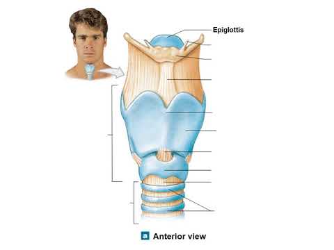
  • 🦴 The Larynx is composed of 6 cartilages: 3 unpaired (Epiglottis, Thyroid, Cricoid) and 3 paired (Arytenoid, Corniculate, Cuneiform).
  • 🔗 The connections within the Larynx are either continuous (Thyrohyoid membrane, Cricothyroid membrane) or discontinuous (synovial joints like Cricoarytenoid articulation).
  • 🏼‍🔬 The Laryngeal walls consist of four layers: Tunica Mucosa, Tela Submucosa, Cartilage and Muscle Layer, and Tunica Adventitia, each with distinct characteristics and functions.
  • 🎙️ The Vocal Cords are a critical part of the Larynx, made up of the Vocal Ligament and lined with epithelium that can withstand strain for speech.
  • 💡 The Laryngeal Cavity is divided into the Laryngeal Vestibule, Glottis, and Infraglottic cavity, with the Glottis further divided into intermembranous and intercartilaginous parts.
  • 👥 The muscles of the Larynx are categorized by their function: opening/narrowing the laryngeal inlet, adjusting the Rima Glottidis, and acting directly on the Vocal Cords.
  • 🔊 The Laryngeal Vestibule and Vocal Cords are key to speech production, with the Laryngeal ventricles acting as resonators to enhance vocal quality.

Q & A

  • What are the primary functions of the larynx?

    -The larynx serves as an air passage, allowing air to flow into the lungs, and it is also known as the voice box because it produces sound through a process called phonation.

  • What are the key cartilages that make up the larynx?

    -The larynx is composed of six types of cartilages: three unpaired (Epiglottis, Thyroid cartilage, Cricoid cartilage) and three paired (Arytenoid cartilage, Corniculate cartilage, Cuneiform cartilage).

  • What is the Laryngeal Prominence, and what is its significance?

    -The Laryngeal Prominence, also known as Adam's apple, is formed by the meeting of the right and left laminas of the thyroid cartilage. It is a noticeable structure in the neck.

  • How does the epiglottis function in relation to the respiratory and digestive systems?

    -The epiglottis closes off the respiratory system during swallowing to prevent food from entering the airway and opens up when breathing to allow air to pass through.

  • What are the two types of articulations found in the larynx?

    -The larynx has continuous and discontinuous articulations. Continuous articulations include cartilaginous and fibrous connections, while discontinuous articulations are synovial joints.

  • What is the purpose of the vocal ligament in the larynx?

    -The vocal ligament, formed by the upper margin of the Conus Elasticus, is a part of the vocal cord and plays a crucial role in phonation by vibrating to produce sound.

  • What is the difference between the intermembranous and intercartilaginous parts of the larynx?

    -The intermembranous part of the larynx is the anterior 3/5 of the glottis, situated between membranes, while the intercartilaginous part is the posterior 2/5, located between cartilages.

  • What is the role of the laryngeal ventricles in voice production?

    -The laryngeal ventricles act as resonators, enhancing the sound produced during phonation by the vocal cords.

  • How does the cricothyroid muscle affect the tension of the vocal cord?

    -The cricothyroid muscle, when contracted, pulls the thyroid cartilage forward, thereby tensing the vocal ligament and affecting the pitch of the voice.

  • What is the significance of the Tunica Adventitia in the larynx?

    -The Tunica Adventitia is a tough connective tissue covering that consists mainly of dense collagen fibers, providing structural support and protection to the larynx.

Outlines

00:00

🗣️ Anatomy of the Larynx

This segment introduces the anatomy of the respiratory system, focusing on the larynx. The larynx is part of the respiratory system and is located between the hyoid bone and the trachea, in front of the esophagus. It starts at the level of the 4th and 5th cervical vertebrae and ends at the 6th and 7th. The larynx serves as an air passage and is also known as the voice box due to its role in sound production through the vocal cords. The orientation of the larynx is discussed, including its anterior and posterior views, and the video promises to cover the cartilages, ligaments, and joints that constitute the larynx, as well as the walls and special features of the laryngeal cavity.

05:03

🦴 Cartilages of the Larynx

The larynx is composed of six types of cartilages: three paired (Arytenoid, Corniculate, and Cuneiform) and three unpaired (Epiglottis, Thyroid, and Cricoid). The video provides a detailed look at each cartilage's characteristics and functions. The thyroid cartilage, with its prominent Adam's apple, is made up of two laminas and has superior and inferior horns. The cricoid cartilage is located below the thyroid and has an arch and a plate with important articular surfaces. The epiglottis is a flap of cartilage that closes off the respiratory system during swallowing. The Arytenoid cartilages are paired and have a vocal process and a muscular process. The Corniculate and Cuneiform cartilages are small and serve as attachment points for muscles.

10:05

🔗 Connections in the Larynx

The connections in the larynx are divided into continuous and discontinuous articulations. Continuous connections include cartilaginous (Synchondroses) and fibrous (Ligaments) connections. The larynx has one cartilaginous connection between the corniculate and arytenoid cartilages and several fibrous connections such as the thyrohyoid membrane, cricothyroid membrane, cricotracheal ligament, thyroepiglottic ligament, and hyoepiglottic ligament. Discontinuous connections are synovial joints, including the cricothyroid and cricoarytenoid articulations. These joints allow for movement of the larynx, which is essential for functions like swallowing and phonation.

15:09

🌐 Layers and Cavity of the Larynx

The larynx is composed of four layers: the Tunica Mucosa, Tela Submucosa, Cartilage and Muscle Layer, and Tunica Adventitia. The Tunica Mucosa is the inner lining and contains respiratory and stratified squamous epithelium, laryngeal glands, and lymph nodules. The Tela Submucosa includes fibroelastic membranes like the Quadrangular Membrane and Conus Elasticus, which are crucial for speech. The Cartilage and Muscle Layer houses the muscles that control the laryngeal inlet, Rima Glottidis, and vocal cord tension. The Tunica Adventitia is a tough connective tissue covering. The Laryngeal Cavity is divided into the Laryngeal Vestibule, Glottis, and Infraglottic cavity, with the Rima Glottis being a key area for phonation. The Glottis contains the intermembranous and intercartilaginous parts and the Laryngeal ventricles, which act as resonators for sound.

Mindmap

Keywords

💡Larynx

The larynx, also known as the voice box, is a crucial organ in the respiratory system situated between the hyoid bone and the trachea, and in front of the esophagus. It plays a vital role in both breathing and phonation. In the video, the larynx is described as having an anterior and posterior view, and it is composed of various cartilages that contribute to its structure and function. The larynx is highlighted for its dual function as an air passage and a sound-producing organ.

💡Cartilages

Cartilages are flexible connective tissues that provide support and structure to various parts of the body, including the larynx. The script mentions six types of cartilages in the larynx: three paired (Arytenoid, Corniculate, and Cuneiform) and three unpaired (Epiglottis, Thyroid, and Cricoid). These cartilages are essential for the larynx's function in breathing and voice production, as they form the framework that allows for the movement necessary in these processes.

💡Phonation

Phonation refers to the process by which the vocal cords produce sound. In the video, it is explained that the larynx produces sound through the vibration of the vocal cords, which is a key aspect of the larynx's function as the voice box. The script describes how the vocal cords rub against each other to produce different pitches, which is central to the theme of understanding the anatomy and function of the larynx.

💡Vocal Cords

Vocal cords, also known as vocal folds, are two bands of muscle tissue in the larynx that vibrate to produce sound. The script discusses the importance of the vocal cords in the process of phonation, explaining that they are lined by stratified squamous epithelium to withstand the strain of speaking and singing. The video also touches on how the vocal cords' tension can be adjusted by certain muscles, which is crucial for pitch control.

💡Respiratory Epithelium

Respiratory epithelium is a type of tissue that lines the vestibular folds in the larynx and is specialized for filtering the air. The script explains that this epithelium contains cilia, which help to catch small particles before they enter the lungs. This epithelium is crucial for the larynx's role in respiration and is an important aspect of the video's discussion on the larynx's anatomy and function.

💡Stratified Squamous Epithelium

Stratified squamous epithelium is a type of tissue that lines the vocal folds and is characterized by its durability. The video script explains that this epithelium is logical for the vocal folds because it allows for loud and prolonged speaking without damage. It is an essential component of the larynx's structure, contributing to its ability to withstand the strain of voice production.

💡Fibroelastic Membranes

Fibroelastic membranes are elastic connective tissues that play a key role in voice production by allowing the necessary movement of the laryngeal structures. The script identifies two such membranes in the larynx: the Quadrangular Membrane and the Conus Elasticus. These membranes are integral to the larynx's function, as they facilitate the movement of the vocal cords and other structures during phonation.

💡Synovial Joints

Synovial joints are a type of joint that allows for smooth movement and are found in the larynx. The video script mentions two synovial joints in the larynx: the cricothyroid and cricoarytenoid articulations. These joints are filled with synovial fluid, which lubricates the joints and allows for the movement necessary for the larynx's function in both breathing and voice production.

💡Tunica Mucosa

The tunica mucosa is the innermost lining of the larynx, which is composed of various types of epithelium and contains glands and lymph nodules. The video script describes how the tunica mucosa is lined by respiratory epithelium at the vestibular folds and stratified squamous epithelium at the vocal folds. This lining is essential for the larynx's role in filtering the air and producing sound.

💡Laryngeal Cavity

The laryngeal cavity, or Cavitas Laryngis, is the internal space of the larynx that extends from the laryngeal inlet to the lower border of the cricoid cartilage. The video script divides this cavity into the Laryngeal Vestibule, Glottis, and Infraglottic cavity, each with specific functions related to breathing and voice production. The laryngeal cavity is crucial for understanding the overall function of the larynx and is a central concept in the video's discussion of laryngeal anatomy.

Highlights

Introduction to the anatomy of the Larynx, part of the respiratory system.

The Larynx is located between the hyoid bone and the trachea, in front of the esophagus.

The Larynx consists of 6 cartilages: 3 unpaired (epiglottis, thyroid, cricoid) and 3 paired (arytenoid, corniculate, cuneiform).

The thyroid cartilage forms the laryngeal prominence, commonly known as the Adam's apple.

The cricoid cartilage is unique in structure and forms joints with both the arytenoid and thyroid cartilages.

The epiglottis functions to close off the respiratory system during swallowing, preventing food from entering the airways.

The arytenoid cartilage plays a crucial role in vocal cord attachment, assisting in voice production.

The conus elasticus and quadrangular membrane are key fibroelastic membranes, important for voice production.

The Larynx has continuous and discontinuous connections, including synovial joints like the cricothyroid and cricoarytenoid articulations.

The laryngeal walls consist of four layers: tunica mucosa, tela submucosa, cartilage and muscle layer, and tunica adventitia.

The vocal folds are lined with stratified squamous epithelium, which is durable and necessary for sustained voice use.

The vocal cords, responsible for phonation, vibrate to produce sound when air passes through them.

The laryngeal cavity is divided into three sections: the vestibule, the glottis, and the infraglottic cavity.

The rima glottidis, located between the vocal cords, plays a key role in both voice production and breathing.

Phonation involves the vocal cords rubbing against each other, while the laryngeal ventricles act as sound resonators.

Transcripts

play00:00

Hey what’s up.

play00:02

Meditay here.

play00:03

Let’s talk about the anatomy of the respiratory system.

play00:08

In this segment, we will be talking about the anatomy of the Larynx.

play00:12

Alright, so the respiratory system consist of all the organs involved in breathing.

play00:17

These are the Nose, Pharynx, Larynx, Trachea, Bronchi and the Lungs.

play00:23

In our last video, we covered the anatomy of the nasal cavity.

play00:27

Now let’s do the anatomy of the Larynx.

play00:29

So in this video, we'll start with orientation by looking at the anterior and posterior view

play00:35

of the Larynx.

play00:36

Then we'll talk briefly about what cartilages make up the larynx.

play00:40

As well as the ligaments and joints that hold the whole thing together.

play00:44

Then we'll do the walls of the Larynx and talk a little bit about what’s special with

play00:48

the Laryngeal cavity.

play00:49

Alright, so the Larynx is located right about here.

play00:53

It lies between the Hyoid Bone, and the Trachea.

play00:56

And right in front of the Esophagus.

play00:58

Topographically, it starts in the region of 4th 5th Cervical Vertebrae, and ends at the

play01:04

region of 6th-7th cervical vertebrae.

play01:06

Let’s list a few of Larynx’s functions.

play01:09

The first is Air passage.

play01:11

It serves as a passage, so that air can go down into your lungs.

play01:15

Another thing is that your Larynx is also called the Voice Box, because it produces

play01:20

sound.

play01:21

And it does that through a process called Phonation, where these vocal cords rub into

play01:25

each other producing different pitch.

play01:27

I’ll go through this at the end of this video.

play01:30

Now let’s continue with the orientation by looking at the Larynx from different angles.

play01:34

So, if you look at it anteriorly This is how it's gonna look like.

play01:39

And posteriorly This is how it's gonna look like.

play01:43

Notice the hyoid bone above the larynx, and the trachea under it.

play01:48

Now let’s start by understanding how the Larynx is built.

play01:51

So the Larynx is made up of 6 types cartilages in total.

play01:55

3 paired and 3 unpaired Cartilages.

play01:58

Cartilages that are unpaired means that you only have one of each, and these are the Epiglottis

play02:04

in Purple, Thyroid cartilage, in blue.

play02:07

And the cricoid cartilage, here in light green.

play02:09

The paired, are the cartilages you have two of.

play02:14

These include the Arytenoid cartilage here in yellow, Corniculate cartilage in red, and

play02:20

the Cuneiform cartilage, here in green.

play02:23

These are the cartilages that make up the Larynx.

play02:25

Now let’s switch up to a model from my anatomy program, just to make it look a little more

play02:28

realistic.

play02:29

We have the Epiglottis, The thyroid cartilage and the Cricoid cartilage.

play02:33

We have the Arytenoid, the Corniculate and the Cuneiform cartilages.

play02:37

Let’s now cover each of these cartilages and look at their characteristics, we’ll

play02:43

start with he thyroid cartilage.

play02:45

So again here’s an anterior view and a posterior view of the Larynx.

play02:49

The thyroid cartilage is highlighted here in blue.

play02:52

And just to have an overview, I think it’s better to have a list of the cartilages we’re

play02:56

about to cover.

play02:57

Now the thyroid cartilage is mainly made up of two plates, or two Laminas.

play03:02

There’s the Right lamina, and the Left lamina.

play03:05

These two meets in the middle, and form the laryngeal Prominence, which is also known

play03:10

as Adam’s apple.

play03:11

You’ll notice there are two processes on the thyroid cartilage as well.

play03:16

There’s one going straight up towards the Hyoid bone, called the superior horn, and

play03:21

a process going down towards the cricoid cartilage, called the inferior horn.

play03:25

So that’s the most important structures of the thyroid cartilage for now, let’s

play03:29

check that one from our list.

play03:31

Next is the cricoid cartilage, here in light green located below the thyroid cartilage

play03:36

The cricoid cartilage looks different, depending on which side you’re looking from.

play03:40

Anteriorly, it has an arch protruding forward, and posteriorly, it has a plate with two important

play03:47

surfaces.

play03:48

There’s the arytenoid articular surface, which is where the arytenoid cartilage binds

play03:53

to the cricoid cartilage forming a joint.

play03:56

And it has a Thyroid articular surface, which form a joint with the inferior horn of the

play04:01

thyroid cartilage.

play04:03

So that’s all for the cricoid cartilage, let’s put a checkmark here.

play04:07

Next is the Epiglottis, which is here in purple.

play04:10

It doesn’t really have any specific structure of importance for now.

play04:13

But it lies behind the thyroid cartilage and the hyoid bone, and if you look at a posterior

play04:19

view.

play04:20

It mainly attaches to the thyroid cartilage through ligaments.

play04:23

Its function is mainly closing off for the respiratory system when you swallow and open

play04:28

up when you’re breathing.

play04:29

So that’s the epiglottis.

play04:31

Next is the Arytenoid cartilages, which are paired.

play04:34

They’re located here in yellow, right above the cricoid cartilage.

play04:39

And if you look at them from a sideview, you’ll notice that it has a triangular shape, which

play04:44

an Apex on top, and a base.

play04:47

The base has two parts, or two processes.

play04:50

The anterior process is called the vocal process, since it attaches to the vocal ligament, I’ll

play04:56

show you this later.

play04:57

The posterior process is called Muscular process, which serves as an attachment point for certain

play05:03

muscles.

play05:04

And the base of the arytenoid cartilage, is called Cricoid Articular Surface, since it

play05:09

sits on the cricoid process.

play05:10

So that’s this one.

play05:11

Next we have the corniculate cartilage, which is here in red, and lie on top of the arytenoid

play05:17

cartilage.

play05:18

It doesn’t really have any specific function on its own, other that serving as an attachment

play05:23

point for certain muscles as well.

play05:25

So, let’s tick that one.

play05:27

The last cartilages are the Cuneiform cartilages, here highlighted in green.

play05:31

They’re two small, elongated pieces of yellow elastic cartilage, which lie in something

play05:37

called the aryepiglottic fold.

play05:39

The aryepiglottic fold is essentially a fold that lines the entrance of the Larynx.

play05:45

And the Cuneiform Cartilage form a tubercle on it, which is visible if you’d examine

play05:50

the larynx of a patient.

play05:52

This tubercle is called Cuneiform tubercle.

play05:54

So, let’s go ahead and tick that one as well.

play05:58

So those are the cartilages that make up the Larynx.

play06:01

Now we need to cover the connections in the Larynx, or the Juncturae Laryngis.

play06:05

But to really understand these connections, we need to have a basic understanding of the

play06:10

different connection forms we have in our body.

play06:14

So the articular system in the Larynx si divided into two types of connection.

play06:19

There are Continuous articulation, and discontinuous articulation.

play06:24

Now what are the differences between those?

play06:27

Continuous connections are uninterrupted articular connection, hence the name continuous.

play06:33

Discontinuous connections are interrupted in that, it contains a cavity within the articulation,

play06:39

which form a joint.

play06:40

Now, I like to use the ribs as an example of the continuous connections in the Larynx.

play06:45

Because one of the connection types is called cartilaginous, or Synchondroses.

play06:50

Which is where you’ll find at the point where the rib meets the costal cartilage,

play06:54

called the costochondral synchondroses.

play06:57

The other type of continuous connection you’ll find in the Larynx is a fibrous type of connection,

play07:02

which form membranes, or Ligaments.

play07:05

Very much like the intercostal membrane you see here.

play07:10

Discontinuous connections are joints.

play07:12

In the Larynx you’ll find two synovial joints, just like this one, which are ligaments forming

play07:18

a sac full of synovial fluid.

play07:20

Now let’s do the continuous connections of the Larynx first, then do the discontinuous

play07:25

connections.

play07:26

The Larynx has only one cartilaginous connection point, and that’s between the corniculate

play07:32

cartilage and the arytenoid cartilage.

play07:35

So here is the Larynx.

play07:36

Here is the Corniculate Cartilage, and here is the Arytenoid Cartilage.

play07:40

Here is the cartilaginous connection.

play07:42

It’s a very dense connection, nearly bony joint.

play07:46

For the fibrous connections.

play07:47

We have the Thyrohyoid membrane, which is a membrane between the thyroid cartilage and

play07:52

the hyoid bone, as you see here, so this in green is the thyrohyoid membrane.

play07:57

Theoretically, it has two parts.

play07:59

A median Thyrohyoid membrane and a lateral thyrohyoid membrane, as you see here.

play08:05

Next we have the Cricothyroid membrane, which lies between the Thyroid cartilage and the

play08:09

Cricoid Cartilage.

play08:10

So it’s this one.

play08:12

The cricothyroid membrane is a membrane that are made up by two ligaments.

play08:17

One of them is the median cricothyroid ligament, and the other one is the lateral cricothyroid

play08:23

ligament, or conus elasticus.

play08:26

The conus elasticus is really important, I’ll talk briefly about it later in this video.

play08:32

Then there’s the cricotracheal ligament, between the cricoid cartilage, and the trachea,

play08:37

as you see here.

play08:38

Then we have the thyroepiglottic ligament, which is this one, connecting the Epiglottis

play08:42

to the Thyroid cartilage.

play08:44

And then we have the Hyoepiglottic ligament, which connects the Epiglottis to the Hyoid

play08:49

bone, as you see here.

play08:51

So all of those are the Continuous connections we have in the Larynx.

play08:56

Now let’s do the Discontinuous connections.

play08:59

We have two synovial joints in the Larynx, and if you really paid attention earlier in

play09:04

this video, you’d know these already from when I talked about the cricoid cartilage.

play09:09

Because the first one is the cricothyroid articulation, which is a synovial joint between

play09:14

the inferior horn of the thyroid cartilage, and the cricoid cartilage.

play09:19

The other one is the Cricoarytenoid articulation, between the base of the arytenoid cartilage

play09:25

and the cricoid cartilage.

play09:27

They look like this, filled with synovial fluid for a nice movement of the larynx.

play09:32

Now.

play09:33

Here you see a lateral view of the larynx.

play09:35

If we make a vertical cut of the larynx, as you see here.

play09:38

And look at it from this view, you’ll see this.

play09:41

So, let’s go over some important landmarks.

play09:44

There’s the Epiglottis, The thyroid cartilage, the cricoid cartilage, and the hyoid bone.

play09:50

And between the cartilages, you’ll find the Thyrohyoid membrane.

play09:53

So, let’s now cover the walls of the Larynx.

play09:57

The larynx has 4 layers.

play09:59

We have the Tunica Mucosa, here in blue.

play10:02

Then underneath that, there’s the Tela submucosa, as you see here.

play10:05

Then you have a layer of cartilages and muscles, as you see here.

play10:08

And lastly, there’s the Tunica adventitia, which are connective tissue covering the surfaces

play10:13

of the larynx.

play10:14

Now, let’s go through each of these layers and look at their characteristics.

play10:19

We’ll start with Tunica Mucosa.

play10:21

So again, the tunica mucosa is here, it’s the innermost lining of the Larynx.

play10:27

At the vestibular folds, which is the upper fold, it’s lined by Respiratory epithelium,

play10:32

which are epithelium specialized in filtrating the air, as they contain cilia that catches

play10:38

small particles before they go further down into your lungs.

play10:42

The Vocal folds, are lined by stratified squamous epithelium, which are essentially logical

play10:47

because the vocal folds are under a lot fo strain, allowing you to speak loudly and for

play10:53

a longer period of time without harming your vocal folds.

play10:57

In chronic smokers and people who use their voices a lot like in singers, the respiratory

play11:03

epithelium lining the vestibular folds are often damaged and replaced by stratified squamous

play11:09

epithelium, which makes them more susceptible for lower respiratory infections, since the

play11:15

cilia of the respiratory epithelium are gone.

play11:18

So that’s the lining, but the tunica mucosa also contains glands, or laryngeal glands,

play11:24

lubricating the surface, and small lymph nodules for immunity.

play11:29

So that’s the tunica mucosa.

play11:32

Next we have the tela submucosa, which are here.

play11:35

Remember earlier when we went through the connections of the Larynx, where we talking

play11:38

about the continuous connections, specifically the fibrous membranes of the Larynx?

play11:43

Well, at some places the Fibrous membranes form a so called Fibroelastic membrane, which

play11:49

are elastic membranes playing a key role in you being able to speak.

play11:55

There are two fibroelastic membranes in the Larynx.

play11:58

The first one is the Quadrangular Membrane, which are here.

play12:01

If we look at a posterior view of the Larynx.

play12:03

It’s much easier to see this membrane.

play12:05

It’s a membrane that goes between the Vestibular Folds and the Epiglottis, as you see here.

play12:11

There’re two margins left that aren’t connected to anything, The upper margin, form

play12:17

the aryepiglottic fold, if you remember from earlier, the aryepiglottic fold is a fold

play12:22

lining the entrance of the larynx.

play12:25

The lower margin forms the vestibular ligament, which is a ligament you see down here, going

play12:30

between the arytenoid cartilage and the thyroid cartilage.

play12:33

The other fibroelastic membrane we have is the Conus Elasticus, or the Lateral Cricothyroid

play12:39

Ligament.

play12:40

It’s this one, if you remember it from earlier.

play12:43

IF we turn it around, you’ll see that it’s connected to the Cricoid cartilage, the Arytenoid

play12:48

cartilage and the thyroid cartilage.

play12:51

But it has one free margin, and that’s the upper margin, which form the Vocal ligament.

play12:57

This ligament is a part of the vocal fold which is the fold that makes it possible to

play13:01

speak once they vibrate, I’ll show you this later.

play13:05

So that is the tela submucosa.

play13:07

Next, we have the Cartilage and Muscle Layer, as you see here.

play13:11

We’ve already covered the cartilages, but the muscles of the Larynx are grouped according

play13:15

to their function.

play13:17

So we have three groups of muscles in the Larynx.

play13:20

The first ones are the muscles that open and narrow the laryngeal inlet, or the entrance,

play13:26

so this one.

play13:27

It opens and narrows this entrance into the Larynx.

play13:30

The next ones are the muscles that open and narrow the Rima Glottidis.

play13:34

And the Rima glottidis is here, situated between the Vocal folds, or the Vocal Cords.

play13:40

The fold superior to the Rima glottidis is called Rima Vestibuli, between the vestibular

play13:45

folds.

play13:46

And again, don’t forget that the Vocal Ligaments are here, as you remember earlier, formed

play13:51

by the superior margin of Conus Elasticus.

play13:53

So, the vocal ligament is a part of the vocal cords.

play13:57

And the Rima glottidis is between the vocal cords.

play14:01

And if we look at the larynx looking from this direction, you’ll see this.

play14:05

With the epiglottis here and the aryepiglottic fold here.

play14:09

So the vocal ligament is located right about here.

play14:12

Always remember that this pointy end is the anterior side, and the rounded one is the

play14:18

posterior side.

play14:19

Now.

play14:20

Rime glottidis is here, between the vocal cords, rima vestibuli, is here, between the

play14:26

vestibular folds.

play14:27

So, the muscles that open and narrow the rime glottidis, work like this.

play14:32

They open, or narrow.

play14:33

The rima glottidis.

play14:35

The last muscle group are the muscles that act on the vocal cord itself.

play14:40

Earlier I told you that the vocal ligament is situated here, but that’s not entirely

play14:46

correct.

play14:47

Look closely at the structures here.

play14:49

The vocal ligament is located within the mucus membranes.

play14:53

So the mucus membrane together who the vocal ligament, is what’s referred to as the vocal

play14:59

cord, so that’s essentially what I mean by the last group of muscles.

play15:03

They act directly on the vocal cord, which essentially is the vocal ligament.

play15:09

There are muscles that tenses the vocal cord, such as the cricothyroid muscle.

play15:14

This muscle is situated here, as you see.

play15:17

And when they contract, they pull the thyroid cartilage to the front, tensing the vocal

play15:22

ligament.

play15:23

If we look posteriorly, and zoom in.

play15:26

When the thyroid cartilage move forward, the vocal cord is tensed.

play15:31

The other muscles decrease the tension of the vocal cord, such as vocalis muscle, situated

play15:36

right next to the vocal cords, as you see here.

play15:39

And when they contract, they pull the arytenoid cartilage forward to decrease the tension

play15:45

of the vocal cord.

play15:47

Lastly, we have the Tunica Adventitia.

play15:50

So here’s the Larynx.

play15:52

Tunica adventitia is a covering around the larynx, which is a tough connective tissue

play15:57

consisting mainly of dense collagen fibers.

play16:00

So that was the Layers of the Laryngeal Wall, the last thing I wanna talk about in this

play16:04

video is the Laryngeal Cavity.

play16:06

So the Laryngeal Cavity, or Cavitas Laryngis, is this whole cavity here.

play16:11

It starts at the Laryngeal Inlet, or entrance, and ends at the lower border of the cricoid

play16:17

cartilage.

play16:18

But we generally divide it into three landmarks.

play16:21

The first one is the Laryngeal Vestibule, going from the laryngeal entrance to the Vestibular

play16:26

Folds.

play16:27

Then there’s Glottis, between the Vestibular Folds and the Vocal Cords, or the Vocal folds

play16:32

it’s a synonym.

play16:34

There’s an important thing we need to discuss here.

play16:37

Remember, the Rima glottis is here.

play16:39

We generally divide this cavity into two parts based on the structures around it.

play16:44

The anterior 3/5 is called intermembranous part, since it’s between membranes.

play16:50

The posterior 2/5 is intercartilaginous part, situated between cartilages.

play16:56

And if we change up to a more realistic one.

play16:59

Here you see that the anterior 3/5 of it is between membranes, and the posterior part

play17:05

lying between cartilages, between the arytenoids here.

play17:08

Now why is this important?

play17:10

Because they give you the possibility to speak!

play17:12

When you’re speaking, the vocal cords rub into each other, in a process called phonation.

play17:18

But when you breath, the folds are open for air to pass through.

play17:23

Now when you look closely at the glottis, notice there is a pouch at either side, called

play17:28

Laryngeal ventricles, which acts as a resonator.

play17:32

It resonates the sound as you speak.

play17:34

From the Vocal Cords, we have the Infraglottic cavity, which is until the lower border of

play17:41

the cricoid cartilage.

play17:42

From this point.

play17:43

That’s where your Trachea is going to be.

play17:45

And that’s a topic for our next video.

Rate This

5.0 / 5 (0 votes)

Related Tags
AnatomyLarynxRespiratory SystemVocal CordsPhonationMedical EducationCartilagesHealth ScienceBiologyEducational Content