What is Gel Electrophoresis | Don't Memorise

Infinity Learn NEET
27 Mar 202008:34

Summary

TLDRThis script delves into the fundamental technique of gel electrophoresis, a method used to separate DNA molecules by size. It explains the process, starting from the negatively charged DNA's movement in an electrical field through an agarose gel, to the use of ethidium bromide for visualization under UV light. The script also covers the use of DNA ladders for size determination and the extraction of specific DNA fragments for further analysis, highlighting gel electrophoresis as a crucial tool in recombinant DNA technology.

Takeaways

  • 🌟 Gel electrophoresis is a technique used to separate DNA molecules based on their size.
  • 🔍 The term 'gel electrophoresis' comes from the use of an electrical field in a gel medium to separate charged molecules.
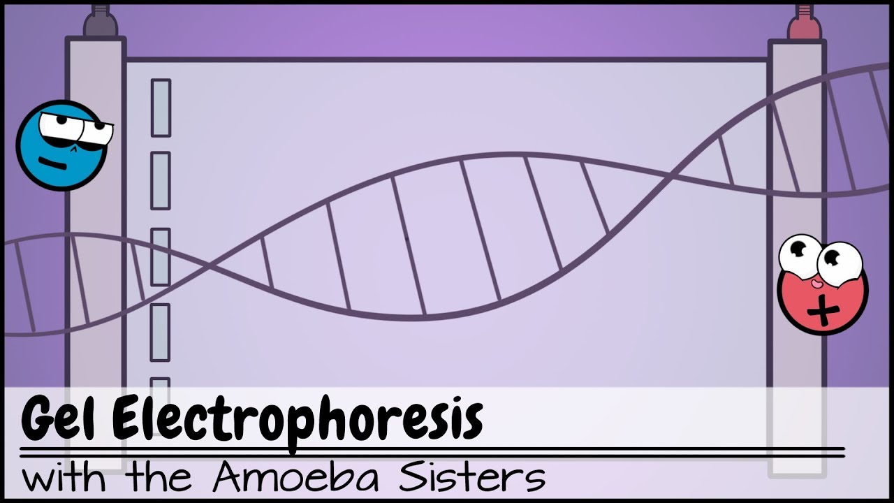
  • 🚀 DNA molecules are negatively charged due to the phosphate groups in their backbone, which facilitates their movement in the gel.
  • 🌊 Agarose gel, derived from seaweed, is commonly used as the medium for DNA separation.
  • 🔬 The separation principle is analogous to a sieve, where smaller particles pass through more easily than larger ones.
  • 🧬 DNA samples with different sized fragments are loaded into wells in the gel for the electrophoresis process.
  • 🔋 An electric field is applied, causing the negatively charged DNA fragments to move towards the positive anode.
  • 🎨 A colored loading dye is used to visualize the movement of DNA through the gel, as DNA itself is colorless.
  • 🌈 Ethidium bromide is used to stain the DNA, allowing the separated fragments to be seen under ultraviolet light as orange bands.
  • 📏 The size of the DNA fragments can be determined by comparing the position of the bands to a DNA ladder.
  • 🔬 The process of cutting out and extracting the desired DNA fragment from the gel is known as 'excision' and is used for further analysis.

Q & A

  • What is gel electrophoresis?

    -Gel electrophoresis is a technique used to separate different DNA molecules based on their sizes by applying an electrical field in a gel medium.

  • Why is the technique called 'gel electrophoresis'?

    -The term 'gel electrophoresis' comes from the fact that the separation of DNA molecules occurs within a gel medium under the influence of an electrical field.

  • What charge do DNA molecules carry?

    -DNA molecules are negatively charged due to the presence of phosphate groups in their backbone, which are negatively charged.

  • What type of gel is commonly used for DNA separation in gel electrophoresis?

    -Agarose gel, which is obtained from seaweeds, is commonly used for DNA separation in gel electrophoresis.

  • How does the size of DNA fragments affect their movement in the gel?

    -Smaller DNA fragments move faster through the gel's pores, while larger fragments find it more difficult to move and lag behind due to the size of the pores and their own size.

  • What is the purpose of using a comb to create wells in the gel?

    -The comb is used to create wells in the gel to load the DNA samples, allowing for the separation of DNA fragments when the electric field is applied.

  • Why are the wells for loading DNA samples formed near the cathode?

    -The wells are formed near the cathode because the negatively charged DNA fragments will move towards the positively charged anode when the electric field is applied.

  • What is the role of the colored loading dye in gel electrophoresis?

    -The colored loading dye is used to track the movement of the DNA through the gel, as DNA is colorless. It travels slightly faster than the DNA segments, indicating their progress.

  • How is the separated DNA visualized after electrophoresis?

    -The separated DNA is visualized by treating the agarose gel with ethidium bromide, which binds to the DNA and fluoresces under ultraviolet light, revealing bright orange bands representing the DNA fragments.

  • How can the size of the DNA fragments be determined after separation?

    -The size of the DNA fragments can be determined by comparing the positions of the bands on the gel with a standard DNA ladder, which provides a reference for fragment sizes.

  • What is the process called when the desired DNA fragment is cut out from the gel and extracted?

    -The process of cutting out and extracting the desired DNA fragment from the gel is called 'excision', and the extracted DNA can be used for further downstream processing.

Outlines

00:00

🔬 DNA Fragment Separation through Gel Electrophoresis

This paragraph introduces the concept of gel electrophoresis, a technique used to separate DNA molecules based on size. It explains that DNA, being negatively charged due to phosphate groups, moves through an agarose gel under the influence of an electric field. The process is likened to a sieve, where smaller particles pass through more easily than larger ones. The paragraph also details the setup for gel electrophoresis, including the use of a casting tray, comb for wells, electric supply, and a DNA sample. It emphasizes the importance of the gel's mesh-like structure at a microscopic level, which allows for the separation of DNA fragments according to their size, with smaller fragments moving faster through the gel's pores.

05:02

🌈 Visualizing DNA Fragments Post-Electrophoresis

The second paragraph delves into the practical steps of conducting gel electrophoresis. It describes the preparation of the apparatus with a buffer for better electrical conductivity and the loading of the DNA mixture into the gel wells. The use of a colored loading dye is highlighted for tracking the movement of DNA through the gel. The paragraph explains that once power is applied, the DNA moves towards the positively charged anode, with the loading dye indicating the progress of the DNA's journey. Upon completion, the DNA fragments are made visible by treating the gel with ethidium bromide, which binds to DNA and fluoresces under ultraviolet light, revealing distinct bands corresponding to different DNA fragment sizes. The bands are then compared to a DNA ladder for size determination, and the desired fragments can be extracted from the gel for further use. The paragraph concludes by emphasizing the significance of gel electrophoresis in recombinant DNA technology and encourages viewers to stay informed about such scientific processes.

Mindmap

Keywords

💡Gel Electrophoresis

Gel electrophoresis is a laboratory technique used to separate DNA, RNA, or protein molecules based on their size and charge. In the context of the video, it is the main focus, explaining how DNA fragments are separated on a gel medium using an electric field. The process is crucial in forensic science and molecular biology for identifying and analyzing genetic material.

💡DNA Fragments

DNA fragments refer to the smaller pieces of DNA that result from the action of restriction enzymes or other processes. In the script, these fragments are the subject of separation through gel electrophoresis, with the video illustrating how different sizes of these fragments move at varying speeds through the gel, leading to their separation.

💡Charged Molecules

Charged molecules are particles that carry an electric charge. DNA molecules are described as negatively charged in the video due to the phosphate groups in their backbone. This charge is essential for gel electrophoresis as it allows the DNA fragments to move towards the positive electrode when an electric field is applied.

💡Agarose Gel

Agarose gel is a substance derived from seaweed and is commonly used as the medium in gel electrophoresis. It forms a mesh-like structure with pores that allow the separation of DNA fragments based on their size. The script mentions agarose gel as the medium facilitating the movement and separation of DNA fragments during the process.

💡Restriction Enzymes

Restriction enzymes are proteins that cut DNA at specific recognition sites. In the video, these enzymes are mentioned as agents that create DNA fragments, which are then separated by size using gel electrophoresis. The activity of these enzymes is foundational to the process being described.

💡Sieve Analogy

The sieve analogy is used in the script to help understand the principle of DNA fragment separation in gel electrophoresis. Just as a sieve separates particles based on size, with smaller particles passing through while larger ones are retained, the gel acts similarly, allowing smaller DNA fragments to move through the pores more easily than larger ones.

💡Electric Field

An electric field is a field that surrounds electrically charged particles and exerts a force on other charged particles within the field. In the context of the video, the electric field is applied across the gel to drive the movement of the negatively charged DNA fragments towards the positive electrode, facilitating their separation.

💡Loading Dye

Loading dye is a colored substance added to the DNA sample before it is loaded onto the gel. It serves as a tracking agent in the video, allowing researchers to visualize the progress of the DNA through the gel. The dye is designed to move slightly faster than the DNA, providing an indication of how far the DNA has traveled.

💡Buffer

A buffer is a solution that helps maintain a stable pH and provides better conductivity for the electric current in the gel electrophoresis setup. In the script, the buffer is mentioned as an essential component that prepares the apparatus for the application of the electric field and the subsequent movement of DNA fragments.

💡Ethidium Bromide

Ethidium bromide is a chemical compound used in molecular biology to stain DNA, making it visible under ultraviolet light. In the video, it is described as a treatment for the agarose gel after electrophoresis, allowing the separated DNA fragments to be observed as bright orange bands, which are indicative of the DNA's presence and size.

💡DNA Ladders

DNA ladders are standard charts with bands of known DNA fragment sizes used for comparison during gel electrophoresis. In the script, they are mentioned as a tool for determining the size of the separated DNA fragments by comparing their positions on the gel to the known sizes on the ladder, which is crucial for identifying the desired DNA.

💡Elution

Elution is the process of extracting a substance, such as DNA, from a matrix, such as an agarose gel, after separation. In the video, it is the final step described for recovering the desired DNA fragment for further use. The term is used to illustrate the practical application of the separated DNA in downstream processing.

Highlights

Introduction to gel electrophoresis as a technique for separating DNA molecules based on size.

Explanation of why the process is called gel electrophoresis, involving the movement of charged molecules in an electrical field within a gel medium.

DNA's negative charge due to phosphate groups in its backbone, influencing its movement in the gel.

Use of agarose gel, derived from seaweed, as the medium for DNA separation.

The principle of DNA fragment separation analogous to a sieve, with smaller particles moving faster through the gel's pores.

Description of the gel's microscopic mesh-like structure and its uniform pore size affecting DNA fragment mobility.

Setup requirements for gel electrophoresis, including a casting tray, gel, comb, electric supply, and DNA sample.

Importance of forming wells in the gel for loading DNA samples near the negative terminal or cathode.

Use of a colored loading dye to track the movement of DNA through the gel, which travels faster than the DNA fragments.

The role of a buffer in providing better conductivity for the electric field during the process.

Process of loading the DNA mixture into the wells and turning on the power supply to initiate DNA movement through the gel.

Observation of DNA movement cessation indicated by the loading dye reaching the anode.

Technique of treating the agarose gel with ethidium bromide to visualize DNA under ultraviolet light.

Identification of DNA bands as bright orange colors, representing separated DNA fragments of varying sizes.

Comparison of DNA fragment bands with a DNA ladder to determine the size of the fragments.

Process of excising and extracting the desired DNA fragment from the gel for further downstream processing.

Emphasis on gel electrophoresis as a crucial technique in recombinant DNA technology.

Invitation to stay tuned to the channel for more interesting scientific processes and to subscribe for updates.

Transcripts

play00:00

[Music]

play00:03

this kind of an image is not new to us

play00:07

especially for people who love forensic

play00:10

science but what exactly is this it's an

play00:14

image of the separated DNA fragments on

play00:17

a piece of gel and the process of

play00:20

separating them is called gel

play00:22

electrophoresis doesn't the sound very

play00:25

difficult separating DNA fragments on a

play00:29

piece of gel but actually the process of

play00:32

gel electrophoresis is not very

play00:35

difficult in fact it's quite easy let's

play00:39

have a look at how the process works and

play00:41

also the principle behind it let's begin

play00:45

with the definition first gel

play00:48

electrophoresis is a technique to

play00:50

separate the different DNA molecules

play00:52

based on their sizes can you guess why

play00:56

is it called gel electrophoresis okay

play00:59

let me help you with this this

play01:02

separation technique is based on the

play01:04

movement of charged molecules when

play01:06

exposed to an electrical field and this

play01:10

movement occurs in a gel medium hence

play01:13

it's known as gel electrophoresis now

play01:17

let's understand the process step by

play01:18

step but before that since the term

play01:21

charge is used by us here can you guess

play01:24

the charge present on a DNA molecule

play01:27

let me tell you that a DNA is negatively

play01:30

charged and this is due to the presence

play01:33

of phosphate groups these phosphate

play01:36

being negatively charged and present in

play01:39

the backbone of the DNA double helix

play01:41

impart a negative charge to the complete

play01:44

molecule and what is the gel used

play01:48

interestingly the gel which is generally

play01:51

used for the DNA is agarose gel and it's

play01:54

obtained from the seaweeds now getting

play01:57

back to a process we said that the DNA

play02:01

molecules get separated on the basis of

play02:03

their sizes so if we have a sample of

play02:06

DNA which is containing fragments formed

play02:09

with the activity of restriction enzymes

play02:11

then separating the fragments based on

play02:14

the difference in their sizes would be

play02:16

possible with gel electrophoresis

play02:20

let's have a look at this in detail do

play02:23

you understand the principle and working

play02:25

let's take the analogy of a sieve we

play02:29

note that if a mixture of different

play02:31

sized particles is poured onto a sieve

play02:33

then only the particles smaller in size

play02:36

than the pores will escape while those

play02:38

bigger in size will be retained back

play02:41

here the size of the particles is the

play02:44

only parameter used for the separation

play02:48

same is the case with DNA fragments when

play02:52

our DNA sample containing various sized

play02:54

fragments is loaded onto a gel then the

play02:57

charge applied across the gel helps in

play02:59

the mobility of these fragments however

play03:02

not all fragments move at the same pace

play03:05

those which are smaller move ahead and

play03:08

those which are larger in size find it

play03:11

difficult to move to understand this

play03:14

better let's zoom into the gel that is

play03:16

prepared for the experiment here at the

play03:19

microscopic level we find that the gel

play03:22

appears to be a mesh-like structure and

play03:24

the pore size is nearly constant

play03:26

throughout so now imagine what will

play03:30

happen if different sized DNA fragments

play03:32

are made to pass through these pores

play03:34

it's obvious that the small sized

play03:37

fragments will escape the pores faster

play03:39

while the larger fragments will find it

play03:42

difficult to come out they will take a

play03:45

lot of time to cross the smaller pores

play03:47

and dust will lag behind this is how we

play03:51

can understand the principle of

play03:53

separation of the DNA fragments based on

play03:55

their sizes now let's see how the

play03:58

procedure works the first requirement is

play04:02

the complete set up this will include

play04:05

the casting tray gel a comb for making

play04:08

wells in the gel electric supply and

play04:10

most importantly DNA sample with

play04:14

different sized DNA fragments here's an

play04:17

illustration of the casting tray

play04:19

containing the gel in which we will load

play04:21

the DNA mixture we usually opt for a

play04:25

comb to form well like structures into

play04:27

the gel and why do we do that because

play04:31

it's in these worlds in the agarose gel

play04:34

that we load the DNA mixture now tell me

play04:37

where should the wells be formed at the

play04:41

ends or at the centre think about it

play04:44

we know that the DNA fragments being

play04:47

negatively charged will travel from a

play04:50

point of negative charge to the point of

play04:52

positive charge

play04:54

does we need to form the wells to load

play04:56

the DNA samples near the negative

play04:59

terminal or the cathode to be precise so

play05:02

that the DNA will move towards the

play05:04

positively charged anode this movement

play05:07

of the DNA molecule will be promoted by

play05:10

the electric field but we cannot track

play05:13

the movement of the DNA in the gel as

play05:15

it's colorless for this we use a colored

play05:19

loading dye to track the movement of the

play05:21

DNA the mixture of the DNA samples with

play05:26

the colored loading dye is now all set

play05:28

to get into the gel but before loading

play05:31

the DNA mixture we will fill the

play05:33

apparatus with a buffer as shown

play05:37

the buffer is used to provide better

play05:39

conductivity of electricity next we will

play05:44

load the DNA mixture into the wells and

play05:46

we're ready to turn on the power supply

play05:48

once the power supply is turned on the

play05:51

DNA mixture will move through the gel

play05:54

which can be tracked with the help of

play05:55

the loading dye the loading dye is

play05:58

selected in such a way that it travels a

play06:01

bit faster than the DNA segments present

play06:04

in the mixture and why is that so that's

play06:08

because we want the loading dye to reach

play06:10

the terminal end faster than the DNA

play06:13

once the dye reaches the anode we get an

play06:16

indication that the DNA must have

play06:18

reached somewhere near and does the

play06:20

power supply has to be turned off

play06:24

moving ahead with the next step now we

play06:27

know that the DNA molecules have got

play06:29

separated but can we really see them no

play06:33

that's not possible so how will that be

play06:37

possible now how do we observe the

play06:39

separated segments to observe the DNA

play06:43

molecules we treat the agarose gel with

play06:46

helium bromide solution the major reason

play06:49

for using ethidium bromide is that it

play06:52

easily binds to the DNA molecules and

play06:54

when the agarose gel containing the DNA

play06:57

is observed under ultraviolet light

play07:00

bright orange colored bands are clearly

play07:03

seen these are nothing but the bands of

play07:06

the DNA the separation has been

play07:09

successful based on the size of the DNA

play07:12

fragments the larger molecule are the

play07:15

ones found here which means these are

play07:17

the ones that moved slowly on the other

play07:20

hand the smaller molecules which moved

play07:22

faster are the ones

play07:24

spotted here now how do we know the size

play07:28

of any DNA fragment the bands obtained

play07:31

are compared with a standard chart known

play07:33

as the DNA ladders on comparing the

play07:37

positions of the bands with the ones

play07:38

from the chart we can easily make out

play07:41

the length of the fragments obtained

play07:43

once we know the length of the fragments

play07:46

we can easily identify the desired DNA

play07:49

now the desired DNA fragment can be

play07:52

first manually cut out from the agarose

play07:54

gel and then extracted this process is

play07:59

called illusion the extraction of the

play08:02

DNA is done in such a way that it can be

play08:05

used for further downstream processing

play08:07

this was the simple explanation of how

play08:11

any gel electrophoresis technique works

play08:14

it's considered as one of the very

play08:16

important techniques used in recombinant

play08:19

DNA technology to learn more about such

play08:23

interesting processes stay tuned to our

play08:26

Channel and do not forget to subscribe

play08:29

happy learning

play08:33

you

Rate This

5.0 / 5 (0 votes)

関連タグ
DNA AnalysisForensic ScienceGel ElectrophoresisMolecular BiologyDNA FragmentsPhosphate GroupsAgarose GelRestriction EnzymesEthidium BromideDNA LadderBiotechnology
英語で要約が必要ですか?