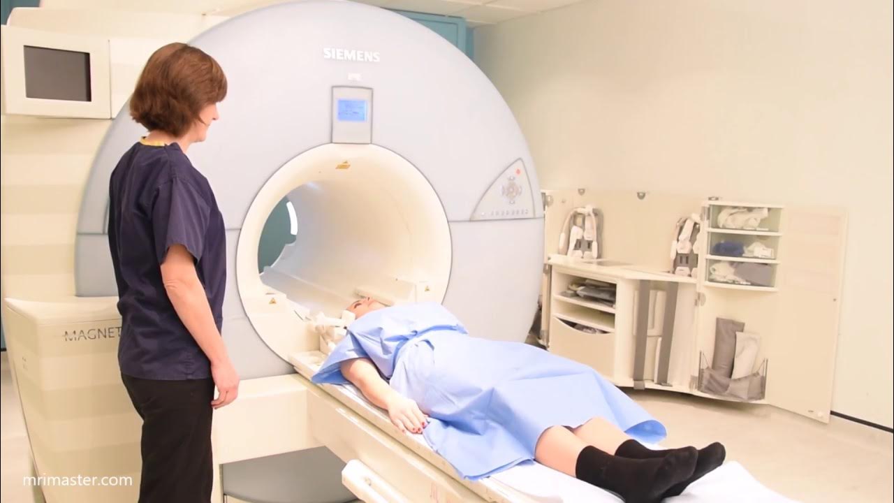
PELAYANAN MRI KONTRAS RSPON Prof. Dr. dr. MAHAR MARDJONO JAKARTA
Summary
TLDRThis video demonstrates the technique of performing an MRI brain scan with contrast in an emergency setting at Rumah Sakit Pusat Otak Nasional. The clinical team guides viewers through the patient preparation process, including removing metal accessories and changing into proper clothing. The video details various MRI sequences used for brain imaging, including Diffusion, FLAIR, T1, and T2 scans, along with 3D navigation techniques for surgical guidance. The team also covers the use of perfusion imaging and contrast injection protocols. Additionally, the video introduces the spectroscopy process for distinguishing normal brain tissue from tumors, providing a comprehensive look at advanced MRI techniques.
Takeaways
- 😀 The MRI examination process is conducted at Rumah Sakit Pusat Otak Nasional with clinical instructors guiding the procedure.
- 😀 The patient is asked to remove all metal accessories and change into appropriate clothing before the MRI.
- 😀 Several MRI sequences are used during the examination, including Diffusion, FLAIR, T1 axial, T1 sagittal, T2 coronal, and T2 axial.
- 😀 3D MP Rage sequence is used for navigation in surgery, replacing the need for manual T1 axial and sagittal images.
- 😀 Accurate positioning of the patient is critical for each sequence, particularly aligning with the corpus callosum.
- 😀 Perfusion imaging involves injecting contrast at a flow rate of 5 mL per second and a total volume of 10 mL, with saline used for additional volume.
- 😀 Perfusion imaging is divided into different phases, with the contrast injection timed to start at phase 8/60.
- 😀 Special care is taken to ensure no part of the patient's head is cut off in the images, especially for 3D navigation.
- 😀 Spectroscopy is used to differentiate between normal brain tissue and tumor tissue, based on the levels of NAA, choline, and creatine.
- 😀 The entire MRI procedure is explained in detail, providing valuable insight for other clinical professionals and practitioners.
Q & A
What is the focus of the MRI examination demonstrated in the video?
-The video demonstrates an MRI head examination with contrast in an emergency setting, focusing on the use of various MRI sequences and techniques for diagnostic and surgical navigation purposes.
What is the first step in preparing the patient for the MRI procedure?
-The first step is to confirm the patient's details, such as name and date of birth, and then ask the patient to change into a gown and remove any metal accessories such as belts and rings.
Which MRI sequences are used in the examination?
-The sequences used include Diffusion, FLAIR, T1 Axial, T1 Sagittal, T2 Coronal, and T2 Axial.
How is the 3D T1 sequence created for surgical navigation?
-The 3D T1 sequence is created by using MP RAGE, which combines axial and sagittal T1 sequences into a 3D image for surgical navigation.
What is the importance of patient positioning for the MRI sequences?
-Proper patient positioning is crucial for accurate imaging. For example, the sagittal sequence must be aligned with the corpus callosum, and the coronal sequence should be perpendicular to it.
What is the procedure for conducting perfusion imaging during the MRI scan?
-Perfusion imaging involves modifying a non-contrast axial sequence to include contrast injection. The contrast is injected using an injector at a flow rate of 5 ml/sec with a volume of 10 ml, and the sequence consists of multiple phases, including wash-in and wash-out phases.
How is contrast injected for the perfusion imaging sequence?
-Contrast is injected using an injector at a flow rate of 5 ml/sec and a volume of 10 ml, while sodium chloride (NaCl) is also injected at a flow rate of 5 ml/sec with a volume of 20 ml.
What is the significance of the 60-phase sequence in perfusion imaging?
-The 60-phase sequence in perfusion imaging tracks the flow of contrast through the brain. It includes phases like wash-in and wash-out, allowing for detailed analysis of blood flow in the brain.
What is the focus of the spectroscopy imaging mentioned in the video?
-Spectroscopy imaging focuses on analyzing brain tissue composition by measuring levels of N-acetylaspartate (NAA) and choline. Healthy brain tissue shows higher NAA levels, while tumors typically show higher choline levels.
What are the precautions taken during the spectroscopy imaging process?
-During spectroscopy imaging, precautions are taken to avoid capturing images of bone, fluid, blood, or air, as these can distort the data and affect the accuracy of the results.
Outlines

このセクションは有料ユーザー限定です。 アクセスするには、アップグレードをお願いします。
今すぐアップグレードMindmap

このセクションは有料ユーザー限定です。 アクセスするには、アップグレードをお願いします。
今すぐアップグレードKeywords

このセクションは有料ユーザー限定です。 アクセスするには、アップグレードをお願いします。
今すぐアップグレードHighlights

このセクションは有料ユーザー限定です。 アクセスするには、アップグレードをお願いします。
今すぐアップグレードTranscripts

このセクションは有料ユーザー限定です。 アクセスするには、アップグレードをお願いします。
今すぐアップグレード関連動画をさらに表示
Brain MRI scan protocols, positioning and planning

#7 Tata Kelola Unit IT SIMRS RSUP Dr Sardjito Yogyakarta Dodi Naftali, S T
#7 Tata Kelola Unit IT SIMRS RSUP Dr. Sardjito Yogyakarta - Dodi Naftali, S.T

Video Profil RS Johannes Leimena

Cardiac (Heart) MRI scan positioning, protocols and planning. Cardiac flow and myocardial T2 mapping

Animasi langkah-langkah RJP/CPR pada orang dewasa yang sangat PENTING untuk diketahui !!!
5.0 / 5 (0 votes)
