Eye Anatomy and Function - Made Easy
Summary
TLDRIn this video, we explore the structure of the human eye, breaking it down into layers and internal structures. The outermost layer consists of the sclera and cornea, providing protection and allowing light to enter. The middle layer, including the choroid, ciliary body, and iris, nourishes the eye, while the inner layer, the retina, converts light into electrochemical signals. The video covers key elements like rods, cones, and the lens, explaining how light travels through the eye and is processed for vision. The detailed anatomy is crucial for understanding how the eye functions.
Takeaways
- 😀 The human eye structure can be divided into two main parts: the layers of the eyeball and the structures inside.
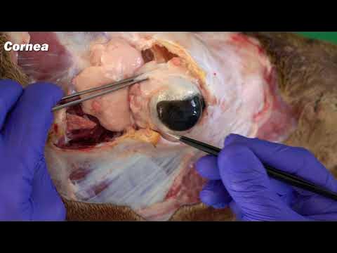
- 😀 The outermost layer of the eyeball, called the sclera, is a tough protective membrane, with a transparent portion at the front called the cornea.
- 😀 The middle layer is the choroid, a highly vascular area that provides nutrition and oxygen to the inner layers of the eye.
- 😀 The ciliary body is a thickened part of the choroid that helps in focusing, while the iris is a vascular diaphragm located in the middle.
- 😀 The inner layer of the eye, the retina, is crucial for converting light into electrical signals and consists of a pigmented layer and a neural layer.
- 😀 The highly pigmented layer of the retina helps absorb light to prevent image distortion, while the rods and cones in the neural layer convert light into electrochemical signals.
- 😀 Rods and cones act as transducers, converting light (electromagnetic energy) into electrochemical energy, which is processed by the central nervous system.
- 😀 The retina's sensitivity to light is limited to the area behind the iris, with no rods and cones extending in front of it.
- 😀 The lens helps focus light onto the retina and is held in position by suspensory ligaments attached to the ciliary body.
- 😀 The eyeball contains two types of fluid: the vitreous humor in the back portion and the aqueous humor in the front, both helping maintain the shape and focus of the eye.
Q & A
What are the three layers of the human eye?
-The three layers of the human eye are the outermost protective layer (sclera and cornea), the middle nutritive layer (choroid, ciliary body, and iris), and the inner sensory layer (retina).
What is the function of the sclera?
-The sclera is the tough, protective outer layer of the eyeball that helps maintain the shape of the eye and protects the internal structures.
What is the role of the cornea?
-The cornea is the transparent part of the outer layer that allows light to enter the eye and provides a protective barrier against dirt and germs.
What is the purpose of the choroid layer in the human eye?
-The choroid layer is highly vascular, providing oxygen and nutrients to the other layers of the eye.
What is the function of the ciliary body and iris?
-The ciliary body thickens and helps control the lens shape, while the iris regulates the size of the pupil to control the amount of light entering the eye.
What is the role of the retina in vision?
-The retina contains rods and cones, which convert light (electromagnetic energy) into electrochemical signals that are sent to the brain for processing, allowing us to see.
What are rods and cones, and how do they work?
-Rods and cones are specialized cells in the retina that convert light into electrical signals. Rods are responsible for low-light vision, and cones are responsible for color vision.
Why is the retina pigmented?
-The retina is pigmented to absorb light and prevent reflection, ensuring that light does not interfere with the image formation, which helps produce clearer vision.
What is the function of the lens in the eye?
-The lens focuses light onto the retina. It works in combination with the cornea to bend and direct light in a way that allows the formation of a clear image.
What is the difference between the vitreous humor and aqueous humor?
-The vitreous humor is a jelly-like substance filling the cavity behind the lens, while the aqueous humor is a watery fluid that fills the anterior compartment of the eye, helping to maintain pressure and shape.
Outlines

Cette section est réservée aux utilisateurs payants. Améliorez votre compte pour accéder à cette section.
Améliorer maintenantMindmap

Cette section est réservée aux utilisateurs payants. Améliorez votre compte pour accéder à cette section.
Améliorer maintenantKeywords

Cette section est réservée aux utilisateurs payants. Améliorez votre compte pour accéder à cette section.
Améliorer maintenantHighlights

Cette section est réservée aux utilisateurs payants. Améliorez votre compte pour accéder à cette section.
Améliorer maintenantTranscripts

Cette section est réservée aux utilisateurs payants. Améliorez votre compte pour accéder à cette section.
Améliorer maintenantVoir Plus de Vidéos Connexes

Medical and Nursing Terminology MADE EASY: Prefixes [Flashcard Tables]

Cahaya dan Alat Optik: Indra Penglihatan Manusia (Bagian-Bagian Mata) - SMP Kelas 8 | Part 1

Anatomy of the Eye, Eye muscles and associated structures

Embryology of the Eye (Easy to Understand)

Los TEJIDOS VEGETALES

Statika/Mekanika Teknik #4: Gaya Dalam
5.0 / 5 (0 votes)
