Glass Dog Anatomy
Summary
TLDRThis script delves into the anatomy of a dog's thorax and abdomen, highlighting the skeletal structure including the skull, vertebrae, ribs, and limb bones. It describes the thoracic and abdominal boundaries formed by the rib cage, diaphragm, and vertebral column. The script details the positioning of vital organs such as the heart, lungs, liver, and kidneys, and guides through the process of identifying them upon removal of the diaphragm and liver. It also mentions the visibility of the gastrointestinal tract, reproductive organs, and urinary bladder when the abdomen is viewed from the ventral aspect.
Takeaways
- 🦴 The anatomy of a dog's thorax and abdomen involves envisioning structures beneath the skin and the relevant parts of the skeleton.
- 💀 The skeletal components include the skull, cervical, thoracic, lumbar, and coxial vertebrae, as well as the rib cage, pelvis, and bones of the thoracic and pelvic limbs.
- 🐾 Thoracic limbs consist of the scapula, humerus, radius, ulna, carpus, metacarpus, and digits.
- 🦿 Pelvic limbs are made up of the femur, tibia, fibula, tarsus, metatarsus, and digits.
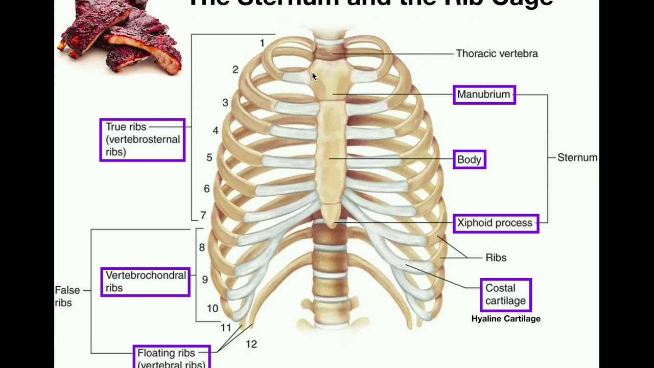
- 🔢 The thoracic vertebrae are associated with 13 pairs of ribs, the sternum, and the diaphragm, which together form the boundaries of the thorax.
- 🧘 The diaphragm, along with the ventral abdominal wall and pelvis, defines the boundaries of the abdomen.
- 🫁 The lungs extend from the level of the first rib to the 10th rib.
- ❤️ The heart is obliquely positioned in the thorax, from the third to the sixth intercostal spaces.
- 🧇 The large, dome-shaped diaphragm separates the thoracic cavity from the abdomen.
- 🍗 Removal of the diaphragm reveals the liver and gallbladder, with the liver spanning the entire width of the dog.
- 👀 After removing the liver, other organs such as the stomach, spleen, and left kidney become more visible on the left side of the abdomen.
- 🔍 On the right side, the duodenum, pancreas, and right kidney are evident, and from the ventral aspect, the cecum, ascending, transverse, and descending colons, rectum, ovaries, uterus, and urinary bladder can be identified.
Q & A
What are the main components of the dog's skeleton that are relevant to the thorax and abdomen?
-The main components include the skull, cervical, thoracic, lumbar, and coxial vertebrae, rib cage, pelvis, and bones of the thoracic and pelvic limbs.
What are the specific bones that make up the thoracic limbs of a dog?
-The thoracic limbs consist of the scapula, humerus, radius, ulna, carpus, metacarpus, phalanges, and digits.
What bones are included in the pelvic limbs of a dog?
-The pelvic limbs include the femur, tibia, fibula, tarsus, metatarsus, and digits.
How many pairs of ribs are associated with the thoracic vertebrae in a dog?
-There are 13 pairs of ribs associated with the thoracic vertebrae.
What structure separates the thoracic cavity from the abdomen in a dog?
-The diaphragm separates the thoracic cavity from the abdomen in a dog.
What is the position of the heart within the dog's thorax?
-The heart is positioned obliquely in the thorax from the third to the sixth intercostal spaces.
What organs are revealed when the diaphragm is removed in a dog?
-When the diaphragm is removed, the underlying liver and gallbladder are revealed.
Which organs are easily identifiable from the left side of the dog's abdomen after removing the liver?
-The stomach, spleen, and left kidney are easily identifiable from the left side of the abdomen.
What organs are evident on the right side of the dog's abdomen?
-The duodenum, pancreas, and right kidney are evident on the right side of the abdomen.
What structures can be identified when viewing the abdomen from its ventral aspect?
-The cecum, ascending colon, transverse colon, descending colon, rectum, ovaries, uterus, and urinary bladder can be identified from the ventral aspect.
What is the anatomical term for the area that extends from the level of the first rib to that of the 10th rib?
-This area is referred to as the lungs, as they extend from the level of the first rib to that of the 10th rib.
Outlines

Esta sección está disponible solo para usuarios con suscripción. Por favor, mejora tu plan para acceder a esta parte.
Mejorar ahoraMindmap

Esta sección está disponible solo para usuarios con suscripción. Por favor, mejora tu plan para acceder a esta parte.
Mejorar ahoraKeywords

Esta sección está disponible solo para usuarios con suscripción. Por favor, mejora tu plan para acceder a esta parte.
Mejorar ahoraHighlights

Esta sección está disponible solo para usuarios con suscripción. Por favor, mejora tu plan para acceder a esta parte.
Mejorar ahoraTranscripts

Esta sección está disponible solo para usuarios con suscripción. Por favor, mejora tu plan para acceder a esta parte.
Mejorar ahoraVer Más Videos Relacionados

Skeletal System (part 1) - Introduction

BAB II | FASE A. Rangka Tubuh | XI BIOLOGI | KURIKULUM MERDEKA
Introduction to the Skeletal System In 7 Minutes

3 regions of the vertebrate skull: splanchnocranium, chondrocranium, dermatocranium

Rangka Aksial pada Sistem Rangka Manusia
Anatomy | The Sternum, Rib Cage, & Vertebrae
5.0 / 5 (0 votes)
