1.1 - Introduction to transmission electron microscopy (TEM)

Kelvin Xie MSEN TAMU
1 Nov 201906:09

Summary

TLDRIn this introductory course on Transmission Electron Microscopy (TEM), Professor Calvin Shear from Texas A&M University offers a comprehensive overview of TEM's principles and applications. He shares his academic background and research experience using TEM in various studies, including materials characterization and phase transformations. The course covers five sections: basics, diffraction, imaging, spectroscopy, and related TEM techniques. It utilizes several key textbooks and resources, emphasizing the structural and chemical information TEM provides. The course also highlights advanced TEM techniques like aberration correction and scanning transmission electron microscopy (STEM).

Takeaways

  • 🤠 The course is an introduction to Transmission Electron Microscopy (TEM), taught by Calvin Shear, an assistant professor in the Department of Material Science and Engineering at Texas A&M University.
  • 🎓 Calvin Shear has a diverse academic background, with undergraduate studies in biomedical engineering and finance at the University of Sydney, and a PhD in mechanical engineering focusing on high-strength low-alloy steel.
  • 🔬 TEM and Atom Probe were key tools in his research during his PhD to study solute clusters, and post-PhD, he worked at Johns Hopkins University, using TEM to study deformation mechanisms in boron carbide and magnesium.
  • 🧑‍🏫 The course will cover a wide range of topics, including electron scattering, instrumentation, sample preparation, diffraction, imaging, spectroscopy, and related TEM techniques.
  • 📚 The main textbook for the course is 'Transmission Electron Microscopy' by Williams and Carter, supported by other resources like 'Microstructural Characterization of Materials' and 'Aberration-Corrected Imaging'.
  • 🖼️ Five main sections of the course include basics, diffraction, imaging, spectroscopy, and TEM-related techniques, with detailed discussions on each topic.
  • 💡 In the basics, the course will explore electron scattering, electron sources, and lenses, as well as sample preparation methods like electro-polishing and lift-out for specific samples.
  • 📐 The diffraction section covers parallel beam diffraction, Kikuchi diffraction, and convergent beam electron diffraction, explaining how each provides different visual patterns (spots, lines, disks).
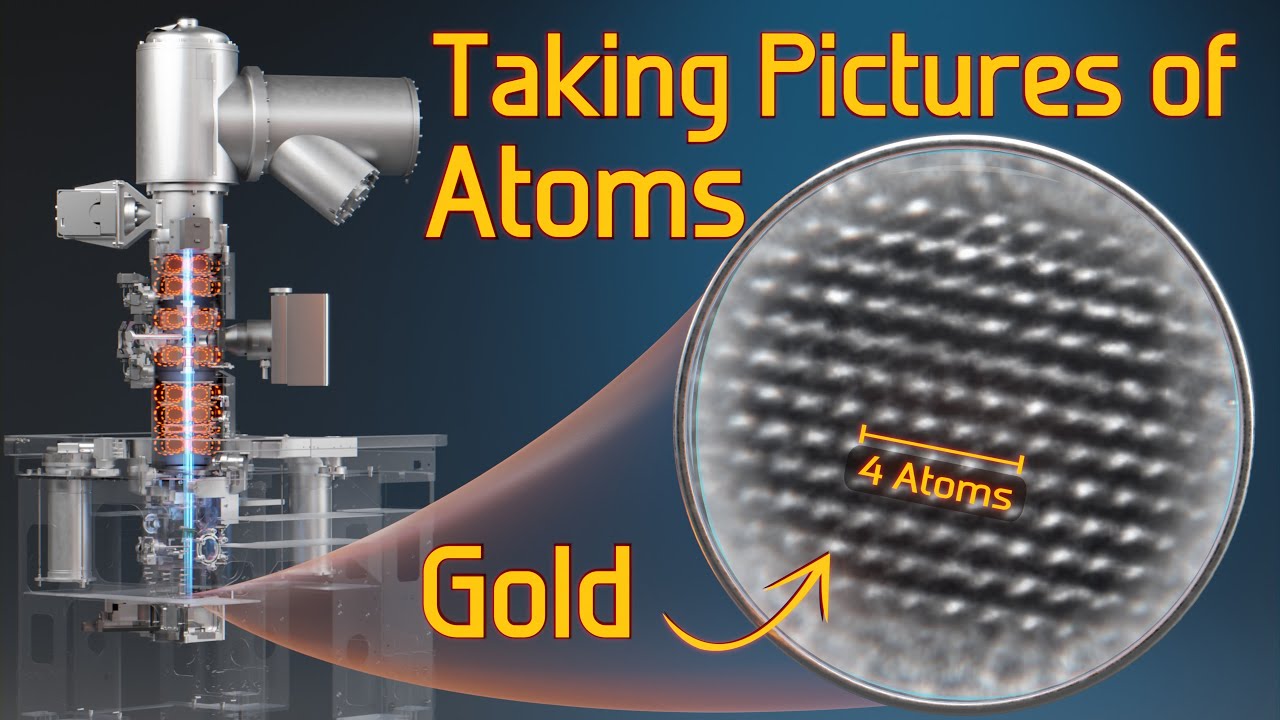
  • 🔍 Imaging will delve into three types of contrast (mass-thickness, diffraction, and phase contrast) to analyze different materials and samples at atomic resolution.
  • 🌈 The spectroscopy section introduces EDS and EELS techniques, enabling students to gather both chemical and bonding information about materials, particularly when combined with scanning TEM and aberration correction.

Q & A

  • What is the main focus of this Transmission Electron Microscopy (TEM) class?

    -The main focus of this class is to provide an overview of Transmission Electron Microscopy (TEM) and its applications in material science.

  • Who is the instructor of the class, and what is his academic background?

    -The instructor is Calvin Shear, an assistant professor in the Department of Material Science and Engineering at Texas A&M University. He has a background in biomedical engineering and finance from the University of Sydney and a PhD in mechanical engineering.

  • What technique is emphasized in this course that is specific to Texas A&M University?

    -The course emphasizes the use of a special TEM technique called precession electron diffraction, which is used to study the formation and phase transformations in materials.

  • Which textbook is primarily used for this course, and what is its significance?

    -The textbook used is 'Transmission Electron Microscopy' by Williams and Carter, often referred to as the 'Bible' for TEM due to its comprehensive coverage of the subject.

  • What are the five main sections of the TEM course as outlined by the instructor?

    -The five main sections of the course are basics, diffraction, imaging, spectroscopy, and TEM-related techniques.

  • What are the three types of contrast discussed in the imaging section of the course?

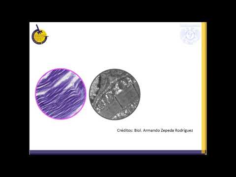
    -The three types of contrast discussed are mass-thickness contrast, diffraction contrast, and phase contrast. These contrasts help in imaging biological samples, crystal defects, and atomic structures, respectively.

  • What are the two spectroscopy techniques covered in the course, and what information do they provide?

    -The two spectroscopy techniques are Energy Dispersive X-ray Spectroscopy (EDS) and Electron Energy Loss Spectroscopy (EELS). EDS provides chemical information, while EELS offers both chemical and chemical bonding information.

  • How does the instructor describe the impact of aberration correction in TEM imaging?

    -The instructor explains that aberration correction in TEM significantly improves image quality. He shows an example where an aberration-corrected image reveals more detailed atomic structures compared to a non-corrected image.

  • What is the significance of combining EDS and EELS with STEM in the context of this course?

    -Combining EDS and EELS with Scanning Transmission Electron Microscopy (STEM) allows for the chemical analysis of individual atomic columns, providing highly detailed structural and chemical information.

  • What is the expected outcome for students by the end of this TEM course?

    -By the end of the course, students are expected to have a good understanding of how TEM works and how it can be applied to research in material science.

Outlines

00:00

👋 Introduction to Transmission Electron Microscopy (TEM)

In this introductory section, Professor Calvin Shear welcomes the audience to his course on Transmission Electron Microscopy (TEM). He provides an overview of his background, including his undergraduate studies in biomedical engineering and finance at the University of Sydney, followed by a PhD in mechanical engineering. His research involved using TEM and atom probe techniques to study high-strength alloy steels. Afterward, he joined Johns Hopkins University to investigate deformation mechanisms in materials like boron carbide and magnesium, eventually becoming a microscopy faculty member at Texas A&M University, specializing in advanced TEM techniques.

05:01

📚 Course Materials and Structure

This section outlines the primary textbook for the course, *Transmission Electron Microscopy* by Williams and Carter, which is considered essential for TEM studies. Additional resources include *Microstructural Characterization of Materials* by Brendan and Kaplan and *Introduction to Scanning Electron Microscopy*. The course will cover five major sections, each corresponding to a YouTube playlist: basics, diffraction, imaging, spectroscopy, and advanced TEM techniques. Each section will dive deep into the various methods and topics in TEM, preparing students for advanced research in material science.

🔬 Basics of Electron Scattering and Instrumentation

In the basics section of the course, Professor Shear will discuss electron scattering and the fundamental components of a TEM instrument, including electron sources, lenses, and the importance of sample preparation. The two examples of sample preparation methods include electro-polishing for metal samples and site-specific lift-out techniques. This foundational knowledge sets the stage for further exploration of how TEM works and how samples are prepared for analysis.

📐 Electron Diffraction Techniques

This paragraph introduces electron diffraction, explaining how and why diffraction occurs within a TEM. Various diffraction techniques are explored, such as parallel beam diffraction, Kikuchi diffraction, and convergent beam electron diffraction (CBED). Each method produces different visual outcomes—spots, lines, or disks—that help researchers analyze material structures. These diffraction techniques are essential for understanding crystallographic information.

🌈 Imaging and Contrast in TEM

In this part of the course, different types of imaging and contrast mechanisms in TEM are discussed. Professor Shear explains three types of contrast—mass thickness contrast, diffraction contrast, and phase contrast—each used to visualize different material features, such as biological samples or crystallographic defects. Examples include high-resolution TEM images showing atomic-level details like nano twins in boron carbide, with applications in understanding material deformation and structure.

📊 Spectroscopy Techniques in TEM

This section covers spectroscopy techniques that provide chemical information about materials. Two key methods—Energy Dispersive X-ray Spectroscopy (EDS) and Electron Energy Loss Spectroscopy (EELS)—are introduced. EDS helps in identifying chemical compositions, while EELS offers insights into chemical bonding. Examples include comparing EELS spectra for diamond, carbon 60, and graphite, showing how these techniques can reveal subtle material differences at the atomic level.

🧪 Advanced TEM Techniques and Applications

The final section delves into advanced TEM techniques, including Scanning Transmission Electron Microscopy (STEM) and precession electron diffraction, which can be compared to Electron Backscatter Diffraction (EBSD). Aberration-corrected TEM is highlighted as a powerful tool for achieving high-resolution images, with examples showing the dramatic improvements in image clarity when aberration is corrected. The course aims to equip students with the skills to understand and apply these advanced techniques in their research.

Mindmap

Keywords

💡Transmission Electron Microscopy (TEM)

Transmission Electron Microscopy (TEM) is a technique that uses a beam of electrons to pass through a sample to create high-resolution images. It allows for detailed structural analysis of materials at the atomic level. In the video, TEM is the central focus, and the course aims to teach its applications, from imaging to diffraction.

💡Electron Diffraction

Electron diffraction refers to the phenomenon where electrons are scattered by the atomic lattice of a material, producing diffraction patterns. These patterns provide information about the material's crystal structure. The video mentions several types of electron diffraction (parallel beam, Kikuchi, converging beam), which are key to analyzing materials in TEM.

💡Sample Preparation

Sample preparation involves techniques to prepare materials for TEM analysis, ensuring that they are thin enough for electrons to pass through. In the video, methods such as electro-polishing and lift-out are highlighted as essential for studying specific material types like metals and site-specific samples.

💡Mass Thickness Contrast

Mass thickness contrast is a type of contrast in TEM imaging that arises from differences in the thickness and atomic mass of the material. Heavier or thicker areas appear darker. The video uses the example of biological samples, like mitochondria, to explain how this contrast helps visualize different material structures.

💡Diffraction Contrast

Diffraction contrast is used in TEM to image crystal defects such as dislocations and stacking faults. It is created when electrons are scattered by different parts of the crystal, leading to contrast. In the video, the instructor shares examples from his own research, showing dislocations in high-strength alloys.

💡Spectroscopy

Spectroscopy in TEM refers to techniques that provide chemical information about the sample, complementing structural data. The video discusses two main types: Energy Dispersive X-ray Spectroscopy (EDS) and Electron Energy Loss Spectroscopy (EELS). These methods are used to identify chemical composition and even chemical bonding at the atomic level.

💡Aberration Correction

Aberration correction is a technique in TEM that improves image resolution by correcting optical distortions caused by lenses. The video contrasts images with and without aberration correction, showing how this technique allows for clearer atomic-level visualization. This is crucial for studying fine structural details.

💡Precession Electron Diffraction

Precession Electron Diffraction is a specialized TEM technique that enhances diffraction data by reducing dynamical effects. It can be seen as analogous to EBSD (Electron Backscatter Diffraction) but inside a TEM. The video explains its importance in material phase transformations and crystallographic studies.

💡High-Resolution TEM (HRTEM)

High-Resolution TEM (HRTEM) allows for atomic-scale imaging, providing detailed information about the arrangement of atoms in a material. The video refers to HRTEM while explaining phase contrast, which enables the visualization of atomic-level defects like nano twins in materials such as boron carbide.

💡Energy Dispersive X-ray Spectroscopy (EDS)

Energy Dispersive X-ray Spectroscopy (EDS) is a TEM-based technique used to determine the elemental composition of materials by detecting X-rays emitted from the sample during electron beam interaction. The video emphasizes its role in combining structural and chemical information, particularly in high-precision material analysis.

Highlights

Introduction to Transmission Electron Microscopy (TEM) by Calvin Shear, Assistant Professor at Texas A&M University.

Calvin Shear's academic background includes a Bachelor’s degree in Biomedical Engineering and Finance from the University of Sydney, and a PhD in Mechanical Engineering.

Shear’s PhD research utilized both TEM and Atom Probe to study solar clusters in high-strength, low-alloy steel.

After his PhD, Shear joined Johns Hopkins University, where he used TEM to study deformation mechanisms in boron carbide and magnesium.

In 2018, Shear became part of Texas A&M University’s Department of Material Science and Engineering, focusing on microscopy.

At Texas A&M, Shear uses a specialized TEM technique called Precession Electron Diffraction to study material formation and phase transformation.

The course’s primary textbook is 'Transmission Electron Microscopy' by Williams and Carter, commonly referred to as the 'Bible' for TEM.

Additional resources include 'Microstructural Characterization of Materials' by Brandon and Kaplan, and 'Aberration-Corrected Imaging in Transmission Electron Microscopy' by Erni.

The TEM course is divided into five sections: basics, diffraction, imaging, spectroscopy, and TEM-related techniques.

The basics section covers electron scattering, electron sources, lenses, and sample preparation techniques like electro-polishing and lift-out methods.

Electron diffraction is explained with various techniques, including Parallel Beam Diffraction (spots), Kikuchi Diffraction (lines), and Converging Beam Electron Diffraction (disks).

The course emphasizes three types of contrast in TEM imaging: mass-thickness contrast, diffraction contrast, and phase contrast.

Spectroscopy section focuses on Energy Dispersive X-ray Spectroscopy (EDS) and Electron Energy Loss Spectroscopy (EELS), providing chemical and bonding information.

Advanced TEM techniques include Scanning Transmission Electron Microscopy (STEM), Precession Electron Diffraction, and Aberration-Corrected TEM.

By the end of the course, students are expected to understand how TEM works and how it can enhance their research.

Transcripts

play00:00

howdy welcome to my introduction to the

play00:03

transmission electron microscopy class

play00:05

my name is Calvin shear and I am an

play00:08

assistant professor in the Department of

play00:10

material science and engineering at

play00:12

Texas A&M University

play00:14

the aim of this online class is to give

play00:17

you an overview of what is TEM and what

play00:20

he young can do or just use one slide to

play00:23

share with you my background I did my

play00:26

undergraduate studies at University of

play00:28

Sydney in Australia my bachelor degrees

play00:31

from biomedical engineering believe it

play00:33

or not I also have a degree in finance

play00:36

aizen State at the University of Sydney

play00:38

and completed my PhD in the Department

play00:41

of Mechanical Engineering my PhD

play00:43

advisors are professors Judy carne and

play00:46

the Simon ringer during my PhD are used

play00:49

both TEM and Adam probe to study the

play00:52

solar clusters in a high strength low

play00:54

alloy steel after my PhD a joint

play00:57

professor Kevin Henkes group at Johns

play01:00

Hopkins University

play01:01

I was the in-house TEM person and using

play01:05

TEM as at all to study the defamation

play01:07

mechanisms in boron carbide and a

play01:09

magnesium in 2018 I joined the

play01:13

department of material science and

play01:15

engineering at Texas A&M as a microscopy

play01:18

faculty at Texas A&M we use a special TM

play01:22

technique called precession electron

play01:24

diffraction to study the formation and

play01:27

the phase transformation in materials

play01:29

the textbook were going to use for this

play01:32

course is transmission electron

play01:34

microscopy by Williams and Carter this

play01:37

is also referred as the Bible for TEM to

play01:41

complement the Williams and Carter books

play01:43

we will also use the resources from a

play01:46

few other books the first one is the

play01:49

micro structural characterization of

play01:51

materials by Brendan and Kaplan a few

play01:54

chapters of the book are devoted to TM

play01:57

and a TM based techniques if you want to

play02:00

have a quick read about TM it's a very

play02:02

nice book we will also use the

play02:05

information from introduction to

play02:07

scanning electron microscopy or stem the

play02:10

last one is the aberration corrected

play02:12

imaging inference electron microscopy by

play02:15

Ernie there will be five sections of

play02:17

this TM course all five playlists on

play02:21

YouTube

play02:21

there will be on the basics diffraction

play02:25

imaging spectroscopy and other TM

play02:28

related techniques in basics we'll start

play02:31

by talking about the electron scattering

play02:33

in TEM the were moved to the

play02:36

instrumentation talking about the

play02:37

electron sources lenses etc we will also

play02:41

talk about sample preparation in these

play02:43

two examples specifically the one on the

play02:46

left is electro polishing to prepare

play02:48

metal samples the one on the right is a

play02:50

lift out for site-specific samples the

play02:54

second section is on electron

play02:55

diffraction both talked about why and

play02:58

how diffraction happens in TEM we will

play03:01

then discuss the parallel beam

play03:03

diffraction and indexing Kikuchi

play03:06

diffraction and convergence beam

play03:08

diffraction in parallel beam diffraction

play03:11

you see spots in Kikuchi diffraction you

play03:13

see lines in converging beam electron

play03:16

diffraction you see disks in the third

play03:19

section of this course or discuss

play03:21

imaging and is the origin of contrast

play03:24

there are three types of contrast mass

play03:27

thickness contrast diffraction contrast

play03:29

and face contrast the mass thickness

play03:32

contrast is used to image stained

play03:35

biological samples the example here

play03:37

shows the mitochondria in a Cell

play03:40

diffraction contrast is used to image

play03:43

crystal graphic defects such as

play03:46

dislocations and stacking faults the

play03:48

examples showing here are from my own

play03:50

research with Professor Kevin honker the

play03:53

dark lines in the left figure and the

play03:56

bright lines in the right figure artists

play03:58

locations the reason we can see the

play04:01

atomic level information in high-res DM

play04:04

images is due to the face contrast again

play04:07

this example was from my own research

play04:10

with Professor Kevin honker showing the

play04:12

nano twins in a boring car by sample the

play04:16

fourth section is spectroscopy imaging

play04:19

and the diffraction will give you the

play04:20

structural information spectroscopy will

play04:22

give you the chemical information the

play04:25

two spectroscopy techniques going to

play04:27

discuss our energy dispersive x-ray

play04:30

spectroscopy EDS and electron energy

play04:33

loss spectroscopy eels using EDS you can

play04:36

get the chemical information using eels

play04:39

in addition to the chemical information

play04:41

you can also get the chemical bonding

play04:44

information from this example here you

play04:46

can see the eros spectrum of diamond

play04:49

looks very different from carbon 60 and

play04:52

graphite combining EDS and eels with

play04:55

stem using an aberration corrected TEM

play04:58

you can get the chemical information of

play05:01

individual atomic columns the figure

play05:04

here is from Cornell University and it's

play05:07

just striking

play05:09

in the last section we'll introduce some

play05:12

tea and related techniques the first one

play05:15

is the scanning transmission electron

play05:17

microscopy stem these stem images were

play05:21

acquired by my student Hershey

play05:23

highlighting the nano precipitates in a

play05:25

shape memory alloy we will also talk

play05:28

about the precession electron

play05:29

diffraction this technique can be

play05:32

somewhat viewed as EB SD inside TEM

play05:36

lastly we'll talk about is the

play05:38

aberration corrected TEM the micrographs

play05:40

were taken from the Williams and Carter

play05:42

book the image on the left has no

play05:45

aberration correction while the one on

play05:47

the right is with the aberration

play05:49

correction you can see the difference

play05:51

straight away at the end of this course

play05:54

I hope you will be able to develop a

play05:56

good understanding on how TM works and

play05:59

what TM can do to help your research in

play06:02

the next video we'll talk about why we

play06:04

use electron inside TEM and the

play06:07

resolution of TEM

Rate This

5.0 / 5 (0 votes)

Etiquetas Relacionadas
TEM basicsMicroscopy techniquesElectron diffractionImaging contrastSpectroscopyEnergy dispersive X-rayAberration correctionSTEM microscopyMaterial scienceTexas A&M
¿Necesitas un resumen en inglés?