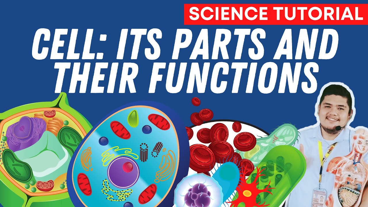
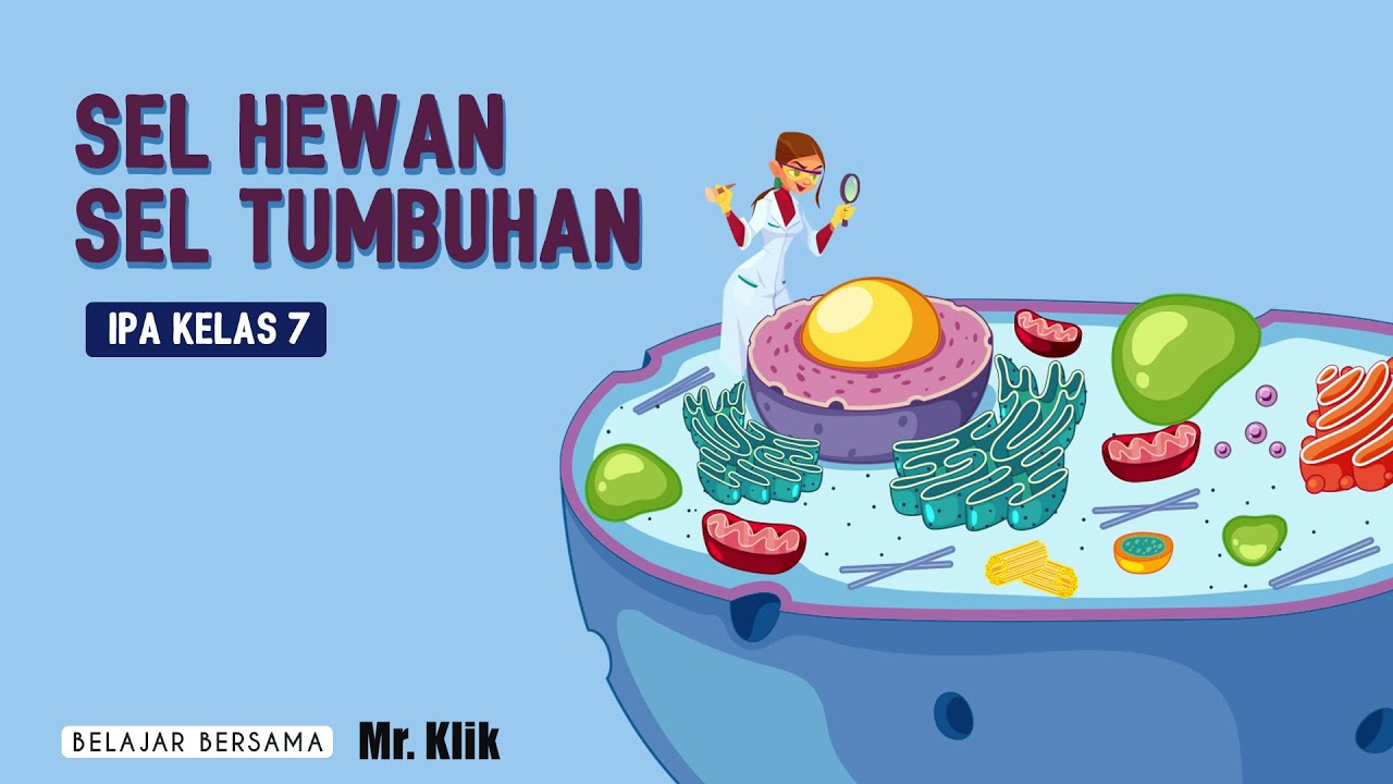
Organelles of the Cell

Beverly Biology
6 Jun 201429:51

Summary

TLDRThis educational video script delves into the intricate components of a eukaryotic cell, emphasizing the plasma membrane, nucleus, and cytoplasm as the cell's foundational sections. It illustrates the cytoplasm's role as a medium for chemical reactions and the plasma membrane's selective permeability. The nucleus, often dubbed the 'control center,' houses DNA and directs protein synthesis. Ribosomes, ER, and the GOI body collaborate in protein production and modification. Mitochondria, known as the 'powerhouse,' generate energy through cellular respiration. The script also touches on the endosymbiosis theory, suggesting mitochondria and chloroplasts were once independent organisms. Lysosomes handle waste and food breakdown, while vacuoles store materials. The video concludes with a review of these concepts, encouraging viewer engagement.

Takeaways

  • 🔬 Organelles are specialized structures within cells that perform specific functions, akin to organs in the body.
  • 🌟 The plasma membrane, or cell membrane, is a lipid bilayer that selectively allows substances to pass in and out of the cell.
  • 🧬 The nucleus acts as the cell's control center, housing DNA and directing the cell's activities through the production of proteins.
  • 🍯 The cytoplasm is a gel-like substance that fills the cell and facilitates chemical reactions, including the dissolution of solutes like carbohydrates and proteins.
  • 🏋️‍♂️ Ribosomes are the cellular structures responsible for protein synthesis, translating genetic information into functional proteins.
  • 📦 The Golgi apparatus modifies, sorts, and packages proteins for transport within the cell or for secretion outside the cell.
  • ⚡ Mitochondria are known as the 'powerhouses' of the cell, generating energy in the form of ATP through cellular respiration.
  • 🔁 Endosymbiosis theory suggests that mitochondria and chloroplasts were once free-living organisms that became incorporated into larger cells.
  • 🌱 Chloroplasts, found in plant cells, carry out photosynthesis, converting sunlight into glucose and oxygen using chlorophyll.
  • 💧 Vacuoles store water, nutrients, waste products, and pigments, and can also regulate the cell's internal environment.

Q & A

  • What is meant by the term 'organelle' in the context of a eukaryotic cell?

    -An organelle refers to the small specialized structures within a eukaryotic cell that perform specific functions necessary for the cell's survival and operation.

  • What are the three basic functions that all cells must perform?

    -All cells must perform the functions of taking in food, getting rid of waste, and reproducing.

  • What is the primary role of the plasma membrane, also known as the cell membrane?

    -The plasma membrane's primary role is to regulate the passage of materials into and out of the cell, acting as a selectively permeable barrier.

  • What is the cytoplasm and what does it do?

    -The cytoplasm is a jellylike substance filling the cell, providing a medium for organelles to float within and facilitating various chemical reactions, including the dissolution of solutes like carbohydrates and proteins.

  • Why is the nucleus often referred to as the 'control center' of the cell?

    -The nucleus is called the 'control center' because it contains DNA in the form of chromatin, which holds the genetic instructions for making proteins that perform most of the work within the cell.

  • What is the function of the nucleolus within the nucleus?

    -The nucleolus is responsible for producing ribosomes, which are essential for protein synthesis within the cell.

  • How does the rough endoplasmic reticulum differ from the smooth endoplasmic reticulum?

    -The rough endoplasmic reticulum is covered in ribosomes, which are involved in protein synthesis, while the smooth endoplasmic reticulum lacks ribosomes and is involved in lipid synthesis and toxin breakdown.

  • What is the role of ribosomes in the cell?

    -Ribosomes are responsible for protein synthesis by linking amino acids together to form proteins, a process also known as translation.

  • What is the function of the Golgi apparatus in the cell?

    -The Golgi apparatus receives proteins from ribosomes, modifies them, sorts, and packages them into vesicles for export from the cell.

  • What is the significance of the mitochondria being referred to as the 'powerhouse' of the cell?

    -Mitochondria are called the 'powerhouse' because they generate ATP, the cell's primary energy molecule, through cellular respiration.

  • What is the endosymbiosis theory and how does it relate to the origin of mitochondria and chloroplasts?

    -The endosymbiosis theory suggests that mitochondria and chloroplasts were once free-living organisms that were engulfed by a larger cell and eventually became integrated as organelles, surviving and reproducing within the host cell.

  • What is the primary role of lysosomes within a cell?

    -Lysosomes contain digestive enzymes that break down various materials, including food particles, pathogens engulfed by immune cells, and even old or damaged cell components.

  • How do cilia and flagella differ in structure and function within cells?

    -Cilia are short, hairlike extensions that aid in movement over short distances, while flagella are long, whip-like structures that can propel cells over greater distances, such as in the case of sperm cells.

  • What is the cell wall and what is its primary function?

    -The cell wall is a tough, outermost layer found in plant, fungal, and bacterial cells, providing structural support and protection.

  • What is the role of chloroplasts in plant cells?

    -Chloroplasts perform photosynthesis, converting sunlight, carbon dioxide, and water into glucose and oxygen, using the green pigment chlorophyll.

  • What is the function of the vacuole in both plant and animal cells?

    -The vacuole serves as a storage area for food, water, waste, and pigments. In plant cells, it is often the largest organelle, pushing other structures to the cell's periphery.

Outlines

00:00

🌐 Overview of Eukaryotic Cell Organelles

The paragraph introduces the concept of organelles within eukaryotic cells, comparing them to organs in the body. It outlines the three fundamental sections of a cell: the plasma membrane, the nucleus, and the cytoplasm. The plasma membrane, also known as the cell membrane, is highlighted as a key part of the cell's structure. The nucleus is referred to as the 'control center' due to its role in housing DNA. The cytoplasm is described as a gel-like substance filling the cell, within which most organelles are suspended. The paragraph also mentions the importance of the cytoplasm in facilitating chemical reactions and providing support to the cell structure.

05:01

🧬 The Nucleus: Control Center and DNA Repository

This section delves into the nucleus, emphasizing its role as the 'headquarters' of the cell due to containing DNA. DNA is depicted as the cell's blueprint, with chromatin being the physical form of DNA within the nucleus. The nucleolus, a distinct structure within the nucleus, is introduced as the site of ribosome production. Ribosomes are then explained as the cellular machinery responsible for protein synthesis, which is central to the cell's functions. The paragraph also touches on the endoplasmic reticulum (ER), distinguishing between the rough ER, studded with ribosomes, and the smooth ER, which lacks ribosomes and is involved in lipid synthesis and toxin breakdown.

10:03

🔬 Ribosomes and Protein Synthesis

The focus shifts to ribosomes, which are described as the cellular structures responsible for protein synthesis. Ribosomes are shown to be composed of RNA and proteins, and their function is to link amino acids together to form proteins. The process of protein synthesis is mentioned as translation, a concept to be further explored. The paragraph also discusses the role of the Golgi apparatus in receiving proteins from ribosomes, modifying them, and packaging them into vesicles for export from the cell. This process is visualized through an animation that shows ribosomes moving through the ER and the Golgi apparatus processing and shipping out proteins.

15:03

🏋️‍♂️ Mitochondria: The Powerhouse of the Cell

This paragraph introduces mitochondria as the 'powerhouses' of the cell, responsible for generating ATP, the cell's energy currency, through cellular respiration. The theory of endosymbiosis is introduced to explain the origin of mitochondria as once free-living organisms that became integrated into cells. The paragraph discusses the unique characteristics of mitochondria, such as having their own DNA and ribosomes, allowing them to replicate independently. The historical context of cell theory development is also briefly touched upon, mentioning the contributions of Robert Hooke and his observations of cell walls in cork.

20:03

🌱 Plant Cells and Their Unique Organelles

The paragraph discusses the cell wall, a feature unique to plant, fungal, and bacterial cells, providing structural support and rigidity. It contrasts with animal cells, which lack a cell wall. The role of cellulose in forming the cell wall is highlighted, along with its toughness and indigestibility. Chloroplasts are introduced as the organelles responsible for photosynthesis in plant cells, containing chlorophyll, which converts sunlight, carbon dioxide, and water into glucose and oxygen. The endosymbiotic theory is also applied to chloroplasts, suggesting they were once free-living organisms that became part of plant cells. The paragraph concludes with a brief mention of vacuoles, which store food, water, waste, and pigments, and are often the largest organelle in plant cells.

25:05

🍄 The Role of Vacuoles and Cell Wall in Plant Cells

This final paragraph emphasizes the role of vacuoles in storing various substances within plant cells, including nutrients, water, and waste. It contrasts the large central vacuoles found in plant cells with the smaller, scattered vacuoles in animal cells. The paragraph also reiterates the importance of the cell wall in providing structural support to plant cells, mentioning the historical significance of Robert Hooke's observations of cell walls in cork. The paragraph serves as a conclusion, summarizing the key organelles and their functions within eukaryotic cells.

Mindmap

Keywords

💡Organelle

An organelle is a specialized subunit within cells that performs a specific function. In the context of the video, organelles are the 'small parts' that enable cells to carry out essential life processes. The video discusses various organelles such as mitochondria, chloroplasts, and the Golgi apparatus, each with unique roles in cellular activities.

💡Plasma Membrane

The plasma membrane, also known as the cell membrane, is a biological membrane that separates the cell from its external environment. It is described in the video as a selectively permeable barrier, composed of a lipid bilayer with embedded proteins, which allows certain molecules to pass through while restricting others. This membrane is crucial for the cell's ability to control the movement of substances in and out.

💡Nucleus

The nucleus serves as the control center of the cell, containing the cell's genetic material (DNA). As explained in the video, it is often referred to as the 'headquarters' because it directs cellular activities by housing DNA, which provides the instructions for making proteins. The video also mentions the nucleolus, a structure within the nucleus responsible for producing ribosomes.

💡Cytoplasm

Cytoplasm is the jelly-like substance that fills a cell, in which organelles are suspended. The video describes it as facilitating chemical reactions within the cell and providing support. It also dissolves solutes, such as carbohydrates and proteins, which are essential for cellular functions. The concept of cytoplasmic streaming is introduced, illustrating the movement of organelles within the cytoplasm.

💡Mitochondria

Mitochondria are referred to as the 'powerhouses' of the cell because they generate most of the cell's supply of adenosine triphosphate (ATP), which is used as a source of chemical energy. The video explains that mitochondria carry out cellular respiration and were once free-living organisms, a concept supported by the endosymbiosis theory.

💡Endoplasmic Reticulum (ER)

The endoplasmic reticulum is a network of membranous tubules and sacs within the cell. It is divided into two types: rough ER, which is studded with ribosomes, and smooth ER, which lacks ribosomes. The rough ER's function, as detailed in the video, includes the synthesis of proteins and lipids, while the smooth ER is involved in lipid metabolism and detoxification.

💡Ribosomes

Ribosomes are cellular structures responsible for protein synthesis. The video explains that ribosomes are produced by the nucleolus, travel through the rough ER, and link amino acids together to form proteins. This process is essential for the cell's ability to produce the proteins necessary for its structure and function.

💡Golgi Apparatus

The Golgi apparatus is an organelle responsible for modifying, sorting, and packaging proteins and lipids for secretion or delivery to other organelles. In the video, it is described as receiving proteins from ribosomes, modifying them, and then packaging them into vesicles for export from the cell, playing a crucial role in the cell's protein production and secretion processes.

💡Cytoskeleton

The cytoskeleton is a network of protein filaments that maintain cell shape, provide structural support, and enable cell movement. The video mentions two types of cytoskeletal structures: cilia, which are short and hair-like, and flagella, which are long and whip-like. These structures are essential for cell motility, such as the movement of sperm cells.

💡Cell Wall

The cell wall is a rigid layer outside the cell membrane in plant, fungal, and bacterial cells. It provides structural support and protection. The video explains that unlike animal cells, plant cells have a cell wall made of cellulose, which is a tough and fibrous material that gives plants their structural integrity.

💡Chloroplasts

Chloroplasts are organelles found in plant cells that carry out photosynthesis, the process by which light energy is converted into chemical energy. The video describes chloroplasts as containing chlorophyll, which absorbs light and carbon dioxide to produce glucose and oxygen. Like mitochondria, chloroplasts are thought to have originated from free-living bacteria through the endosymbiosis theory.

💡Vacuoles

Vacuoles are membrane-bound organelles that store water, nutrients, waste products, and pigments. The video explains that in plant cells, the central vacuole can be quite large and push other organelles to the cell's periphery. In contrast, animal cells have smaller vacuoles scattered throughout the cytoplasm, serving similar storage functions.

Highlights

An organelle is a small part within a cell, akin to how organs are small parts within the body.

Cells have three main sections: the plasma membrane, the nucleus, and the cytoplasm.

The plasma membrane, also known as the cell membrane, regulates the entry and exit of materials.

The nucleus acts as the control center of the cell, containing DNA.

The cytoplasm is a jellylike substance within the cell where organelles float.

Cytoplasmic streaming is the movement of organelles within the cytoplasm.

The cytoplasm facilitates chemical reactions and dissolves particles like carbohydrates and proteins.

The plasma membrane is composed of a bilayer of lipids and proteins, and is selectively permeable.

The nucleus contains chromatin, which is DNA that provides instructions for protein synthesis.

The nucleolus within the nucleus is responsible for producing ribosomes.

Ribosomes, made by the nucleolus, are transported by the rough endoplasmic reticulum to make proteins.

The Golgi apparatus receives, modifies, sorts, and packages proteins for export from the cell.

Mitochondria are known as the powerhouse of the cell, generating energy through cellular respiration.

The theory of endosymbiosis suggests that mitochondria were once free-living organisms that became part of the cell.

Lysosomes contain digestive enzymes to break down food, pathogens, and even old cells through a process called autophagy.

Cilia and flagella are cell parts used for movement, with cilia being short and flagella being long and whip-like.

The cell wall is the outermost layer in plant, fungal, and bacterial cells, providing support and structure.

Chloroplasts, found in plant cells, perform photosynthesis using chlorophyll, a green chemical.

Like mitochondria, chloroplasts are thought to have evolved from free-living bacteria through endosymbiosis.

Vacuoles store food, water, waste, and color pigments, and are often the largest part in plant cells.

Transcripts

play00:01

okay so in this video we're going to go

play00:03

over some of the common organel of a

play00:05

eukaryotic cell now an organel is simply

play00:08

a word that means small part so just

play00:10

like the body has small parts called

play00:12

organs cells have small parts called

play00:16

organel well there's a lot of them so

play00:18

let's go ahead and get

play00:20

started first thing I want to mention

play00:22

are the three basic sections to the cell

play00:25

and all cells need to take in food get

play00:27

rid of waste and reproduce well cells

play00:31

have three main sections to them and

play00:33

inside of those three sections are parts

play00:36

that allow them to take in food get rid

play00:38

of waste and reproduce so the first

play00:41

section you can see highlighted by the

play00:42

flashing black line the plasma membrane

play00:45

you might also hear the plasma membrane

play00:47

simply called the cell membrane we'll

play00:48

talk more about this in a little

play00:51

bit pulsing in the picture you can see

play00:54

the second main section the nucleus uh

play00:57

you often hear the nucleus called the

play00:58

control center we'll explain why in a

play01:01

little

play01:02

bit and lastly we have the cytoplasm now

play01:05

the arrow in the picture looks like it's

play01:07

kind of just pointing to empty space

play01:09

well cells are not filled with empty

play01:10

space they're filled with a part called

play01:12

the cytoplasm we'll go into that in more

play01:14

detail let's actually do the cytoplasm

play01:16

first here we

play01:19

go so the cytoplasm the first thing I

play01:22

want to mention is that it's the

play01:23

jellylike material on the inside of a

play01:26

cell you know when you look at diagrams

play01:28

sometimes in a diagram a cell might look

play01:30

like it's empty or filled with Hollow

play01:32

air Hollow air space and that's not true

play01:35

cells are filled with a jellylike

play01:36

liquidy material called the cytoplasm

play01:39

and in that cytoplasm most organel float

play01:42

within you can see here's a list of a a

play01:45

few of the organel that are floating

play01:47

around in the cytoplasm we're going to

play01:48

go and talk about all of these

play01:50

throughout this video we'll talk about

play01:51

what mitochondria is what lomes

play01:56

are and because the cytoplasm is a jelly

play02:00

material and things are floating around

play02:01

inside of it you often under a

play02:04

microscope can actually see Parts

play02:06

floating around in this animation here

play02:09

you can see what's known as cytoplasmic

play02:11

streaming the green circles that are

play02:14

drifting around inside of the square

play02:16

cells the rectangular shaped cells those

play02:19

green circles are plant cell

play02:21

chloroplasts you can see in every

play02:24

rectangular shaped plant cell there's a

play02:26

few dozen

play02:28

chloroplasts in in each of these cells

play02:30

here and this is known as cytoplasmic

play02:35

streaming okay well what about the job

play02:37

what does the cytoplasm actually do well

play02:40

a lot of chemical reactions take place

play02:43

within the cell and chemical reactions

play02:45

take place in the cytoplasm so that's

play02:47

kind of what the job of the cytoplasm is

play02:49

it helps to facilitate chemical

play02:51

reactions and it also helps to dissolve

play02:54

uh solutes uh particles that are in the

play02:57

cell carbohydrates proteins these are

play02:59

all dissolved within the actual

play03:01

cytoplasm of the cell so that's really

play03:04

what the main function of the cytoplasm

play03:07

is it also adds support to the cell a a

play03:10

pressure a support for the

play03:14

cell so the next part I want to mention

play03:17

is called the plasma membrane also known

play03:19

as the cell membrane and first of all

play03:21

it's composition means what is it made

play03:23

from it's a by layer of lipids and

play03:26

proteins earlier in the school year uh

play03:28

we we learned lipids lipid and this is a

play03:31

drawing of a very simple lipid here the

play03:34

round part represents the head two tails

play03:36

are the fatty acids and what we mean by

play03:39

a by layer it's not just one layer but

play03:41

two layers on top of one another this is

play03:44

a by layer of lipids right here in

play03:47

between the by layer of lipids are also

play03:49

proteins I've labeled this protein

play03:52

Channel and you're going to see its

play03:53

importance in a moment well the job of

play03:56

the plasma membrane is to allow

play03:57

materials to p uh to pass to enter and

play04:00

exit you know small molecules like water

play04:03

and oxygen H2O for water O2 for oxygen

play04:08

small molecules like this can simply

play04:10

pass through the lipid by layer however

play04:13

larger molecules larger molecules like

play04:16

glucose have to pass through these

play04:18

protein channels and that's what you see

play04:20

happening in the animation and so that's

play04:22

really the job of the cell membrane but

play04:24

the cell membrane is what we call

play04:26

semi-permeable or selectively permeable

play04:29

that means that it doesn't allow

play04:30

everything in and out it only allows um

play04:33

certain specific molecules and ions and

play04:38

uh and and charged particles to enter or

play04:41

exit through the plasma

play04:47

membrane so when we come back to this

play04:49

picture we saw earlier here we have the

play04:51

next part I want to mention you can see

play04:53

the black Square highlighting the

play04:55

nucleus let's zoom on in for a closer

play04:58

look when we zoom on in here we have the

play05:00

nucleus and you've heard maybe in middle

play05:03

school that the job of the nucleus is to

play05:05

control the cell's activity you often

play05:07

hear it given the nickname of the

play05:08

headquarters what does that mean well

play05:11

the reason we call the nucleus the

play05:14

headquarters is because it contains DNA

play05:17

and in the picture we see five uh

play05:20

Twisted strands of what is called

play05:22

chromatin chromatin are long strands of

play05:25

DNA and DNA is the instructions the

play05:29

blueprint the manual on how to make

play05:32

proteins and this is why we call the

play05:34

nucleus the control center is because

play05:36

the chromatin will be sending out

play05:39

information to make proteins and

play05:41

proteins do a lot of the work the vast

play05:43

majority of the work inside of our cells

play05:45

inside of our bodies are performed by

play05:47

proteins so this is why the nucleus gets

play05:50

the nickname of the control

play05:52

center also inside the nucleus there's a

play05:55

round object known as the nucleolus and

play05:57

the job of the nucleis is to make a

play05:59

another part of a cell another organel

play06:02

called the MIT excuse me called the

play06:04

ribosome so the job of the nucleis is to

play06:08

make ribosomes and we're going to see

play06:09

what ribosomes do in just a

play06:13

moment when we look at this picture

play06:16

right here we can see a nice electron

play06:18

microscope picture the overall round

play06:21

object is the nucleus you can see the

play06:23

dark spot is the nuclea a really nice

play06:26

picture here of the nucleus and the

play06:29

nucle

play06:33

olis so we come back here we have the

play06:36

box highlighting the rough ER and the

play06:38

smooth ER let's zoom on in and take a

play06:40

closer look at the endoplasmic

play06:43

reticulum okay first of all there's two

play06:46

kinds of er let's look at the rough ER

play06:49

first the ER stands for endoplasmic

play06:52

reticulum the rough ER the reason it's

play06:54

called rough is that's covered in

play06:56

ribosomes so we mentioned a moment ago

play06:59

that the nucleolus makes ribosomes the

play07:02

ribosomes then exit the nucleus and

play07:06

travel along this tunnel system known as

play07:08

the rough ER that's kind of the job of

play07:11

the rough ER is to receive the ribosomes

play07:15

and transport and carry around the

play07:18

ribosomes from one part of the cell to

play07:21

another as you can see we can uh we

play07:23

finally see what the job of a ribosome

play07:26

is protein making ribosomes so so that's

play07:29

the job of a ribosome is to make a

play07:31

protein so the rough ER will transport

play07:34

the ribosomes as the ribosomes are

play07:36

making a

play07:39

protein well if we look at this picture

play07:41

here we have the purple nucleus identify

play07:44

part A in the nucleus I hope you know

play07:46

that's the

play07:48

nucleis the the the lines the purple

play07:52

lines identified by letter B that's the

play07:54

DNA I hope you remember the name of the

play07:56

DNA that's called chromatin and then the

play07:59

outside of the nucleus in picture C I

play08:01

never mentioned the name earlier but

play08:02

that's called the nuclear envelope

play08:05

notice how the blue is the rough ER and

play08:08

there's gray dots all over it well look

play08:10

in the animation you see gray dots

play08:13

exiting the nucleolus in the animation

play08:16

and those gray dots are traveling

play08:18

through the blue rough ER that means

play08:20

those gray dots have to be the ribosomes

play08:24

the job of ribosomes are to make

play08:26

proteins we're going to go into more on

play08:28

that in just a few moments

play08:34

so what about the smooth ER well if the

play08:37

rough ER is covered in ribosomes then a

play08:39

little Common Sense the smooth ER is not

play08:42

covered in ribosomes and you can see it

play08:44

has a different job the job of the

play08:46

smooth ER is to help make lipids and

play08:48

fats and to help break down certain

play08:50

toxins but the reason it gets its name

play08:52

smooth ER is it's not covered in

play08:54

ribosomes the rough ER is covered in

play08:57

ribosomes and that's why it kind of has

play08:58

a rough appearance to

play09:02

it so let's talk a few moment a few

play09:05

extra moments about those things called

play09:07

ribosomes they're created by the nucleis

play09:10

remember the nucleis is inside the

play09:13

nucleus they're transported the

play09:15

ribosomes are transported by the rough

play09:17

ER and as the ribosomes are being

play09:20

transported by the rough ER we now come

play09:22

to their job their Duty their function

play09:25

is to make proteins

play09:29

and so if we highlight a few a few

play09:31

ribosomes with that flashing Black Box

play09:34

let's zoom on

play09:35

into a ribosome that's attached to the

play09:40

Ruff well here we have a ribosome and

play09:43

the reason what what I mentioned a

play09:46

moment ago is its job is to make a

play09:47

protein and what it does is it will

play09:50

gather individual amino acids and

play09:52

earlier in the school year I hope you

play09:54

learn you remember that amino acids are

play09:57

the building blocks of protein

play09:59

and so here we see a ribosome is

play10:02

gathering and connecting a bunch of

play10:05

amino acids into a long chain this long

play10:09

chain of amino acids is what we call a

play10:11

protein and that's the job of a ribosome

play10:13

is to gather and build these amino acids

play10:17

into a larger protein this process by

play10:19

the way is known as translation we're

play10:22

going to learn about it later in the

play10:23

school year

play10:27

though so if we come back back out here

play10:29

to our diagram I've highlighted the

play10:31

flashing box of the GOI body let's zoom

play10:34

on in to the GOI body you might often

play10:37

hear it called the GGI apparatus or the

play10:39

GGI complex it's always going to be

play10:41

called GOI because it's named after the

play10:43

scientists with the last name of GOI who

play10:45

discovered it and so the job of the GOI

play10:48

body is to receive proteins that

play10:52

ribosomes made receive proteins from

play10:55

that ribosomes made modify them sort

play10:57

them and package them so they can be

play10:59

exported from the cell and the GOI body

play11:03

will put the proteins into a protective

play11:06

little envelope known as a vesicle and

play11:08

so let's go ahead and review what we've

play11:10

learned really

play11:13

quick when we look at the process of

play11:16

shipping out of protein in our animation

play11:19

we can see the nucleolus is releasing a

play11:21

black dot that black dot represents RI a

play11:24

ribosome so the nucleis makes a ribosome

play11:27

notice that ribosome then travels

play11:30

through that rough ER that system of T

play11:33

tubes and tunnels so the ribosome is

play11:35

traveling through the rough ER making a

play11:37

protein let's zoom on in for a closer

play11:40

look so there's the

play11:44

ribosome okay now that we can see the

play11:47

ribosome remember what the job of a

play11:48

ribosome is it it makes a protein and so

play11:52

there you see the word protein now it's

play11:53

spelled p r o t i n but it's not really

play11:57

put together properly so this protein is

play11:59

not quite finished even though the

play12:01

ribosome made it well Watch What Happens

play12:03

Next remember this is all happening at

play12:06

the rough ER the protein is going to be

play12:08

placed inside a of a protective vesicle

play12:11

and travel away where is it going I hope

play12:14

you know where it's

play12:18

going it's going to a GGI body and so

play12:22

the protein arrives at the gii body what

play12:24

happens to the vesicle the vesicle fuses

play12:27

and becomes a part of the gii body

play12:29

well Watch What Happens the protein

play12:31

moves through the gii body and is

play12:33

eventually processed and modified and

play12:37

ultimately is going to be released so

play12:39

the GOI body kind of makes the final

play12:41

changes to the protein well when the GOI

play12:44

body releases the protein it also will

play12:46

put it in a protective vesicle so the

play12:50

vesicle is going to be a protective

play12:52

little envelope watch this the protein

play12:55

is put into a protective vesicle and

play12:57

it's released the vesicle fuses with the

play13:00

cell membrane and then the protein is

play13:02

simply released from the cell goes where

play13:05

it's

play13:08

needed all right so if we were in class

play13:10

I would have you turn to your neighbor

play13:12

and try to work on these review

play13:13

questions so we've kind of covered a lot

play13:15

of information let's take a quick moment

play13:17

you know pause the video pause the video

play13:20

try to answer these seven questions I'm

play13:22

going to go over the answers in three 2

play13:25

one so which organel builds proteins I

play13:27

hope you know that a ribosome which

play13:30

organel stores DNA well that's the

play13:32

that's the control center called the

play13:34

nucleus which organel produces ribosomes

play13:38

that's the nucleolus it's found inside

play13:41

the

play13:41

nucleus which organel creates fats and

play13:44

breaks down toxins well that's the

play13:46

smooth ER which organel number five

play13:49

packages and releases proteins from a

play13:51

cell that's the goldi body remember

play13:53

they're put inside of a protective

play13:55

vesicle number six which organel allows

play13:58

material to enter and exit a cell that's

play14:01

the cell membrane also called the plasma

play14:03

membrane number seven which organel

play14:05

links chains of amino acids together

play14:09

that's the same thing as question number

play14:11

one question number one and question

play14:12

number seven it's the same question just

play14:15

worded a little differently because

play14:16

amino acids make proteins let's try this

play14:22

one same thing in class I would have you

play14:25

work on this with a neighbor for a

play14:26

minute but pause the video try to put

play14:28

these four step steps in order what

play14:29

comes first second third fourth I'm

play14:32

going to show the answer in 3 2 1

play14:38

okay so the answer is I hope you chose

play14:41

step C first the nucleolus makes

play14:44

ribosomes and then I hope you chose D

play14:47

ribosomes will exit the nucleus and then

play14:50

I hope you chose B the ribosomes are

play14:52

going to travel along the rough ER and

play14:54

create proteins and then finally I hope

play14:57

you chose a the gold body will package

play14:59

and Export the finished protein inside

play15:02

of a protective vesicle let's move

play15:06

on so now we're back to our diagram and

play15:09

I've highlighted the flashing box the

play15:11

mitochondria let's zoom on in for a

play15:13

closer look here we have the

play15:14

mitochondria artist drawing on top

play15:17

actual picture on the bottom and so the

play15:20

job of the mitochondria you know in

play15:22

middle school they give cute little

play15:23

nicknames to cell parts in Middle School

play15:26

you may have been told this is the

play15:27

PowerHouse but you may never have told

play15:29

why well it's the PowerHouse because its

play15:32

job is to make an energy molecule by the

play15:35

name of a Denine

play15:36

triphosphate ATP so the mitochondria

play15:40

does a very important process known as

play15:42

cellular respiration and during the

play15:44

process of cellular respiration it will

play15:47

create an an energy molecule by the name

play15:49

of ATP ATP is a molecule used by your

play15:53

cells for energy so the analogy is the

play15:56

mitochondria is kind of like the battery

play15:58

of your cell

play16:00

cells what's interesting about the

play16:02

mitochondria is we're pretty certain the

play16:04

mitochondria used to be a living

play16:06

organism it used to be a basically a

play16:08

bacteria cell but it has become part of

play16:12

today's modern cells and this theory of

play16:15

of how we think this happened is called

play16:18

endosymbiosis I'll go over in more

play16:20

detail in just a

play16:22

moment well when we look at some of the

play16:25

evidence for why we think the

play16:26

mitochondria used to be its own living

play16:29

organism is because it has its own DNA

play16:32

it has its own ribosomes because it has

play16:35

its own ribosomes it can manufacture its

play16:37

own proteins and the mitochondria does

play16:40

something very important to life the

play16:42

mitochondria can reproduce and replicate

play16:45

so when you look at this evidence it's

play16:46

pretty C we're pretty certain the

play16:48

mitochondria used to be a free living

play16:51

organism a bacteria species but it's

play16:54

become part of cells how did that happen

play16:57

that's what endosymbiosis tries to

play16:59

explain I hope I can visualize that on

play17:01

the next

play17:06

slide okay so I really want to try to

play17:08

visualize the endosymbiosis Theory here

play17:11

we have a large Predator cell in Orange

play17:13

and it's going to devour that red

play17:15

mitochondria ancestor Watch the watch

play17:18

this so there it goes it just devoured

play17:20

the mitochondria ancestor but the way we

play17:23

think it were it happened is that red

play17:26

mitochondria survived and divided and

play17:29

multiplied it's just trapped inside of

play17:32

the large Predator cell well as the two

play17:36

go about their lives the large Predator

play17:39

cell Al eventually is going to multiply

play17:41

and divide as well and watch this when

play17:44

cells divide they often grow and then

play17:46

they split right down the middle and now

play17:48

you have two Cells Two cells with the

play17:51

mitochondria ancestors trapped inside of

play17:54

them and then the process simply

play17:57

continues so the the mitochondria

play18:00

ancestors keep multiplying and then the

play18:02

orange Predator cell divides in half and

play18:05

over time over millions and millions of

play18:07

years the two have become so dependent

play18:09

on one another that they can't survive

play18:11

without them so this is kind of what the

play18:13

uh what the Endo symbiosis Theory uh it

play18:16

tries to explain is how cells became

play18:18

more complex how did we obtain certain

play18:21

cell parts and this is the explanation

play18:24

for how we think the mitochondria was

play18:26

obtained

play18:31

okay let's zoom on in Let's uh focus on

play18:33

the lomes back in our diagram here and

play18:35

let's zoom on in for a closer look at

play18:37

the

play18:38

lomes and so the lomes contain inside of

play18:42

them very powerful digestive enzymes and

play18:45

that's what my little animated scissors

play18:47

here are showing so the blue circle is

play18:49

the lome with the animated scissors

play18:52

representing the enzymes

play18:54

inside and so lomes have various jobs

play18:57

number one is to break break down food

play19:00

so here we have a protein molecule

play19:02

that's being taken into the cell and the

play19:04

protein is being broken down into

play19:07

smaller parts I hope you know those

play19:08

smaller parts are called amino

play19:12

acids here we have an example of a white

play19:15

blood cell you probably know white blood

play19:17

cell's job is to fight off infections

play19:20

like bacteria and viruses well that's

play19:22

what we call pathogens things that cause

play19:24

disease are called pathogens and so here

play19:27

is a virus or a bacteria and and notice

play19:30

how it just landed on a white blood cell

play19:32

well what happens is the white blood

play19:34

cell will swallow up that pathogen put

play19:37

it inside of a lome and those powerful

play19:40

digestive enzymes will destroy it so

play19:42

that's one way that our immune system

play19:44

Keeps Us

play19:47

healthy and then there's the process

play19:49

called autois where uh it's basically um

play19:53

an aging cell will destroy itself and so

play19:57

in our in the middle of our picture here

play19:59

we have a group of cells and I have the

play20:01

animated Scissors because what happens

play20:03

is when cells are dying and aging they

play20:06

will kind of self-destruct in a process

play20:09

called autois where the lomes will

play20:12

release all the digestive enzymes and

play20:16

kill that cell it leaves behind a gap

play20:19

but don't worry that Gap will simply be

play20:21

filled in as cells multiply so you've

play20:23

heard the expression out with the old in

play20:26

with the new

play20:30

another a couple other cell parts I want

play20:32

to mention are used in movement and

play20:34

that's the job of cyia and fella now not

play20:36

all cells move so not all cells have

play20:39

cyia and fagella but if cells move they

play20:42

probably have either cyia or

play20:45

fella so the picture on the left shows

play20:48

the cyia short hairlike extensions

play20:51

there's numerous hundreds and thousands

play20:53

of them per cell way too many to count

play20:56

very short stubby almost look like

play20:58

eyelash ashes they're so short and

play21:00

stubby the picture on the right shows a

play21:03

sperm cell with a real long fella fella

play21:06

is actually the plural version fum is

play21:10

singular and so the difference is really

play21:13

their length the cyia short stubby fella

play21:17

are long and whip-like and usually

play21:19

there's only a few of them maybe only

play21:21

one flagellum maybe two maybe three

play21:24

maybe four but not nearly as many as the

play21:26

cyia

play21:28

well in that picture on the right we

play21:30

have a sperm cell about to penetrate a

play21:33

female egg cell to create a fertilized

play21:36

egg also known as a zygote let's have a

play21:38

little fun with

play21:41

this so after the fertili after the

play21:46

sperm fertilizes the egg that fertilized

play21:48

egg is going to grow into a fetus here

play21:50

we have a nice little ultrasound showing

play21:52

a child growing inside of a pregnant

play21:56

woman well eventually 9 months later

play21:59

that child's going to be born well let's

play22:01

follow the life of this

play22:04

child 17 years go by 17 years later hey

play22:07

it's your first day at high school and

play22:10

so here you go welcome to high

play22:12

school and hopefully four years after

play22:16

that you know you'll be celebrating your

play22:18

graduation from high school so this all

play22:20

started because of that flagella that

play22:22

provided the swimming movement to

play22:25

fertilize an egg

play22:29

so we look at the next part I want to

play22:31

mention is the cell wall and so this is

play22:34

the outermost layer of plants fungus and

play22:37

bacteria cells so from left to right we

play22:39

have plants fungi and bacteria notice

play22:42

animals like humans do not have a cell

play22:47

wall well we also mentioned that the

play22:50

cell wall is the outermost layer so when

play22:53

we look at this picture we have a yellow

play22:55

layer and a Green Layer so I hope you

play22:57

know that yellow layer represents the

play22:59

plasma membrane or the cell membrane and

play23:02

the definition in the definition we see

play23:04

that the outermost layer is the cell

play23:06

wall so that's represented by the Green

play23:10

Layer what about its job one of the jobs

play23:12

of the cell wall is to add support it's

play23:15

made from a real tough sturdy material

play23:18

known as cellulose and so this adds some

play23:21

of the support to plants and fungus and

play23:24

bacteria cells that have

play23:28

and so as I just mentioned the cell wall

play23:31

is made from a real tough material by

play23:33

the name of cellulose it's very hard to

play23:35

digest the reason for the picture is

play23:38

celery has a lot of cellulose in it and

play23:41

I'm sure you've had bites of celery it's

play23:43

a very tough very fibrous plant very

play23:46

hard to digest well that's because

play23:48

you're having to chew through a lot of

play23:50

cellulose which is a main ingredient of

play23:53

the cell

play23:55

wall and the cell wall really started

play23:58

our understanding of cells earlier in

play24:01

another video we learned that Robert

play24:02

Hook in 1665 again there's there's no

play24:05

real good pictures of Robert Hook so

play24:08

here we just have a a glass stained

play24:10

glass picture in a cathedral Robert Hook

play24:13

in 1665 examined

play24:15

Cork and so he used a very primitive

play24:19

microscope like this one in this picture

play24:21

here and as I mentioned a moment ago he

play24:23

examined cork now cork we might be

play24:26

familiar with cork from you know wine

play24:28

bottles

play24:28

but cork is actually tree bark and he

play24:32

examined a real thin slice of tree bark

play24:35

called Cork and this is what he

play24:37

saw in this picture

play24:40

here is a drawing that he made it's kind

play24:43

of shocking to believe that this picture

play24:44

right here is a drawing made by hand but

play24:46

that's how good of an artist he was and

play24:49

what you see are hundreds of little tiny

play24:52

cells but the outline of the cell is all

play24:55

that's left he did a drawing of the cell

play24:58

w there was no nucleus Cork and tree

play25:02

bark is dead there was no nucleus when

play25:04

he made the drawing there were no

play25:05

mitochondria no vacul all that's left

play25:09

and all you can see in the picture is

play25:10

the drawing of the cell wall the cell

play25:13

wall is very tough very strong and can

play25:16

last for years and years after the cells

play25:19

have

play25:23

died so uh the next part I want to

play25:25

mention is called the chloroplasts and

play25:27

so you might might know the job of the

play25:29

chloroplast is is to perform

play25:32

photosynthesis well here are several

play25:34

dozen plant cells taken with a

play25:37

microscope let's zoom on in for a closer

play25:40

look when we zoom on in we can see that

play25:43

inside of every cell are dozens of those

play25:47

green round shaped objects called

play25:49

chloroplasts they're inside every single

play25:52

cell there's dozens of them per

play25:55

cell so you know that the job of the

play25:59

chloroplast is to perform photosynthesis

play26:01

well inside of the chloroplast is a

play26:04

chemical A green chemical by the name of

play26:07

chlorophyll the job of chlorophyll is to

play26:10

actually perform photosynthesis it will

play26:13

absorb sunlight absorb carbon dioxide

play26:16

absorb water and turn those into oxygen

play26:21

and sugar particularly in the form of

play26:23

glucose so what photosynthesis does is

play26:26

it uses three ingredients

play26:28

water carbon dioxide sunlight and will

play26:32

release two products oxygen and sugar

play26:37

sugar called

play26:40

glucose just like the mitochondria the

play26:44

chloroplast is thought to have evolved

play26:46

according and become a cell part

play26:48

according to the endosymbiosis Theory we

play26:50

think the chloroplast was also a free

play26:53

living organism a bacteria in our

play26:55

animation here we have a pred ether cell

play26:58

devouring the chloroplast but not really

play27:01

being digested that chloroplast ancestor

play27:04

in green multiplies just like we saw a

play27:07

moment ago with the mitochondria

play27:10

ancestors and notice how the two are

play27:13

just living together the green

play27:15

chloroplast ancestors are just trapped

play27:17

inside of the larger one well as time

play27:21

goes by the larger cell divides and

play27:23

splits into two and as years and go by

play27:26

these two have become so dependent on

play27:28

one another that they are now a part of

play27:32

each other and the evidence to support

play27:35

this is again the the chloroplasts just

play27:38

like the mitochondria have their own DNA

play27:40

and ribosomes they make their own

play27:42

proteins they can replicate and multiply

play27:44

and divide these are all real strong

play27:47

indicators that the chloroplasts were

play27:49

probably once a free living organism all

play27:52

by itself but has become taken into the

play27:56

cell and become part of mod

play28:02

cells so the final one the last part I

play28:05

want to mention is called the vacu the

play28:07

job of the vacu is to store food store

play28:10

Water waste color pigments and in the

play28:13

picture here you can see it's the big

play28:16

blue round object in the middle of the

play28:18

cell often in plants that is it's often

play28:21

the largest part of a plant

play28:24

cell so in Plants it's often the largest

play28:28

Central meaning in the middle so we

play28:30

often think of the nucleus being in the

play28:33

middle of a cell often the vacu is

play28:36

because it's so big it pushes all the

play28:38

other parts to the

play28:41

edges when we look at this little

play28:43

animation right here again I mentioned

play28:45

cytoplasm ex streaming earlier here the

play28:48

green circles represent chloroplasts but

play28:50

they're all kind of revolving around the

play28:53

Central vacu and so the central vacu is

play28:56

often so big it will put the other

play28:58

organel to the edge of the

play29:03

cell so in Plants there tends to be a

play29:06

large Central vacle but it's not like

play29:09

that with animal cells here's a diagram

play29:11

a drawing of animal cell of an animal

play29:13

cell and the vacu are much smaller and

play29:17

they're usually randomly scattered

play29:19

throughout the cytoplasm but they still

play29:21

serve the same purpose they still store

play29:23

food store water store waste and so

play29:26

that's really the job of the vacuo right

play29:31

there well so there you have it I mean

play29:33

there's a lot of cell parts that we

play29:35

covered in this video here so try and

play29:37

pause the video and work through these

play29:39

questions if you're in my biology class

play29:42

bring your answers to on a separate

play29:44

sheet of paper I'd be happy to check

play29:46

your accuracy either before school or

play29:48

after school good luck

Rate This

5.0 / 5 (0 votes)

Ähnliche Tags
Cell BiologyOrganellesCytoplasmPlasma MembraneNucleusCellular RespirationMitochondriaEndosymbiosisChloroplastsVacuoles
Benötigen Sie eine Zusammenfassung auf Englisch?