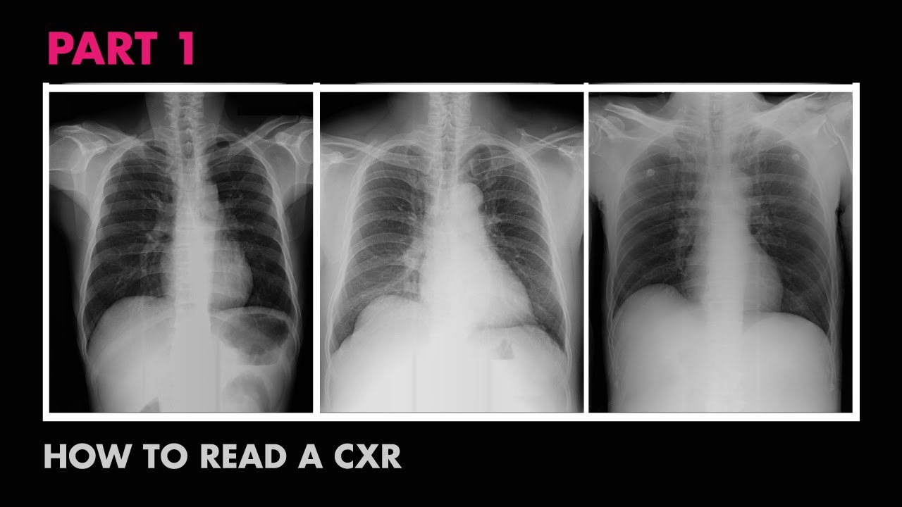
Cardiac Silhouette - How to Read a Chest X-Ray (Part 7) - MEDZCOOL
Summary
TLDRThis educational video script focuses on the 'C' in the ABCDE mnemonic for reading chest X-rays, emphasizing the assessment of the cardiac silhouette. It discusses the normal size limits on PA and AP films, and what an enlarged silhouette might indicate, such as cardiomegaly, pericardial effusions, or left ventricular hypertrophy. The script also touches on heart shape, calcifications, prosthetic valves, and the heart's position in the thoracic cavity, inviting viewers to analyze a final X-ray and share their observations.
Takeaways
- 📚 The ABCDE mnemonic is used for reading chest X-rays, with 'C' standing for 'Cardiac'.
- 🔍 When assessing the cardiac silhouette, consider the size and position of the heart in the thoracic cavity.
- 📏 The cardiac silhouette should not exceed half the size of the patient's thoracic cavity, with specific measurements for PA and AP films.
- 📹 The appearance of the cardiac silhouette can be affected by the type of X-ray view and the patient's breathing phase.
- 💔 An enlarged cardiac silhouette may indicate cardiomegaly, with various underlying causes including pericardial effusions and hypertension.
- 👀 Pay attention to the heart's shape, which can appear more rounded in severe cardiomegaly.
- 🛠 Look for signs of calcifications and the presence of prosthetic valves in the cardiac silhouette.
- 📐 Normally, two-thirds of the heart is positioned to the left of the thoracic cavity on a chest X-ray.
- 🤔 The last X-ray in the video prompts viewers to observe and comment on the cardiac silhouette's characteristics.
- 🔔 Encourages viewers to subscribe for updates and engage with the channel on social media and Patreon for more content.
Q & A
What does the mnemonic 'ABCDE' represent in the context of reading chest X-rays?
-In the context of the script, 'ABCDE' is a mnemonic used for systematically assessing chest X-rays, where 'C' stands for 'cardiac'.
What should the size of the cardiac silhouette be on a PA film according to the video?
-On a PA (posteroanterior) film, the cardiac silhouette should be no larger than half the size of the patient's thoracic cavity, specifically less than 50% of the chest diameter.
What is the acceptable size of the cardiac silhouette on an AP film?
-On an AP (anteroposterior) film, the cardiac silhouette can be less than 60% of the chest diameter, as the heart may appear artificially larger due to the filming technique.
What condition is commonly implied by a heart size larger than the mentioned limits?
-A heart size larger than the normal limits can imply cardiomegaly, which is an enlarged heart.
What are some possible causes of cardiomegaly mentioned in the video?
-Some possible causes of cardiomegaly include pericardial effusions, mediastinal masses, a prominent epicardial fat pad, and left ventricular hypertrophy due to long-standing hypertension.
How can the position of the heart during expiration affect the appearance of the cardiac silhouette on a chest X-ray?
-If a chest X-ray is obtained during expiration, the thoracic diameter would be smaller compared to the heart, making the heart appear relatively larger.
What is the significance of the heart shape in assessing chest X-rays?
-The heart shape can provide clues to certain conditions. For example, in cases of severe cardiomegaly, the heart may appear more rounded.
What are some features to note when assessing the cardiac silhouette for calcifications and prosthetic valves?
-When assessing the cardiac silhouette, one should look for the presence of calcifications, which can indicate conditions like atherosclerosis, and the presence of prosthetic valves, which may be visible as metallic densities.
What is the normal position of the heart in relation to the thoracic cavity on a chest X-ray?
-Normally, two-thirds of the heart lies to the left of the thoracic cavity, and roughly one-third lies to the right on a chest X-ray.
How can viewers engage with the content and support the creators of the video?
-Viewers can engage by subscribing to the channel for updates, following on social media, and supporting the creators through Patreon by making a pledge.
What does the video encourage viewers to do after watching?
-The video encourages viewers to leave their thoughts in the comments section about the last chest X-ray presented, and to subscribe and follow for more content.
Outlines

Dieser Bereich ist nur für Premium-Benutzer verfügbar. Bitte führen Sie ein Upgrade durch, um auf diesen Abschnitt zuzugreifen.
Upgrade durchführenMindmap

Dieser Bereich ist nur für Premium-Benutzer verfügbar. Bitte führen Sie ein Upgrade durch, um auf diesen Abschnitt zuzugreifen.
Upgrade durchführenKeywords

Dieser Bereich ist nur für Premium-Benutzer verfügbar. Bitte führen Sie ein Upgrade durch, um auf diesen Abschnitt zuzugreifen.
Upgrade durchführenHighlights

Dieser Bereich ist nur für Premium-Benutzer verfügbar. Bitte führen Sie ein Upgrade durch, um auf diesen Abschnitt zuzugreifen.
Upgrade durchführenTranscripts

Dieser Bereich ist nur für Premium-Benutzer verfügbar. Bitte führen Sie ein Upgrade durch, um auf diesen Abschnitt zuzugreifen.
Upgrade durchführenWeitere ähnliche Videos ansehen

Radiology of Thorax (Chest)

How to Interpret a Chest X-Ray (Lesson 2 - A Systematic Method and Anatomy)

Diaphragms and Pleural Effusion - How to Read a Chest X-Ray (Part 8) - MEDZCOOL

How to Interpret a Chest X-Ray (Lesson 5 - Cardiac Silhouette and Mediastinum)

ABCs of Reading a Chest X-ray - How to Read a Chest X-Ray (Part 2) - MEDZCOOL
Anatomy of a Chest X-Ray - How to Read a Chest X-Ray (Part 1)
5.0 / 5 (0 votes)
