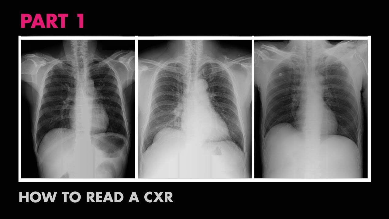
How to Interpret a Chest X-Ray (Lesson 5 - Cardiac Silhouette and Mediastinum)
Summary
TLDRThis educational video, the fifth in a series on interpreting chest X-rays, focuses on analyzing the cardiac silhouette and mediastinum. It outlines how to assess these structures and recognize common abnormalities. The video clarifies the impact of PA and AP films on heart size perception and details specific cardiac conditions like cardiomegaly, left atrial enlargement, and pericardial effusions. It also discusses mediastinal regions, abnormalities like widened mediastinum and hilar enlargement, and uses the hilum overlay sign to differentiate hilar masses. The script provides a comprehensive guide for medical professionals to diagnose conditions affecting the heart and mediastinum.
Takeaways
- ๐ This video is part of a series on interpreting chest x-rays, focusing on the cardiac silhouette and mediastinum.
- ๐ The learning objectives include assessing these structures on x-ray and recognizing common abnormalities.
- ๐ง It's crucial to remember the anatomy of the cardiac silhouette and mediastinum from lesson two.
- ๐ The video is divided into two halves: the first covers heart abnormalities, and the second covers abnormalities of the hila and mediastinum.
- ๐ A key difference between PA and AP films is highlighted, emphasizing that heart size is only accurately assessed on PA films.
- โค๏ธ Cardiomegaly, or an enlarged heart, is a common abnormality and can be quantified using the cardiothoracic ratio.
- ๐ Left atrial enlargement can be identified by splaying of the coronal angle beyond 90 degrees and the double density sign.
- ๐ซ Right ventricular enlargement is indicated by filling of the retrosternal space on lateral views.
- ๐ง Pericardial effusions can present as a large cardiac silhouette and may have a 'water-bottle' shape or 'Oreo cookie' sign.
- ๐ Dextrocardia, a congenital anomaly, is mentioned as a rare condition where the heart is positioned opposite to normal.
- ๐ฅ A widened mediastinum can indicate mediastinal masses, which are categorized into anterior, middle, and posterior regions.
Q & A
What is the main topic of the fifth video in the series on interpreting chest x-rays?
-The main topic of the fifth video is the cardiac silhouette and mediastinum, focusing on how to assess these structures on x-ray and understanding common abnormalities seen in them.
What is the importance of remembering the structures that comprise the cardiac silhouette?
-Remembering the structures of the cardiac silhouette is important for accurately assessing abnormalities on x-rays, as these structures can indicate various cardiac conditions.
How does the divergence of x-ray beams affect the appearance of the heart on a PA film?
-On a PA film, the divergence of x-ray beams does not significantly affect the heart's appearance because the heart is a relatively anterior structure and close to the film, so the shadow it casts is an accurate representation of its true size.
Why is the heart size on an AP film not as accurate as on a PA film?
-The heart size on an AP film is less accurate because the greater separation between the heart and the film causes the diverging x-ray beams to create a larger shadow, exaggerating the size of the heart.
What is the definition of cardiomegaly in the context of chest x-rays?
-Cardiomegaly is defined as the overall size of the heart being larger than normal, commonly assessed using the cardiothoracic ratio, which is the maximum horizontal cardiac width divided by the maximum horizontal thoracic width. Cardiomegaly is present if this ratio exceeds 50% on a PA film.
What is the cardiothoracic ratio and how is it measured?
-The cardiothoracic ratio is the ratio of the maximum horizontal cardiac width to the maximum horizontal thoracic width, measured between the inner surfaces of the ribcage. A ratio exceeding 50% on a PA film indicates cardiomegaly.
What are the two cardiac chambers that can be individually identified as enlarged on an x-ray?
-The two cardiac chambers that can be individually identified as enlarged on an x-ray are the left atrium and the right ventricle.
What is the 'double density sign' and what does it indicate?
-The 'double density sign' is an x-ray finding where the left atrial enlargement causes a second shadow along the right heart border, indicating the left atrium is enlarged.
What is the significance of the 'Oreo cookie sign' in x-ray interpretation?
-The 'Oreo cookie sign' is seen on lateral films and indicates a pericardial effusion. It consists of three layers: a radiolucent posterior layer (pericardial fat), a radiodense middle layer (effusion), and an anterior radiolucent layer (epicardial fat).
What is dextrocardia and how is it depicted on an x-ray?
-Dextrocardia is a congenital anomaly where the heart loops around in the opposite direction during early embryologic development, resulting in the heart being on the right side of the chest instead of the left. On an x-ray, it appears as if the heart is reversed in orientation.
What is the definition of a widened mediastinum on an x-ray?
-A widened mediastinum is generally defined as being greater than 8 centimeters on either a PA or an AP film. However, many cases of an apparently widened mediastinum are due to patient rotation, poor inspiratory effort, or an AP view.
What are the four regions of the mediastinum and what types of masses can be found in each?
-The four regions of the mediastinum are the anterior, superior, middle, and posterior mediastinum. Masses in the anterior and superior mediastinum can include lymphoma, goiter, thymus tumors, and teratomas. Middle mediastinum masses can be lymphadenopathy, aortic aneurysm, pericardial cysts, dilated esophagus, or hiatal hernia. Posterior mediastinum masses typically include neurogenic tumors and spinal mass extensions.
Outlines
๐ Introduction to Cardiac Silhouette and Mediastinum
This paragraph introduces the fifth lesson in a series about interpreting chest X-rays, focusing on the cardiac silhouette and mediastinum. The lesson aims to teach how to assess these structures on X-ray and recognize common abnormalities. It reviews the anatomy of the cardiac silhouette and mediastinum, emphasizing the importance of remembering the structures that make up the silhouette. The video is divided into two halves: the first covers heart abnormalities, and the second covers abnormalities of the hila and the rest of the mediastinum. The paragraph also discusses the difference between PA and AP films, explaining how the diverging X-ray beams in PA films can affect the perceived size of the heart on the image.
๐ Abnormalities of the Cardiac Silhouette
This paragraph delves into specific abnormalities of the cardiac silhouette, starting with cardiomegaly, which is an enlargement of the heart's overall size. The cardiothoracic ratio is introduced as a common way to define cardiomegaly on chest X-rays, with a ratio exceeding 50% on PA films indicating its presence. The ideologies of cardiomegaly are discussed, including left or right-sided heart failure. The paragraph also covers the identification of enlarged cardiac chambers, such as the left atrium and right ventricle, and their respective ideologies. Additionally, it touches on pericardial effusions, their appearances on X-ray, and the various causes of these effusions.
๐งฌ Congenital Anomaly and Mediastinal Abnormalities
The paragraph discusses a rare congenital anomaly called dextrocardia, where the heart loops abnormally during early development, resulting in the heart being on the right side of the chest. It then moves on to abnormalities of the mediastinum and hila, with a focus on a widened mediastinum, generally defined as greater than 8 centimeters. The four regions of the mediastinum are outlined, and the differential diagnosis for masses in these regions is discussed. Examples of masses in different mediastinal regions are provided, including a multinodular goiter and a pericardial cyst. The paragraph concludes with a discussion of the hilum overlay sign, which helps distinguish the location of a mass relative to the hilum.
๐ฅ Hilar Abnormalities and Differential Diagnosis
This paragraph focuses on abnormalities of the hila, such as bilateral hilar enlargement, which can be caused by a variety of conditions including malignancy, infection, and other diseases like sarcoidosis and silicosis. The differential diagnosis for hilar enlargement is broad and can include lung cancer, lymphoma, metastatic disease, tuberculosis, and viral infections, among others. The paragraph provides examples of hilar enlargement in patients with sarcoidosis and pulmonary hypertension. It also discusses the hilum overlay sign to differentiate hilar masses from those anterior or posterior to it, using an example of a saccular aortic aneurysm.
Mindmap
Keywords
๐กCardiac Silhouette
๐กMediastinum
๐กCardiomegaly
๐กCardiothoracic Ratio
๐กLeft Atrial Enlargement
๐กRight Ventricular Enlargement
๐กPericardial Effusion
๐กDextrocardia
๐กMediastinal Masses
๐กHilar Enlargement
๐กHilum Overlay Sign
Highlights
The video discusses interpreting chest x-rays, focusing on the cardiac silhouette and mediastinum.
Learning objectives include assessing these structures on x-ray and recognizing common abnormalities.
Anatomy of the cardiac silhouette and mediastinum is reviewed, emphasizing their segments.
The difference between PA and AP films is highlighted, particularly how it affects the heart's size appearance.
Cardiomegaly, or an enlarged heart, is the most common abnormal finding, defined using the cardiothoracic ratio.
Left atrial enlargement can be identified by splaying of the coronal angle and the double density sign.
Right ventricular enlargement may present as filling of the retrosternal space on lateral views.
Pericardial effusions can cause a large cardiac silhouette and may present with a water-bottle morphology or the Oreo cookie sign.
Dextrocardia, a congenital anomaly where the heart is on the right side, is mentioned as a rare condition.
A widened mediastinum, generally greater than 8 cm, can indicate various abnormalities.
Mediastinal masses are categorized into anterior, superior, middle, and posterior regions, each with different potential causes.
Hilar enlargement can result from malignancies, infections, or other conditions like sarcoidosis or pulmonary hypertension.
The hilum overlay sign helps differentiate hilar masses from those anterior or posterior to it.
An example of using the hilum overlay sign to identify the location of a mass is provided.
The video concludes with an overview of upcoming topics, including the assessment of the diaphragm and pleura.
Transcripts
this is the fifth video in this series
on how to interpret a chest x-ray and
the topic is the cardiac silhouette and
mediastinum learning objectives are to
be able to assess those structures on
x-ray and to know the common ideologies
of abnormalities seen in them let's take
a look at the anatomy of the cardiac
silhouette and mediastinum that was
first introduced in lesson two for this
lesson it will be important to remember
the structures listed here which
comprise the different segments of that
silhouette this video will be divided in
half the first half will cover
abnormalities of the heart the second
will cover abnormalities of the hila and
the remainder of the mediastinum before
I discuss specific findings I need to
also review another very important
difference between PA and AP films that
I haven't talked about yet let's start
with a PA film and here is an axial
cross-section through the thorax at the
level of the heart the patient's front
is up against a photographic plate at
the top in some of the prior videos I've
sort of implied that the x-ray beams
which are responsible for creating the
x-ray image are all parallel to one
another but this isn't actually true the
x-ray source is a fixed point or close
to a fixed point and therefore the x-ray
beams actually diverge outward as they
get further from their origin for a PA
film this makes not much difference
since the heart is a relatively anterior
structure on the chest it is close to
the film where the x-ray is taken and
therefore the shadow it casts on the
x-ray is an accurate representation of
its true size however how does this
change if the same exact patient has an
AP film taken now there is greater
separation between the heart and the
film as a consequence the diverging
x-ray beams will create a larger shadow
that will exaggerate the size of the
heart
in short the heart size is only accurate
when assessed on APA film some
structures within the mediastinum suffer
from the same effect but to lesser
extent as normal mediastinal structures
tend to be more centrally located within
the thorax to see just how much of a
differences can make here are two x-rays
taken up the same patient minutes apart
notice how much larger the heart appears
on the AP film the upper mediastinum is
also affected but not as much I'll now
talk about some specific abnormalities
of the cardiac silhouette the most
common abnormal finding is cardiomegaly
cardiomegaly simply means that the
overall size of the heart is larger than
normal there are a couple of ways it can
be defined in radiology but by far the
most common way to define it for
analysis of chest x-rays uses the
cardiothoracic ratio this ratio is the
maximum horizontal cardiac width divided
by the maximum horizontal thoracic width
as measured between the inner surfaces
of the ribcage cardiomegaly is said to
be present if this ratio exceeds 50% on
the PA film here are two x-rays looking
at the one on the Left first the red
line represents the maximum horizontal
cardiac width and the purple line
represents the maximum horizontal
thoracic width this is a ratio of 40% or
0.4 which is normal looking at the same
on the right film the heart is obviously
much larger this is a ratio of 60% or
0.6 identifying cardiomegaly on x-ray is
really that simple
as far as the ideologies of cardiomegaly
it can essentially be the consequence of
any cause of left or right side of heart
failure it's important to realize that
pericardial effusions which I'll discuss
in a minute can be indistinguishable
from the cardiomegaly that's a
consequence of an enlarged heart while
the finding of cardiomegaly concerns the
heart as a whole there are two cardiac
chambers
that can be identified as being enlarged
on x-ray individually the first is the
left atrium as discussed in the last
lesson one finding of left atrial
enlargement is splaying of the coronal
angle to a value greater than 90 degrees
the second finding is something called
the double density sign usually the
right border of the left atrium is not
visible on x-ray because it is
contiguous with the right atrium and it
lies right in the middle of the chest
where it usually gets obscured by a
number of other structures however as
the left atrium increases in size it
actually will stretch well across the
midline and create a second shadow along
the right heart border this dark red
line here is the right atrium the pink
line here is the left atrium ideologies
of left atrial enlargement include any
cause of left-sided heart failure also
included is mitral valve disease such as
mitral stenosis mitral regurgitation or
mitral valve prolapse for this
particular x-ray there is a clue as to
why this patient has a large left atrium
while the overall size of the heart is
enlarged suggesting the presence of
heart failure assuming this is a PA film
there's another important finding it is
this round structure right here what is
that
it's the ring of a mitral valve
replacement so this patient has had some
form of mitral valve disease that at
least contributed to the enlarged left
atria the second cardiac chamber whose
enlargement can sometimes be
individually spotted on x-ray is the
right ventricle the main finding of this
is filling of the retrosternal space as
seen on the lateral view consider these
two lateral films have included a normal
one for comparison and examine the
retrosternal space that is the one or
two centimeters directly behind the
sternum in the mid chest region in the
normal film this will be relatively
Lucent however as the right ventricle is
the most anterior of the cardiac
chambers as it enlarges it begins to
occupy the space ideologies of right
ventricular enlargement include any
cause of pulmonary
attention and pulmonary valve disease
the second of which is much less common
in the adult population moving on to
pericardial effusions not all clinically
relevant diffusions are visible on x-ray
particularly if they developed acutely
that's because pericardial tamponade
a condition where diastolic cardiac
filling is impaired due to high intra
pericardial pressure is dependent on
both the volume of fluid within the
pericardium as well as the speed with
which the fluid accumulated the primary
finding of an effusion is a large
cardiac silhouette other findings
include what is frequently called the
water-bottle morphology of the
silhouette as well as something called
the Oreo cookie sign here are two
examples of the water-bottle shape of a
very large pericardial effusion you may
be wondering how in the world these look
like water bottles but the name was
given when water bottles weren't made
out of aluminum or rigid plastic but
rather soft sided materials like leather
next the Oreo cookie sign is a little
amusing it's seen on the lateral film
I'm going to zoom in on this part right
here to understand what you're looking
at you'll need to know what an Oreo
cookie looks like I'm sure there are
parts of the world that are not familiar
with this fantastic mass-produced
artificially flavored treat so here's a
picture the important aspect is that it
consists of a layer of white Cream
Sandwich between two dark supposedly
chocolate flavored discs if you look
really carefully you can actually see
the same configuration in front of the
heart just above the diaphragm it's not
normal to have three layers discernible
here so what's responsible for each the
posterior chocolate layer which is
relatively radiolucent is the
pericardial fat the middle cream layer
which is relatively radio dense is a
pericardial effusion and the anterior
chocolate layer is the epicardial fat
this sign exists because fluid absorbs
ever so slightly more x-rays than fat
does there are many ideologies of
pericardial effusions which can be
divided into those who
cause acute effusions and those which
cause sub acute to chronic effusions
acute effusions can be from trauma viral
pericarditis a complication from
myocardial infarctions such as a
catastrophic free wall rupture or a post
mi inflammatory process called Dressler
syndrome where it can be iatrogenic from
a right ventricular biopsy or from any
one of a number of EP procedures sub
acute and chronic infusions are seen in
malignancies such as lymphoma breast and
lung but can also be due to renal
failure collagen vascular disease like
lupus and rheumatoid arthritis
hypothyroidism and finally tuberculosis
which is the major cause of pericardial
effusions in some parts of the world
before moving on to the mediastinum in
hila i want to briefly mention a
congenital anomaly which although it's
relatively rare tends to be mentioned
during rounds or on tests out of
proportion to its prevalence at first
you might assume this x-ray was loaded
into the system backwards if it wasn't
for the capital R in the upper corner
indicating that in fact the x-ray is in
the correct orientation
this person has dextrocardia which
occurs when the heart loops around in
the opposite direction as normal during
early embryo logic development its
incidence is about 1 in 12,000
pregnancies it may be isolated and
discovered incidentally or it can be
associated with life-threatening
additional congenital malformations now
I'll move on to abnormalities of the
mediastinum and hila the most important
of these abnormalities is a widened
mediastinum this is generally defined as
greater than 8 centimeters on either a
PA or an AP film however most cases of
an apparently widened mediastinum are
due to rotation of the patient poor
inspiratory effort or an AP view to best
understand mediastinal masses which are
responsible for truly widened
mediastinum
one must be familiar with the four
regions of the mediastinum these regions
are not precisely defined by any actual
tissue planes
and their definition varies slightly
between sources but this is
approximately where the divisions live
the first two regions are the anterior
mediastinum which is everything anterior
to the pericardium and inferior to the
sternal angle and superior mediastinum
which is everything superior to the
sternal angle the differential diagnosis
of masses in these two regions overlap
greatly so I'll consider them together
they include lymphoma enlarged thyroid
such as the goiter thymus tumors tumors
called teratomas and when occurring in
just the superior mediastinum an aortic
aneurysm the middle mediastinum includes
everything within the pericardium and is
variably defined to also include the sub
kernel para tracheal and hilar lymph
nodes and the esophagus the differential
diagnosis of masses here include lymph
adenopathy from any cause and a arctic
aneurysm pericardial cysts dilated
esophagus or a hiatal hernia finally the
posterior mediastinum includes
everything behind those structures which
ends up being largely just a
paravertebral space thus the
differential diagnosis of mass is
located here include neurogenic tumors
and extension of spinal masses such as
tumors and infections
I'll show you one or two examples from
each region beginning in the superior
mediastinum we see that there is a mass
here that is actually causing rightward
shift of the trachea although you could
not know it just from this x-ray this
particular patient has a multinodular
goiter this patient has an even larger
mass in the superior mediastinum this
one is from lymphoma anterior
mediastinum masses can easily be missed
especially on p.a films since their
shadow can get lost in the cardiac
silhouette this is a pretty unusual case
in which an anterior mass was not only
visible it was so large it resembled the
water-bottle morphology of a large
pericardial effusion the hint to the
correct diagnosis is from the lateral
film what do you notice here there's an
obliteration of the retrosternal space
while this could be from concurrent
right ventricular hypertrophy as we saw
a few minutes ago in this case it's from
a massive thigh mo lipoma that is
literally wrapped around the anterior
surface of the heart here we see an
unusual bump coming off the left upper
heart border even if I didn't tell you
that this was a middle mediastinal mass
you could still tell that from the lack
of a clear boundary between a normal
cardiac silhouette and the mass here's
the corresponding lateral film to
confirm its placement in the middle
mediastinum its appearance is consistent
with a pericardial cyst which could be
confirmed with a chest CT or
echocardiogram finally here is a
posterior mass you can tell it's not
middle from the PA film because there is
still a very well-defined left heart
border the lateral film confirms this
mass is arising from the posterior
mediastinum in this case biopsy
demonstrated a schwannoma here's an
interesting collection of mediastinal
masses which are critical to accurately
identify these are all a or t'k
aneurysms the one in the middle is
particularly easy to identify given the
calcified walls since only vascular
structures or cysts tend to do that I'll
move on from the mediastinum to talk
about the hila here are two examples of
bilateral hilar enlargement in this case
both patients had stage 1 sarcoidosis
the differential diagnosis of hilar
enlargement is very long and can be
divided into three categories first
malignancy which includes primary lung
cancer lymphoma and metastatic disease
the second category is infection
particularly tuberculosis and viruses
but a whole host of rarer diseases are
included here as well depending on where
in the world you are practicing and
watching this video there may be many
more diseases on your list than listed
here finally in the other category are
sarcoidosis silicosis pulmonary
hypertension a pulmonary artery aneurysm
and a bronchogenic cyst here's another
example of hilar enlargement in this
case the patient had severe pulmonary
hypertension in this case there is
unilateral right-sided enlargement
unfortunately it's associated with this
opacity in the right upper lung that
looks suspicious for a primary tumor
overall the most likely diagnosis here
is a primary lung cancer with a hilar
node metastasis I'll end this lesson by
talking about how one can use the hilum
overlay sign to distinguish a hilar mass
from one anterior or posterior to it
consider this film which has an obvious
mass of some kind in the vicinity of the
left hilum when a mass arises from the
hilum the adjacent pulmonary vessels
will become obscured
if the pulmonary vessels are still
visible through the mass the mass is not
in the hilum if we zoom in on the mass
like this you can still see the outline
of a left pulmonary artery and if we
check the lateral film we see that
indeed the mass is posterior to the
hilum to diagnose the cause of the mass
look here there is a very thin rim of
peripheral calcification that means
either a vascular structure or assist in
this case it was a saccular a or t'k
aneurysm that concludes this video on
the cardiac silhouette and mediastinum
the next video will cover assessment of
the diaphragm and pleura
you
Browse More Related Video
Cardiac Silhouette - How to Read a Chest X-Ray (Part 7) - MEDZCOOL
Assessment of CXR Positioning & Views - How to Read a Chest X-Ray (Part 4) - MEDZCOOL
Radiology of Thorax (Chest)
Chest X-ray: Introduction and Approach
How to Interpret a Chest X-Ray (Lesson 2 - A Systematic Method and Anatomy)
How to Interpret a Chest X-Ray (Lesson 1 - An Introduction)
5.0 / 5 (0 votes)