Radiology of Thorax (Chest)
Summary
TLDRIn this educational video, the presenter delves into the radiological anatomy of the thorax, emphasizing the importance of reading chest X-rays. They explain the standard posterior-anterior (PA) view, detailing its advantages over the anterior-posterior (AP) view for accurate cardiac assessment. The script outlines key structures to identify, including the trachea, lungs, and heart, and discusses the significance of the costophrenic angle in detecting pleural effusions. It also covers the identification of bones, the heart's silhouette, and potential deformities, providing a comprehensive guide to understanding chest X-rays.
Takeaways
- 📚 The video discusses the radiological anatomy of the thorax, focusing on how to read chest X-rays.
- 🎥 It suggests watching a previous video for understanding the radiological anatomy of the upper limb.
- 🔍 The X-ray type discussed is a plain film, requiring no special positioning or radio-opaque dye.
- 🏥 The standard view for chest X-rays is the posterior-anterior (PA) view, which is preferred for accurate cardiac size assessment.
- 📏 The PA view minimizes magnification, showing the heart's true size, unlike the anterior-posterior (AP) view which can overstate it.
- 🌟 The video introduces the 'abcd' method for remembering structures to identify: Airway (trachea), Breathing (lungs), Bones, and Circulation (heart).
- 🌈 The color effect in X-rays depends on tissue absorption of the X-ray beam, with less absorption appearing darker, as seen in the lungs.
- 💨 The trachea is identified as a midline dark shadow in the upper part of the mediastinum.
- 🫁 The lungs are identified by their air content, which appears dark, and the cardiac notch helps differentiate the left lung from the right.
- 🦴 Ribs, clavicle, sternum, and cervical vertebrae are among the bones visible in a chest X-ray.
- 🧡 The heart's shadow in the mediastinum helps identify the right and left borders, and any enlargement may indicate cardiomegaly.
- 🔑 The costophrenic angle is clinically important for diagnosing pleural effusion, as fluid accumulation can obliterate this angle.
Q & A
What is the main topic of the video 'Viva Wars of Anatomy'?
-The main topic of the video is the radiological anatomy of the thorax, focusing on how to read chest X-rays.
What is the first step in analyzing a chest X-ray according to the video?
-The first step is to describe the type of radiological film, which in this case is a plain film because no special position is required and no radio-opaque dye is used.
What is the standard view for a chest X-ray and why is it preferred over the AP view?
-The standard view for a chest X-ray is the posterior-anterior (PA) view. It is preferred over the AP view because it allows for accurate assessment of the heart size due to minimum magnification as the heart lies closer to the X-ray plate at an optimum distance.
What does the acronym 'ABCD' stand for in the context of describing the structures seen in a chest X-ray?
-In the context of the chest X-ray, 'ABCD' stands for Airway (trachea), Breathing (lungs), Circulation (heart), and Deformity (any bone fractures or soft tissue abnormalities).
Why does the trachea appear as a midline dark shadow in the upper part of the mediastinum on a chest X-ray?
-The trachea appears as a midline dark shadow because it contains air, which absorbs the least amount of the X-ray beam, resulting in a darker appearance on the radiograph.
How can you differentiate between the left and right lungs on a chest X-ray?
-The left and right lungs can be differentiated by the presence of the cardiac notch on the left lung, which is a feature that helps identify the left lung from the right lung.
What is the clinical importance of the costophrenic angle on a chest X-ray?
-The costophrenic angle is clinically important because it contains the costophrenic recess, which is the most dependent part of the lung. In cases of pleural effusion, fluid accumulates in this area, and the angle may appear as a white shadow or obliterated on the X-ray.
What does the term 'cardiomegaly' refer to and how can it be diagnosed from a chest X-ray?
-Cardiomegaly refers to an enlarged heart. It can be diagnosed from a chest X-ray if the maximum horizontal diameter of the heart is more than half the width of the thoracic cavity.
What structures can be seen in the lower part of the mediastinum on a chest X-ray?
-In the lower part of the mediastinum on a chest X-ray, you can see the heart shadow, including the right and left borders of the heart, and the aortic arch, which is known as the aortic knuckle.
How can you identify the presence of a pleural effusion on a chest X-ray?
-A pleural effusion can be identified on a chest X-ray by the obliteration or whitening of the costophrenic angle, which indicates the accumulation of fluid in the pleural cavity.
What is the significance of the aortic knuckle in a chest X-ray and why is it more prominent in older individuals?
-The aortic knuckle, or the arch of the aorta, is a convex shadow seen in the left border of the heart on a chest X-ray. It is more prominent in older individuals due to the calcification of the aortic wall, which makes it more visible on the X-ray.
Outlines

This section is available to paid users only. Please upgrade to access this part.
Upgrade NowMindmap

This section is available to paid users only. Please upgrade to access this part.
Upgrade NowKeywords

This section is available to paid users only. Please upgrade to access this part.
Upgrade NowHighlights

This section is available to paid users only. Please upgrade to access this part.
Upgrade NowTranscripts

This section is available to paid users only. Please upgrade to access this part.
Upgrade NowBrowse More Related Video

How to Interpret a Chest X-Ray (Lesson 2 - A Systematic Method and Anatomy)

Chest X-ray: Introduction and Approach
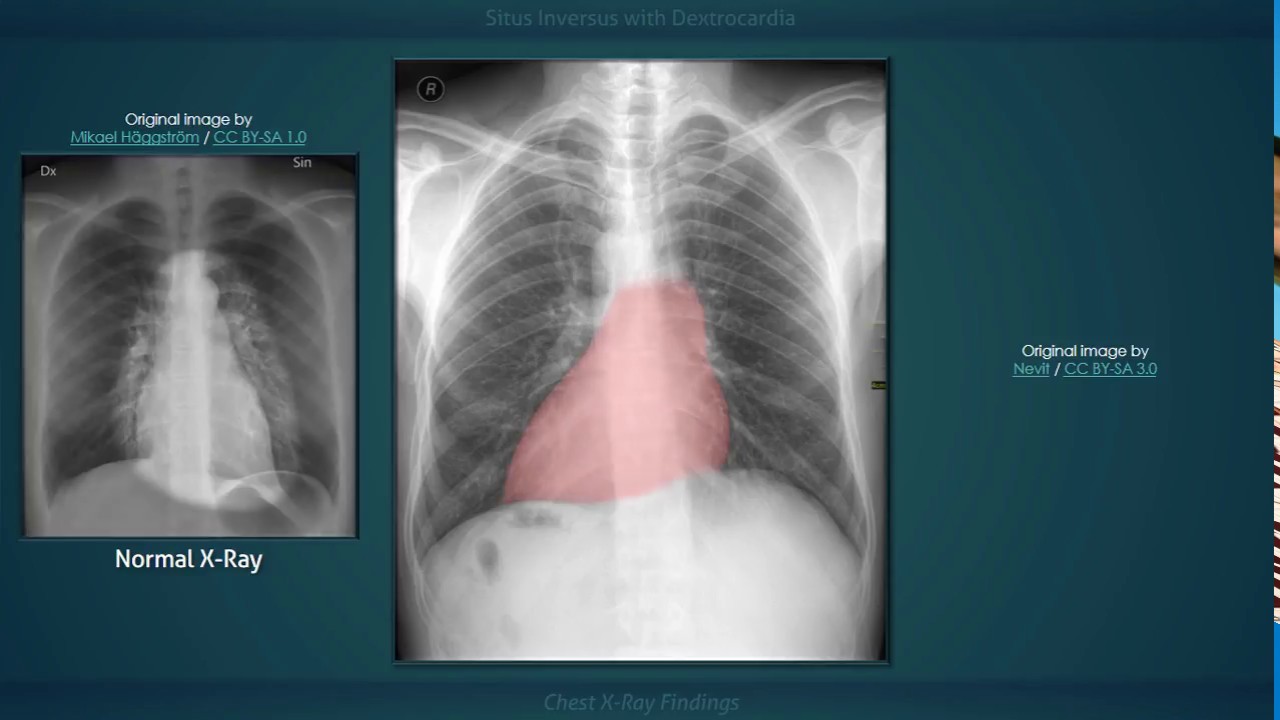
Situs Inversus with Dextrocardia: Explanation of Chest X-ray Findings
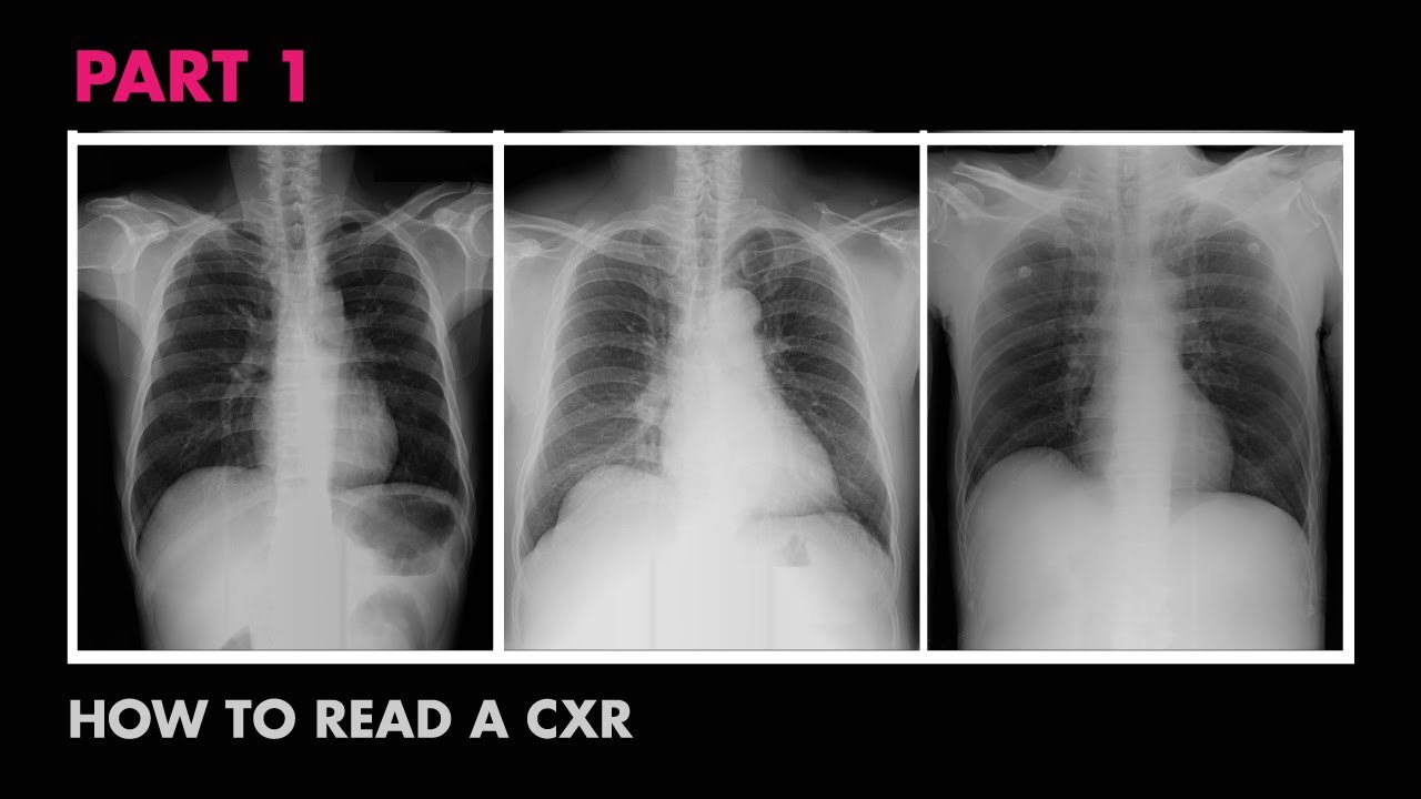
Anatomy of a Chest X-Ray - How to Read a Chest X-Ray (Part 1)

Cardiac Silhouette - How to Read a Chest X-Ray (Part 7) - MEDZCOOL

Grasshopper Dissection
5.0 / 5 (0 votes)