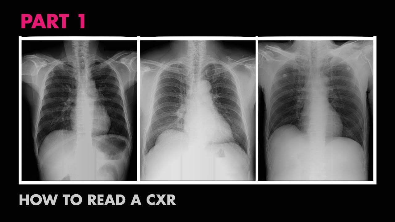
Reading a chest X-ray
Summary
TLDRThis video explains how to analyze a chest X-ray using a simple checklist based on the first seven letters of the alphabet. The checklist helps ensure accurate diagnosis by focusing on key aspects like patient data, image quality, bones, heart size, diaphragm appearance, equipment placement, lung fields, and great vessels. Key considerations include checking for abnormalities like fractures, masses, and fluid collections, as well as identifying issues like air where it shouldn’t be. This systematic approach helps clinicians detect critical pathologies such as pneumothorax, pleural effusion, and cardiac enlargement.
Takeaways
- 😀 X-rays are high-energy photons that penetrate body tissues to help visualize internal structures, similar to how visible light works.
- 😀 Dense materials like bones block more photons, which results in the white areas seen on an X-ray, while areas like the lungs appear darker.
- 😀 The ABCDEFG checklist helps systematically assess chest X-rays, with each letter standing for a critical element in evaluation.
- 😀 'A' stands for assessment of the patient and exam data, ensuring you are reviewing the right study and that the image quality is appropriate for diagnosis.
- 😀 'B' is for bones and the body wall. It's crucial to check clavicles, ribs, and the soft tissues for any fractures, deformities, or masses.
- 😀 'C' refers to the cardiac silhouette and size, ensuring the heart's dimensions are normal—less than 50% of the ribcage diameter.
- 😀 'D' stands for diaphragms, which should be symmetric and not overly flat. This is assessed in the lateral view of the chest X-ray.
- 😀 'E' is for equipment, including life support devices. Proper placement and function of tubes, wires, and lines should be verified.
- 😀 'F' covers lung fields, which should appear symmetric with no haziness or blotches. A frontal and lateral X-ray helps identify abnormalities in specific lung lobes.
- 😀 'G' focuses on the great vessels (superior vena cava, inferior vena cava, aorta, etc.), ensuring they are correctly positioned and of normal size.
- 😀 Proper X-ray technique, such as ensuring the patient is in the right position, the lungs are fully expanded, and exposure is correct, is essential for accurate diagnostics.
Q & A
What is the purpose of X-rays in medical imaging?
-X-rays are used to capture images of the inside of the body by using high-energy photons. These photons penetrate tissues, allowing us to see structures like bones and lungs. X-rays are useful for diagnosing conditions by revealing issues that are otherwise hidden inside the body.
Why are bones visible as white areas on a chest X-ray?
-Bones appear white on a chest X-ray because they are denser than other body tissues. X-rays have difficulty passing through dense materials, so bones block most of the photons, creating bright white spots on the image.
What does the ABCDEFG checklist help with in chest X-rays?
-The ABCDEFG checklist is a mnemonic to help radiologists systematically analyze chest X-rays. Each letter stands for an important step or structure to assess, from assessing patient data and image quality (A) to checking great vessels (G).
What does 'A' in the ABCDEFG checklist represent?
-A stands for 'Assessment,' which involves verifying the patient’s data and ensuring the image quality is adequate. This includes checking for rotation, ensuring full lung expansion, and verifying the exposure levels to detect any issues with the X-ray.
How can you assess the quality of a chest X-ray?
-To assess chest X-ray quality, check for proper rotation, good lung inspiration (e.g., at least the 10th or 11th posterior ribs visible), and proper exposure levels. Fine markings in the lung fields should be visible to ensure accurate diagnosis.
What are examples of 'air where it shouldn't be' that can be detected in a chest X-ray?
-Examples include pneumothorax (air in the pleural space), pneumomediastinum (air in the mediastinum), pneumoperitoneum (air in the abdomen), and subcutaneous emphysema (air in the soft tissues). These conditions are surgical emergencies and can be identified through chest X-rays.
What does 'B' in the ABCDEFG checklist stand for?
-B stands for 'Bones,' which involves checking the clavicles, ribs, and body wall for fractures, deformities, or missing bones. It also includes assessing the soft tissues outside the chest for swelling or masses.
How do you assess the size of the heart in a chest X-ray?
-To assess the heart size, measure the cardiac silhouette on a chest X-ray. The heart should be less than 50% of the greatest diameter of the ribcage. A larger heart may indicate cardiac pathology.
What does 'D' in the ABCDEFG checklist stand for?
-D stands for 'Diaphragms,' referring to the appearance of the diaphragms in the X-ray. They should be symmetric, and the hemidiaphragm should be about 1.5 cm above the line connecting the costophrenic and sterno-phrenic angles.
What role does 'E' in the ABCDEFG checklist serve?
-E stands for 'Equipment,' which refers to checking medical devices like tubes, wires, and life support equipment on the chest X-ray. It's essential to ensure that these devices are properly positioned and functional, such as confirming that the endotracheal tube is centered and the tip is appropriately placed.
What is a pleural effusion, and how can it be detected on a chest X-ray?
-A pleural effusion is a condition where fluid accumulates in the pleural cavity, causing blunting of the normally sharp costophrenic angle on a chest X-ray. It’s a common but subtle pathology that can be detected with careful image analysis.
What does 'G' in the ABCDEFG checklist stand for?
-G stands for 'Great vessels,' which includes major blood vessels like the superior vena cava, inferior vena cava, aortic arch, and pulmonary arteries. It's important to ensure that these structures are in the right location and size on the chest X-ray to rule out any abnormalities.
Outlines

هذا القسم متوفر فقط للمشتركين. يرجى الترقية للوصول إلى هذه الميزة.
قم بالترقية الآنMindmap

هذا القسم متوفر فقط للمشتركين. يرجى الترقية للوصول إلى هذه الميزة.
قم بالترقية الآنKeywords

هذا القسم متوفر فقط للمشتركين. يرجى الترقية للوصول إلى هذه الميزة.
قم بالترقية الآنHighlights

هذا القسم متوفر فقط للمشتركين. يرجى الترقية للوصول إلى هذه الميزة.
قم بالترقية الآنTranscripts

هذا القسم متوفر فقط للمشتركين. يرجى الترقية للوصول إلى هذه الميزة.
قم بالترقية الآنتصفح المزيد من مقاطع الفيديو ذات الصلة

ABCs of Reading a Chest X-ray - How to Read a Chest X-Ray (Part 2) - MEDZCOOL
Anatomy of a Chest X-Ray - How to Read a Chest X-Ray (Part 1)

How I Read a Lateral CXR

VAS101_Topic015
UAD - Kuliah Online 1475530 Karakterisasi Material Lanjut (Lecture 1b)

How to Interpret a Chest X-Ray (Lesson 1 - An Introduction)
5.0 / 5 (0 votes)
