How I Read a Lateral CXR
Summary
TLDRIn this video, Rishi Agrawal provides a concise guide on how to interpret a lateral chest X-ray, covering essential landmarks and key structures. He explains the significance of areas like the hila, retrosternal clear space, and retrocardiac space. Agrawal also emphasizes the importance of identifying pulmonary arteries, heart borders, and diaphragm characteristics. He walks through the process of detecting abnormalities such as pleural effusion, vascular anomalies, and organ enlargement, while also discussing how to identify bone fractures. The video serves as a practical resource for those looking to understand the fundamentals of lateral chest X-ray interpretation.
Takeaways
- 😀 The lateral chest X-ray is useful for localizing lesions, even though most abnormalities are seen on the frontal view.
- 😀 The left upper lobe bronchus is an important landmark, visible as a lucency on the lateral X-ray, helping to identify nearby structures.
- 😀 The right and left pulmonary arteries can be identified in the lateral view, with the right pulmonary artery positioned at 9:00 and the left at 10:00-3:00.
- 😀 The infrahilar window beneath the left upper lobe bronchus should be lucent; opacity in this area indicates lymphadenopathy.
- 😀 There are three key clear spaces on a lateral chest X-ray: retrosternal (behind the sternum), retrotracheal (behind the trachea), and retrocardiac (behind the heart).
- 😀 Abnormalities in the retrosternal space could suggest airspace disease or an anterior mediastinal mass.
- 😀 Vascular anomalies or issues with the esophagus may appear in the retrotracheal space, which lies behind the trachea.
- 😀 The retrocardiac space is where lower lobe consolidation, masses, and hiatal hernias can be seen.
- 😀 Right ventricular hypertrophy is indicated when the right ventricle extends significantly into the retrosternal space on the lateral view.
- 😀 The lateral view is not ideal for evaluating the ascending aorta or pulmonary arteries, as they overlap, but it is useful for detecting abnormalities in the descending aorta.
Q & A
What is the primary purpose of analyzing a lateral chest X-ray?
-The primary purpose is to localize lesions, as abnormalities are usually more obvious in the frontal view, but sometimes lateral views can reveal lesions not visible on the frontal X-ray.
What anatomical structures are used as landmarks in the lateral chest X-ray?
-The left upper lobe bronchus, right pulmonary artery, and left pulmonary artery serve as important landmarks in the lateral chest X-ray.
Why is the left upper lobe bronchus important in a lateral chest X-ray?
-The left upper lobe bronchus serves as a key landmark for identifying surrounding structures such as the right pulmonary artery and the left pulmonary artery.
What is the significance of the infrahilar window in a lateral chest X-ray?
-The infrahilar window is typically lucent, and any opacity in this area can indicate hilar or mediastinal lymphadenopathy.
What are the three clear spaces identified in a lateral chest X-ray?
-The three clear spaces are the retrosternal clear space, retrotracheal clear space (or Raider's triangle), and retrocardiac clear space.
What abnormalities might be indicated by changes in the retrosternal clear space?
-Abnormalities in the retrosternal clear space can indicate airspace disease in the upper lobes or an anterior mediastinal mass.
What is the significance of the retrotracheal clear space on a lateral chest X-ray?
-The retrotracheal clear space can reveal vascular anomalies, such as an aberrant right subclavian artery, or abnormalities with the esophagus.
How does left ventricular hypertrophy affect the lateral chest X-ray?
-In left ventricular hypertrophy, the left ventricle pushes into the retrocardiac clear space, causing it to become smaller.
Why is it difficult to evaluate the pulmonary artery and ascending aorta on a lateral chest X-ray?
-The pulmonary artery and ascending aorta are partially superimposed on each other in a lateral chest X-ray, making it challenging to evaluate them accurately.
What role does the diaphragm play in the lateral chest X-ray evaluation?
-The diaphragm is assessed for abnormalities, and the costophrenic angles are important for detecting pleural effusions, which are more easily seen on a lateral view than a frontal one.
Outlines

This section is available to paid users only. Please upgrade to access this part.
Upgrade NowMindmap

This section is available to paid users only. Please upgrade to access this part.
Upgrade NowKeywords

This section is available to paid users only. Please upgrade to access this part.
Upgrade NowHighlights

This section is available to paid users only. Please upgrade to access this part.
Upgrade NowTranscripts

This section is available to paid users only. Please upgrade to access this part.
Upgrade NowBrowse More Related Video

CARA MEMBACA RONTGEN PARU / THORAX ‼️ HAPAL SAMPAI KAPANPUN 😱😱

Wheelchair Chest

How To Read A Chest X-ray

Assessment of CXR Positioning & Views - How to Read a Chest X-Ray (Part 4) - MEDZCOOL
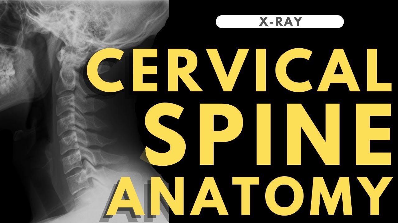
Cervical spine anatomy | Radiology anatomy part 1 prep | C-spine X-ray interpretation
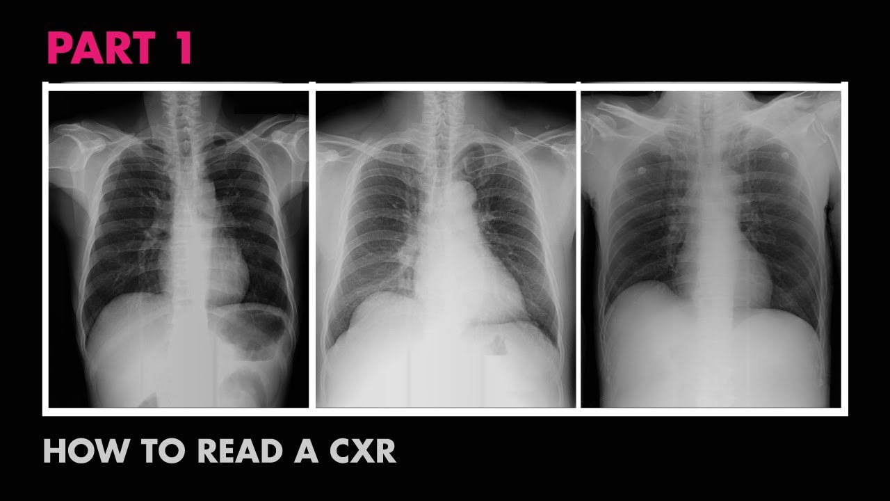
Anatomy of a Chest X-Ray - How to Read a Chest X-Ray (Part 1)
5.0 / 5 (0 votes)