Skeletal system
Summary
TLDRThis module delves into the intricacies of the human muscular skeletal system, emphasizing its role in providing structural support, protecting vital organs, and facilitating movement. It outlines the composition of the system, including bones, cartilage, ligaments, and tendons, and categorizes bones based on their shape and function. The module also explores different types of joints, highlighting their movement capabilities and the unique roles of connective tissues like tendons and ligaments in transmitting muscle force and providing joint stability. Understanding these components is crucial for movement analysis and overall human body functionality.
Takeaways
- 🏃♂️ The human body is a complex machine with the muscular skeletal system playing a key role in movement and structural integrity.
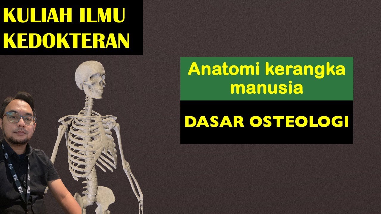
- 🦴 The skeletal system is composed of 206 bones, categorized into axial and appendicular parts for easier understanding.
- 💀 The axial skeleton includes the skull, rib cage, and vertebral column, providing form, function, and protection for vital organs.
- 🤸♀️ The appendicular skeleton consists of the limbs and girdles, facilitating movement, stability, and interaction with the environment.
- 🦵 Long bones like the femur and humerus support body weight and enable movement, while short and flat bones provide stability and protection.
- 🔗 Tendons and ligaments are crucial connective tissues; tendons attach muscle to bone for force transmission, and ligaments connect bones to provide joint stability.
- 🛡️ Cartilage acts as a cushion in joints, allowing for smooth movement and reducing friction, whereas fascia surrounds muscles and provides support and protection.
- 🔄 Joints are classified by their movement capability, ranging from immovable (synarthrosis) to freely movable (diarthrosis or synovial joints).
- 🔻 Pivot, saddle, hinge, and ball-and-socket joints are examples of different types, each allowing for specific ranges and directions of motion.
- 🏋️♂️ Understanding the skeletal system and joint types is fundamental for human movement science, aiding in the analysis and improvement of movement patterns.
- 🩹 Injuries to the skeletal system and connective tissues, such as tendonitis, sprains, and fascia tightness, can result from overuse, improper technique, or trauma.
Q & A
What is the primary function of the human skeletal system?
-The primary function of the human skeletal system is to provide structural integrity, protect vital organs, and assist in movement.
How many bones are there in the human body, and what are the main components of the skeletal system?
-There are 206 bones in the human body. The skeletal system is composed of bones, cartilage, ligaments, and tendons.
What are the two main divisions of the human skeleton?
-The two main divisions of the human skeleton are the axial skeleton and the appendicular skeleton.
What are the key structures of the axial skeleton and what are their roles?
-The key structures of the axial skeleton include the skull, rib cage, and vertebral column. The skull protects the brain and sensory organs, the rib cage provides protection for the lungs and heart, and the vertebral column supports the spinal cord.
What is the role of the appendicular skeleton in the body?
-The appendicular skeleton facilitates movement, stability, and interaction within the environment. It includes the upper and lower limbs, which attach to the girdles and connect to the axial skeleton.
What are the different types of bones based on their shape, and what are their functions?
-Bones are categorized into long bones, which support body weight and enable movement; short bones, which provide stability and support; flat bones, which protect organs and provide muscle attachment surfaces; irregular bones, which have complex shapes and include the vertebrae; and sesamoid bones, which reduce friction and protect tendons.
How are joints classified based on their movement capability?
-Joints are classified as immovable (synarthrosis), slightly movable (amphiarthrosis), and freely movable (diarthrosis or synovial joints) based on their movement capability.
What is the role of tendons in the skeletal system?
-Tendons are connective tissues that attach muscle to bone. They transmit the force generated by the muscle to the bones, which is crucial for efficient movement and stability.
What is the function of ligaments in the body?
-Ligaments connect bones to other bones and provide stability to the joints. They help maintain natural alignment and prevent excessive and abnormal joint movement.
What is the main function of cartilage in the skeletal system?
-Cartilage is a tough, flexible connective tissue that cushions the joints, providing structural support and allowing for smooth movement between bones.
What is fascia, and how does it contribute to the skeletal system?
-Fascia is a connective tissue that surrounds muscles, blood vessels, and nerves. It provides support and protection to these structures and helps compartmentalize different muscle groups.
Outlines
💪 Introduction to the Human Body and Musculoskeletal System
This paragraph introduces the human body as a complex machine, focusing on the musculoskeletal system. It emphasizes the importance of understanding the mechanics of movement and the role of the muscular and skeletal systems in providing structural integrity, protecting vital organs, and enabling movement. The paragraph outlines the structure of the upcoming modules, which will delve into the anatomy of human joints and the functions of the muscular skeletal system, including the axial and appendicular skeletons.
🦴 Composition and Functions of the Musculoskeletal System
The paragraph discusses the composition of the musculoskeletal system, highlighting the 206 bones in the human body, the role of cartilage, ligaments, and tendons, and the division of the skeleton into axial and appendicular parts. It explains the functions of the axial skeleton, including the skull, rib cage, and vertebral column, and touches on the importance of understanding the structure and function of each part for movement analysis. The appendicular skeleton's role in facilitating movement, stability, and environmental interaction is also described.
🦵 Types and Functions of Bones
This section categorizes bones into five types based on their shape and function: long bones (e.g., femur and humerus), short bones (e.g., wrist and ankle bones), flat bones (e.g., skull and sternum), irregular bones (e.g., vertebrae), and sesamoid bones (e.g., patella). It explains how these bones contribute to body weight support, movement, stability, and muscle attachment. The dynamic nature of bones, their constant remodeling, and the importance of nutrition and exercise for bone health are also discussed.
🔗 Understanding Joints and Their Classification
The paragraph explains what joints are and how they are classified based on their movement capability. It introduces different types of joints, including immovable (synarthrosis), freely movable (diarthrosis or synovial joints), and limited mobility (amphiarthrosis) joints. The role of joints in providing stability, support, and allowing for movement is emphasized, with examples such as the sutures of the skull, the shoulder and hip joints, and the joints between vertebrae.
🔄 Types of Joint Movements and Their Functions
This section delves into the specifics of joint movements, starting with the pivot joint found in the neck that allows for rotational movement around a central axis. It then describes the saddle joint, which due to its shape, allows for a wide range of motion and fine movements, such as those of the thumb. The plain joint, characterized by flat and smooth surfaces, is designed for stability and a wide range of motion. Hinge joints, like the elbow and knee, allow for movement in one direction. The condyloid joint, with its oval and ball-head shapes, provides stability and versatility for movements like waving. Lastly, the ball and socket joint, the most mobile joint in the body, is exemplified by the hip and shoulder joints, offering stability and a wide range of movements in multiple directions.
🤕 Connective Tissues: Tendons, Ligaments, Cartilage, and Fascia
The paragraph discusses the crucial role of connective tissues in the musculoskeletal system. Tendons, which attach muscles to bones, transmit the force generated by muscles for efficient movement. Ligaments connect bones to each other, providing joint stability. Cartilage cushions joints, allowing for smooth movement between bones. Fascia surrounds muscles, blood vessels, and nerves, offering support and protection. Injuries to these tissues, such as tendonitis, ligament sprains or tears, and fascia tightness, can result from overuse, improper technique, or trauma and can lead to discomfort or reduced mobility.
📝 Recap of the Human Skeletal System and Connective Tissues
The final paragraph recaps the main points discussed in the module. It summarizes the human skeletal system's role in providing structural integrity, protecting vital organs, and assisting in movements. It reiterates the composition of the system, including bones, cartilage, ligaments, and tendons, and how bones are classified based on their shape. The paragraph also reviews the different types of joints based on movement capability and the unique roles of connective tissues in supporting and enabling movement.
Mindmap
Keywords
💡Musculoskeletal System
💡Axial Skeleton
💡Appendicular Skeleton
💡Joints
💡Tendons
💡Ligaments
💡Cartilage
💡Fascia
💡Bone Remolding
💡Skeletal Structures
Highlights
The human body is a complex machine with different systems working in harmony.
The muscular skeletal system is integral for movement, structural integrity, and protection of vital organs.
The human skeleton is made up of 206 bones, divided into the axial and appendicular skeleton.
The axial skeleton provides structural support and protects vital organs, including the brain, heart, and lungs.
The appendicular skeleton facilitates movement, stability, and interaction with the environment.
The pectoral girdle (shoulder girdle) connects the upper limbs to the axial skeleton, providing mobility and flexibility.
The pelvic girdle supports body weight and provides stability, attaching the lower limbs to the axial skeleton.
Bones are dynamic connective tissues that undergo constant remodeling through resorption and formation.
Bones are categorized into long, short, flat, and irregular bones based on their shape and function.
Joints are connections between bones that allow for movement, classified by their movement capability.
Synarthroses are immovable joints, like the sutures of the skull, providing stability and support.
Diarthroses, or synovial joints, are freely movable joints that are the most common type in the human body.
Amphiarthroses have limited mobility and provide stability while allowing for slight movement.
Tendons are connective tissues that attach muscles to bones, transmitting the force generated by muscles for movement.
Ligaments connect bones to bones, providing stability to joints and maintaining natural alignment.
Cartilage is a tough, flexible connective tissue that cushions joints and allows for smooth movement.
Fascia is a connective tissue that surrounds muscles, providing support and protection, and compartmentalizing muscle groups.
Injuries to the skeletal system, such as tendonitis, sprains, and tears, can result from overuse, trauma, or improper technique.
Understanding the skeletal system and its components is crucial for human movement science and overall well-being.
Transcripts
[Music]
hello and welcome to module 2 the human
body we've previously discussed an
introduction into human movement science
we've understood what the field entails
we've also looked at a couple of
interviews of experts in the field field
and how they utilize U movement analysis
or the study of human movement science
in their day-to-day professional lives
so before we understand the mechanics of
the movement which will follow in the uh
next few modules let's understand the
human body the human body is a very
fascinating complex machine as you all
know it consists of different systems in
the body but for first and importantly
let's look at the muscular skill
skeletal system so in this module we'll
be looking at the human body
specifically the muscular skeletal
system functions of the muscular
skeletal
system skeletal structures so the
important structures that are there
within the muscular skeletal system that
allow for movement that are designed for
movement we'll also be looking at the
anatomy of human joints so how exactly
do we move where do these bones connect
right so so the muscular skeletal system
it's an intricate network of of bones
and muscles you also have skeletal
structures of tendons ligaments and
joints that collectively support the
body's structure they enable movement
and they also protect vital organs so
it's very important that the muscles and
Bones Work in integration a harmonious
integration to provide structural
integrity and also to allow for movement
which is key for overall functional and
well-being of the human body so the
skeletal system system is made out of
206 bones in our body the human skeletal
system provides structural support as we
learned earlier it also helps us protect
vital organs and assist in movement so
it's composed of Bones cartilage
ligaments and tendons so all of these
are
different connective tissues that we
will be
studying as we progress in this module
and the quite
key um to know they're quite key to
understand movement so this system is
divided into axial and appendicular
skeleton so just for the for our purpose
of understanding and for easier
understanding per se it's divided into
axial and appendicular skeleton so let's
look at this uh nomenclature and the
division so the axial skeleton plays a
fundamental Ro role in maintaining the
body's Form and Function so it's very
important we
know what its role is and what does it
consist of so so the axial skeleton
consist of the
skull it consists of the rib cage that
protects your lungs and your heart so
give structural support and then also
our vertebral column that supports the
spinal cord
inside
so in the vertebral
column we
have cervical
vertebrae we
have thoracic
vertebrae we have
lumbar
vertebrae
and sacrum
or
sacral
vertebrae so just um a note that
although we are covering the human
muscular skeletal system uh for human
movement science purposes of course you
don't dwell too much into the detail of
what these uh vertebrae look like um
it's key to understand what it's each
function is how they are designed um to
know what movement is allowed at each of
these vertebrae and for you to know more
we do have other modules as well that
dwell into anatomy and physiology so I
would highly recommend to get into the
details but for this module we'll be
sticking to um brief details of the
axial skeleton its function and what
does it consist
of um specifically let's look at the
skull so what does the skull do the
skull protects the brain and the sensory
organs within it
so let's do
[Music]
brain and it
protects so it provides stability there
as well then we have the vertebral
column that supports and encases the
spinal cord as we spoke about earlier
and the rib cage that protects the
thoracic organs like heart and the
lungs the appendicular skeleton
facilitates movement stability and the
interaction within the environment so
this term appendicular here
means
parts
that
append
or
attach so what that means
is you have the upper Limbs and lower
limbs of the body that attach to the
girdles right let's look at these
girdles in details in the next slide and
that connect them to the axial skeleton
so what are these girdles that we we are
talking
about so you have the pelvic girdle
sorry you have the pelvic girdle and you
have the shoulder girdle so these upper
limbs what do they do their function
it's enable intricate movement including
reaching grasp manipulating
objects um are some of the essential
movements of the upper limbs so the
lower limbs on the other hand support
body weight balance and also facilitate
Locomotion or
movement so let's look at these girdles
so we have something called as the
pectoral girdle or the shoulder girdle
so it connects these this Upper Limb to
the connects the upper limbs to the
axial skeleton so what does it consist
of it consist of this shoulder
blade so this shoulder blade called as
scapula and then you have
your collar
bone that is
your
clavicle
right so that's your pectoral girdle or
shoulder girdle so its key function is
to provide mobility and flexibility to
the arms that it is attaching to the
actual
skeleton so the pelvic
girdle it attaches the lower limbs to
the axial
skeleton so that's
the pelvic
gurle
so its function is to support body
weight and provide stability so as we
know that the hip bones take most of the
weight of the body and the pelvic girdle
uh that it attaches to and it provides
support and
stability so let's look at some of the
skeletal structures in the body
now so one of the key or the main
skeletal structure is the bone so what
is the bone and as we spoke about
earlier it's a connective tissue that
provides structural
support protects the internal organs and
it also serves as an attachment site for
the muscles so very important to know
this is it serves as an attachment site
for the
muscles so bones they're quite dynamic
they undergo constant remodeling you
know through bone reabsorb absorption
there's bone formation so whenever you
have an injury and you know uh you're
building a new bone or when you have a
fracture and the bone is broken down it
has the capacity of healing so they are
quite Dynamic and they undergo constant
remodeling through the processes of bone
resorption and Bone formation so bone
health is quite crucial for structural
Integrity the overall Integrity of the
human
body and it requires good diet or good
nutrition good balance of exercise
hormonal regulation quite key for bone
health so how are these bones
categorized into you know it's very
important to know them we have 206 of
them so let's look at how these bones
are categorized and based on the
categorization what's their
function so the first category is long
bones so they called long because they
are longer than they are wide so imagine
the femur bone in the body so that's
your
femur which is your thigh
bone you can also look at
humorus which is that
bone so your upper arm bone so these are
longer so they're longer
then they
are
wider so what is its function they
support body weight and they enable
movement the next category of
Bones is short
bones so as opposed to the long bones
they're nearly equal in length and width
so as you can see it's pretty much equal
in lead and width so you can look at our
wrist B bones or our ankle bones that
are equal in length and width so these
are short bones and they provide
stability and support also in addition
they they are also attachment sides for
your ankle and wrist
muscles flat Bones on the other hand
which is our third category they are
thin and they are quite flattened right
so example the skull bones
quite
thin
flattened right also your
scapula
sorry another example of a flat
bone and your
sternum so your sternum is where your
rib attach right so they as you can see
here they protect the organs and provide
surface for muscle attachments so all of
your rib muscles which are called as
inter coal muscles so they attach here
your shoulder muscles or rotator cuff
attached to the scapula and then of
course the skull protects the
brain inside
it our fourth category is something
called as irregular bones so we've
looked at long short and flat but
something of an irregular bone is they
don't have a particular shape so they
are they are complexed shaped so for
example vertebra as you can
see um they're neither long neither
short neither flat it's an irregular
bone and what they do is they form the
vertebral column so they form this
vertebral column and they provide
support and protection to the spinal
cord as we've looked at earlier so based
on just an additional information for
you based on where these are
located so based on
location the shape of these bones shape
of
these
ver they change
right so let's look at our fifth
category which is the sesamoid bones so
sesamoid bones are they quite small and
they usually embedded within the tendons
so let's let's look at tendons in the
next few slides but for you to remember
they're small and they're embedded in
the tendons so for example your kneecap
that's that's the best example so that's
your
kneecap and you have the patella bone
that is embeded in the
kneecap so what is the function of the
seso bone they reduce friction and
protect tendons from excessive vear and
tear so all of your ACL injuries PCL
injuries very very very important to
keep a healthy knee joint that is
protecting the patella bone and the tiia
and the femur that is attached to
it so let's look at type of joints now
that we know the different type of Bones
and how they're categorized based on
shapes let's look at type of joints so
what is what is a joint a joint is a
connection between two or more
bones so this is particularly to allow
movement so for us to understand
movement where it is occuring how does
it occur it is very important for us to
understand the type of
joints so these type of joints they are
classified by movement capability so
let's look at the first
one so sin arthrosis is an immovable
joint so sin here
means held together firmly coming
together firmly and arth is means it's a
medical
term it's a
medical
term
for
joints just makes
the understanding and remembrance of
this word easier so your sin
means
together for
firmly and arthrosis means joints so sin
arthrosis is an immovable joint so
example the sutures of the skull so they
don't really move so it's a joint where
as we said bones are held together
firmly and do not allow for movement so
their role is to provide stability and
support to the structures they connect
so we've we've looked at it enough now
that the skull protects the brain and
the structures within it and provides
stability as well to your head so let's
look at
diarthrosis which is our second type of
joint so
D is either
two
or
both and arthrosis as we know is a joint
so these are freely movable
joints for example elbow shoulder and
hip right so this in this joint two
articulating surfaces so two surfaces
that are attaching together are within a
joint
cavity that allow for a wide range of
movement right they're also known as
synovial joints so synovial joints is
because of this liquid which is
a
sovial fluid
that is
important
for shock
absorption so important
for shock
absorption right and they are the most
common type of joints in the human
body so they're characterized as we said
by a joint capsule so there's usually a
capsule the
fluid and an articular
cartilage
right so we'll we'll see more about
cartilages and tendons in the following
few slides but you all you have to
remember is they are categorized by a
joint capsule sovial fluid and articular
C cartilage that allow for smooth and
flexible movement right it's very
important that synovial joints as they
are the most common type of joints in
the human body so another type of joints
is amphi arthrosis amphia
means
both which means that it has limited
Mobility so it does not have much
mobility and it also has Mobility which
is why it's called amphy arthrosis as we
looked at is joints so examp example the
adjoining vertebraes so they have
limited mobility within them right so
joint which allows for a slight or
limited movement on both
sides so it's important so imagine
yourself U side bending or doing a slide
flexion so you have these adjoining
vertebras that are providing this
movement on both
sides so they provide stability and
support while all owing for limited
movement as we looked
at so now that we looked at the
different categorization of the joints
let's look at how are these named and
based on their type or the shape of
these joints how exactly the function
happens So based on the nature and the
type of the joint movement availability
varies how the movement availability
varies and what movement m is available
at each joint we will be looking at that
in module three for now let's look at
the type of joints so the first type of
joint is a pivot joint right so that's a
first type of joint so what do you mean
by the word pivot so this joint got its
name for categorization from uh
something that pivots so which has a
rotational movement and it has around a
central axis so around that axis you are
having this rotational movement so
example of that is a neck joint so here
at your cervical vertebra so again for
your more understanding at C1 C2
vertebra you have the neck joint so what
does it allow for it provides necessary
range of motion which is nothing but
movement while providing stability and
support so that's your pivot
joint the next type of joint is saddle
joint so as this word suggest
saddle the joint is shaped like a saddle
so as you can look
here it has concave and convex surface
so this is
your concave and convex surfaces so
where are these present they are present
in the carpo metac car
joint of the thumb so it's present in
the carpo metac carple joint of the
thumb now based on its shape it allows
for wide range of motion so we have wide
range of motion at the thumb and its
function is to look at fine movements so
the fine movements that we do with the
thumb like writing you know like holding
grasping all of those movements it also
provides for a unique motion which is
you know the ability for you to touch
your thumb and your pinky finger or all
your all your fingers right so it allows
for Unique motion because of its shape
so that's the saddle
joint the next type of joint is the
plain joint so plain joint here as the
name suggests is characterized by flat
and relatively smooth surfaces so
imagine these flat and relatively smooth
surfaces that
can
slide and
glide so your two
surfaces that can slide and glide over
each other
over
right that's the plane joint so what are
they designed for they designed for
again stability most of the joints of
course are designed for stability and
they're also quite versatile as they
provide wide range of motion so these
are for example your inasal joint here
right so the next type of joint let's
look at is hinge joint so as the name
suggest here hinge again imagine like a
mechanical hinge on the
door right which which allows for
um movement in One Direction so again
like the previous joint we have a convex
and a concave interlock same as the
hinge so you have
the convex and the concave
interlock
interlock
like
door
like the door
hinge right so it primary allows for One
Direction movement same like the door so
how the door it opens and
closes and provides stability as we know
all joints do and control for specific
movements so in this case here imagine
bending your arm and straightening it
what is this motion called we will look
at it in the next coming modules so that
is at
the
elbow
joint and another example is
the
knee knee
joint both of them are hinged
joints now let's look at cond deloid
joint now this condid word here comes
from one of the sides of the joint being
being shaped like an oval which is also
called an
ellipsoid and the other one has a ball
head so one side has the ball
head the other one has
a
ellipsoidal um you can say
cavity or a depression where it
attaches
right so its function is again to
provide St stability and versatility for
wide range of
movements and the example here is a
wrist joint right so your radiocarpal
joint you have a lot of movements at the
wrist again detail of these uh movements
we look at it in the coming
modules and also your knuckle joints as
well so let's again this joint provides
unique actions like waving your hand
fingers in
multi-direction
right now let's look at the last type of
joint which is your ball and socket
joint so again here you have a concave
socket that's your concave socket and
then you have the bone with the ball
head right so that when these two attach
you get a ball
then
socket
joint
right so these allow for wide range of
movements in multiple directions and
because of that they are the most mobile
joints in the body so example of this is
your hip joint and then also
your shoulder joint
right so your hip joint that's the
hip joint and
your shoulder joint right so they
provide stability Mobility flexibility
and vers they're quite
versatile as they provide wide range of
movements
now that we've looked at bones type of
Bones let's look at some of the
structures that we mentioned earlier and
then we came across earlier which are
tendons ligaments cartilage and fascia
so as you can see here in the image
tendons cartilage ligaments and fascia
are quite crucial connective tissues so
they all have a very distinct role that
they play but commonly they support and
enable movement and provide structural
Integrity similar to
the as the bone
does so tendons let's look at them first
they are connective tissues that attach
muscle to the Bone so they either wrap
around bones or they pass over joints so
this here is a
tendon it's attaching the muscle to the
Bone
so what is their function they transmit
the force that is generated by the
muscle so the force then gets
transmitted by the muscle to the
bones and hence they are quite important
for efficient movement and
stability right so injuries such as
tendonitis which is an inflamed tendon
or any kind of tear here so any kind of
tear here uh may result from either
overuse very common if we don't uh pay
attention to how our workouts and
everything are designed if you do it if
you overwork out if there's any trauma
improper Technique we will talk about
this in module 6 improper technique
during physical activities or sport so
it's quite important to maintain good
tendon health so that the force that is
transmitted by the muscles can reach the
bone that allows for efficient
movement the next uh skeletal structure
we looking at is a ligament let's not
get too overwhelmed with this um picture
of the knee we just have couple of
things that we spoke about earlier we
have the femur bone which is
your thigh
bone and then this is your tiia this is
p
as we spoke about earlier which is a
seso bone and then we have this as your
tendon like we looked at in the previous
slide so it's attaching the quadriceps
muscle to the patella so it's called a
quadriceps
tendon so this is the articular
cartilage that we talk about let's look
at it in the next slide and the ligament
here it connects bones to Bones right so
as you can see it in the picture it
connects bones to bones and provides
stability to the Joint so for example
this one here which is your anterior
cruciate ligament which is nothing but
your ACL and your posterior cruciate
liament which is nothing but your PCL
two very important ligaments of the knee
we will be looking at how to manage um
injuries reduce injuries with related to
these two um ligaments in sport so for
now a ligament is a skeletal structure
that connects bones to bones to provide
stability another ligament here which is
got
your patella tendon ligament that is
connecting this small bone of patella to
the tibia so it's very important um that
your
ligaments help your or knee to maintain
a natural alignment and prevent
excessive and abnormal joint movement so
injuries to the ligaments like sprains
or tears are generally a result of
sudden twisting motion or excessive
force on the joint so in sport like
soccer and basketball ACL injuries are
quite common we'll be looking at the
injury mechanics in the following
modules but for you to remember that
sprains and tears of the ligament are uh
due to the sudden twisting motion or
excessive force on the knee
joint another skeletal uh structure that
we finally come to is the cartilage so
cartilage is a tough it's a very tough
flexible connective tissue and its main
function is to cushion the joints so it
provides structural support and also
allows for smooth movement between the
bones so for example if you have a look
at here
you have your articular cartilage so
this cartilage here for example it's
very
important that it cushions the joint so
it cushions the joints and also allows
for the movement so damage to the
cartilage such as an
osteoarthritis which is a clinical
condition can lead to join pain and
decreased
Mobility
another important skeletal uh structure
is what we know as fascia so fascia is a
connective tissue that surrounds muscles
so we've looked at muscle to bone bone
to bone another important connective
tissue is the cartilage that we looked
at in the earlier slides but fascia here
is a connective tissue that surrounds
muscles or group of muscles right or
blood vessels and nerves so what it does
is it provides support and protection to
these structures so as we can see
here that is
a
fascia right so it's providing it's
surrounding this
muscle so it also helps
compartmentalized different muscle
groups so imagine the muscle groups in
your calf muscles fascia helps comp
departmentalize these different muscle
groups and it provides pathway for blood
vessels and nerves to
travel through the body so it's an
important connective
tissue so injuries to the fascia can
occur when it becomes extremely tight
and it also develops trigger points so
as you must have um you know going to
your
um physio with tight trigger points or
fascia being tight it can lead to
discomfort or pain in the muscles and
the tissue so that's a minor injury to
the
fascia so in this entire module let's
just recap and look at the summary so
what did we learn about we learned about
the human skeletal system its major role
which is to provide structural Integrity
protect vital organs and also assist in
movements what is it composed of it's
composed of your bones skeletal
structures like cartilage ligaments and
tendons a bone as we looked at is a
specialized connective tissue and they
are classified based on their shape when
two bones come together to form a
connection uh to allow for movement it
is known as a joint and these are
categorized based on movement capability
we also looked at tendons cartilage
ligaments and fascia which are crucial
connective tissues that each have a very
unique role that help in providing
structural Integrity again support and
also allowing for
[Music]
movement
استعرض المزيد من الفيديوهات ذات الصلة
![](https://i.ytimg.com/vi/z-lbu2p2tmI/hq720.jpg)
Chapter 1 Mechanical principles (force and tension) | Kinesiology | Physio Class
![](https://i.ytimg.com/vi/hxvep2Y8ShI/hq720.jpg)
Neurology | Basal Ganglia Anatomy & Function | Direct & Indirect Pathways
![](https://i.ytimg.com/vi/R9XCsQSoATE/hq720.jpg)
Connective Tissue Fibers Histology [Connective Tissue Histology Part 1 of 3]
![](https://i.ytimg.com/vi/N6oly45yK6A/hq720.jpg)
BIOLOGI Kelas 11 - Sistem Ekskresi Manusia | GIA Academy
![](https://i.ytimg.com/vi/SrbQlSZr50c/hq720.jpg?v=608336fd)
Le Cellule Gliali | NEUROSCIENZE - Lezione 3
![](https://i.ytimg.com/vi/-SeKcGX-c7I/hq720.jpg)
ACIDI NUCLEICI e NUCLEOTIDI
5.0 / 5 (0 votes)