EKG 2
Summary
TLDRThis presentation explains the components and interpretation of a normal electrocardiogram (EKG). It covers the P wave, QRS complex, T wave, and other important intervals such as the PR, QT, and ST segments, which are critical in diagnosing cardiac rhythms and abnormalities. The video also explains the importance of EKG in identifying arrhythmias, myocardial ischemia, electrolyte imbalances, and other cardiac conditions. The use of 12 leads and their placement on the body is detailed to understand heart activity in different areas. EKG is a vital diagnostic tool for assessing heart health and ensuring timely interventions.
Takeaways
- 😀 EKG (Electrocardiogram) records the electrical activity of the heart and is essential for diagnosing various cardiac conditions.
- 😀 The key components of a normal EKG include the P wave, QRS complex, T wave, PR interval, and QT interval, each representing different phases of heart activity.
- 😀 The P wave represents atrial depolarization, the QRS complex represents ventricular depolarization, and the T wave represents ventricular repolarization.
- 😀 The PR interval is the time between the onset of the P wave and the QRS complex, and the QT interval represents the total duration of ventricular depolarization and repolarization.
- 😀 The normal duration of key waves and intervals includes: P wave (0.06-0.10 seconds), PR interval (0.12-0.20 seconds), QRS complex (0.06-0.10 seconds), and QT interval (0.36-0.44 seconds).
- 😀 Abnormalities in wave duration can indicate arrhythmias or other heart issues, such as ischemia or hypertrophy.
- 😀 EKG is also used to identify conditions like myocardial ischemia, infarction, pericarditis, and electrolyte imbalances (e.g., hyperkalemia, hypokalemia).
- 😀 There are 12 leads in an EKG: 3 bipolar limb leads (I, II, III), 3 augmented voltage limb leads (aVR, aVL, aVF), and 6 unipolar chest leads (V1-V6).
- 😀 Each lead in an EKG corresponds to specific regions of the heart, with leads like V1-V6 representing the ventricles and limb leads representing different heart areas.
- 😀 EKG is critical for detecting abnormalities in heart rate (bradycardia and tachycardia) and rhythm, as well as changes in the morphology of waves, such as ST segment elevation or depression.
Q & A
What are the main components of a normal electrocardiogram (ECG)?
-The main components of a normal ECG include the P wave (atrial depolarization), QRS complex (ventricular depolarization), T wave (ventricular repolarization), PR interval, QT interval, and sometimes a U wave.
Why is atrial repolarization not visible on an ECG?
-Atrial repolarization is not visible on an ECG because it is buried within the larger electrical activity of the QRS complex, which represents ventricular depolarization.
What does the PR interval represent, and what is its normal duration?
-The PR interval represents the time between the onset of atrial depolarization (P wave) and the onset of ventricular depolarization (QRS complex). Its normal duration is between 0.12 and 0.20 seconds.
What is the significance of the QT interval on an ECG?
-The QT interval represents the total duration of ventricular depolarization and repolarization. A prolonged QT interval can indicate various cardiac conditions, such as an increased risk of arrhythmias.
How do electrolyte imbalances affect the ECG?
-Electrolyte imbalances, such as hyperkalemia (high potassium) or hypokalemia (low potassium), can alter the morphology of the ECG, affecting the appearance and duration of waves and intervals, potentially leading to arrhythmias.
What are some of the diagnostic uses of an ECG?
-An ECG is used to diagnose arrhythmias, myocardial ischemia, myocardial infarction, pericarditis, chamber hypertrophy, and to assess electrolyte imbalances or drug-induced abnormalities like prolonged QT interval.
What is the role of the U wave in an ECG?
-The U wave, when present, represents repolarization of the papillary muscles or can be associated with conditions like old myocardial infarction. Its presence is not always observed and may indicate pathological conditions.
What is the significance of lead placement in ECG recordings?
-Lead placement in an ECG determines which part of the heart's electrical activity is recorded. For example, the limb leads provide a general view of the heart’s electrical activity, while chest leads focus on specific areas like the right ventricle or left ventricle.
How can ECG help in identifying heart rate abnormalities?
-ECG helps identify heart rate abnormalities by measuring the duration of the waves and intervals. A rate slower than 60 beats per minute is called bradycardia, while a rate faster than 100 beats per minute is called tachycardia.
What are the implications of abnormal ST segment findings on an ECG?
-Abnormal ST segment findings, such as ST depression or ST elevation, can indicate myocardial ischemia or infarction, which are critical conditions requiring immediate attention to prevent permanent cardiac damage.
Outlines

此内容仅限付费用户访问。 请升级后访问。
立即升级Mindmap

此内容仅限付费用户访问。 请升级后访问。
立即升级Keywords

此内容仅限付费用户访问。 请升级后访问。
立即升级Highlights

此内容仅限付费用户访问。 请升级后访问。
立即升级Transcripts

此内容仅限付费用户访问。 请升级后访问。
立即升级浏览更多相关视频
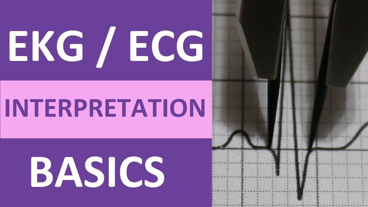
EKG/ECG Interpretation Basics Nursing NCLEX | QRS Complex, P Wave, T Wave, PR Interval

Pemeriksaan Elektrokardiografi (EKG)

Performing an ECG on a Teenage Girl

Dasar-Dasar EKG (Prinsip EKG) : #1 ELEKTROKARDIOGRAM
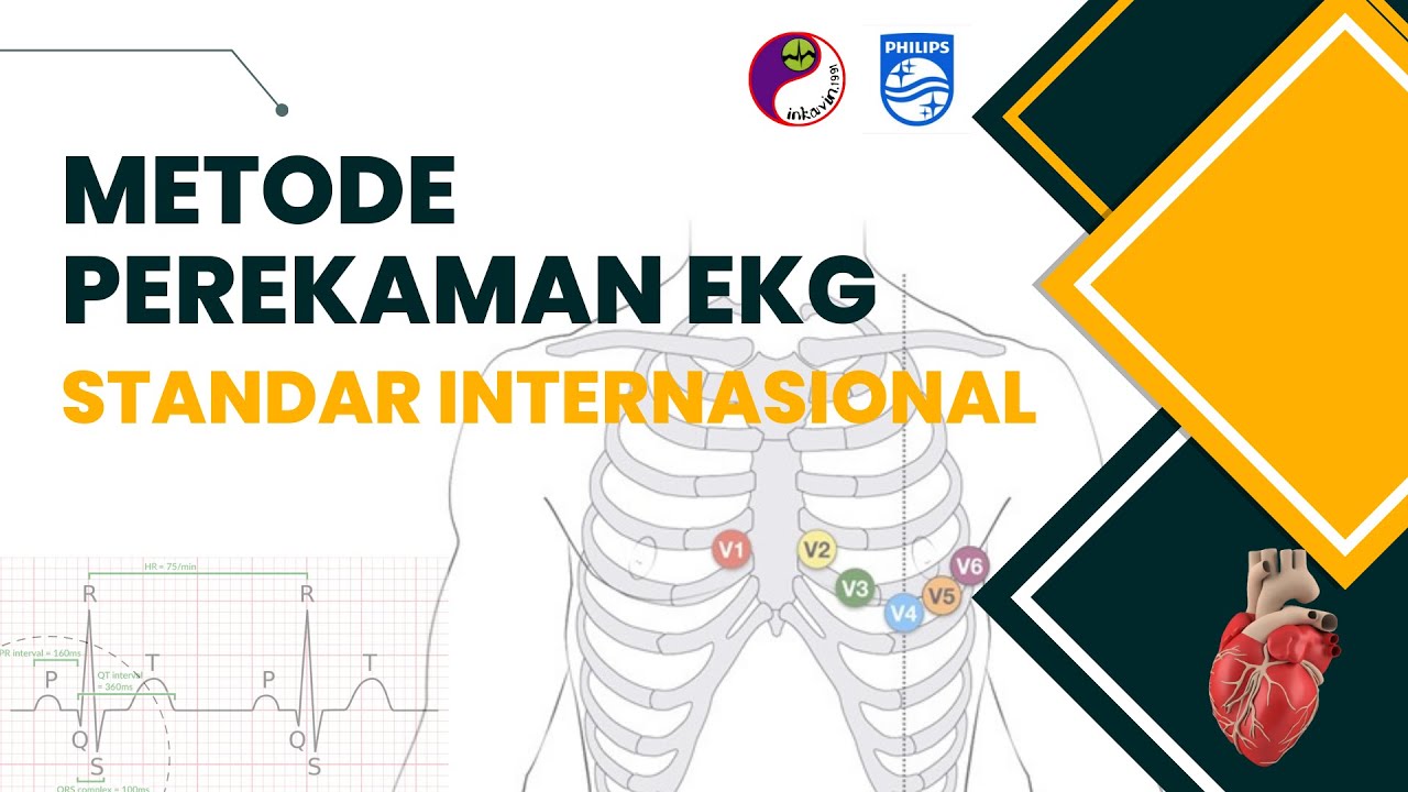
Metode Perekaman EKG Standar Internasional | ECG Recording Technique

Guyton and Hall Medical Physiology (Chapter 11) REVIEW The Normal Electrocardiogram || Study This!
5.0 / 5 (0 votes)
