Structural organization of skeletal muscle | Biomechanics of human skeletal muscle
Summary
TLDRThis video script explains the structure and function of skeletal muscles in the human body. It covers the organization of muscle fibers, their types, and their architectural features, such as myofibrils and proteins like actin and myosin. The process of muscle contraction is discussed, focusing on how the sarcomere and bands like A-band, I-band, and M-line contribute to muscle movement. The script also introduces key terms, such as endomysium and perimysium, which surround muscle fibers and bundles. Additionally, it touches on how the number of muscle fibers is determined genetically and the impact of injuries on muscle structure.
Takeaways
- 😀 The human body contains a total of 434 skeletal muscles, which are present in pairs, i.e., one on the right side and one on the left side.
- 😀 Skeletal muscles are responsible for controlling posture, movement of limbs, and other functions such as swallowing and eye movements.
- 😀 Muscle fibers are the building blocks of muscles and are elongated, thread-like cells that contain nuclei and cytoplasm, also known as sarcoplasm.
- 😀 The sarcoplasm within muscle fibers contains myofibrils, which are responsible for muscle contraction and movement.
- 😀 There are two main types of proteins in muscle fibers: actin and myosin. Their interaction helps with the contraction of muscles.
- 😀 Skeletal muscles are also referred to as striated muscles due to their striped appearance, which is visible under a microscope.
- 😀 Muscle fibers are surrounded by connective tissue called endomysium, and multiple fibers together form a fascicle, which is wrapped in perimysium.
- 😀 A group of fascicles together forms a whole muscle, which is encased in a connective tissue called epimysium.
- 😀 The muscle fibers are structured with bands like I-bands (isotropic) and A-bands (anisotropic), which help in muscle contraction and relaxation.
- 😀 The interaction of actin and myosin filaments causes muscle contraction by sliding over each other, which shortens the muscle fiber and reduces its length.
- 😀 Muscle fiber characteristics, such as the number of fibers and their diameter, are genetically determined and remain mostly constant throughout life, except in cases of injury.
Q & A
What is the total number of skeletal muscles in the human body?
-The human body contains a total of 434 skeletal muscles, which are present in pairs on the left and right sides of the body.
What are the main functions of skeletal muscles?
-Skeletal muscles are primarily responsible for controlling posture, movement of limbs, swallowing, and eye movements.
Why are skeletal muscle fibers referred to as fibers?
-Skeletal muscle fibers are called 'fibers' because they are thread-like in structure, resembling threads.
What is the structure of a muscle fiber?
-A muscle fiber is a single cell that contains a nucleus, cytoplasm (called sarcoplasm), and mitochondria, similar to other cells in the body.
What is myofibril and why is it important?
-Myofibrils are specialized protein structures within muscle fibers, composed of proteins that play a crucial role in muscle contraction.
What are the two types of proteins present in muscle fibers?
-The two types of proteins present in muscle fibers are actin and myosin, which are responsible for the contraction mechanism.
Why are skeletal muscles also called 'striated muscles'?
-Skeletal muscles are called 'striated muscles' because they have a striped appearance under a microscope due to the arrangement of actin and myosin filaments.
What is the role of the I-band and A-band in muscle contraction?
-The I-band (isotropic) is where actin filaments are present, while the A-band (anisotropic) contains myosin filaments. These bands shift during muscle contraction, leading to shortening of the muscle fiber.
What is the M-line and why is it important?
-The M-line is located at the center of the sarcomere and serves as an anchor for myosin filaments. It plays a critical role in maintaining the structural integrity of the muscle during contraction.
What is the function of the transverse tubules (T-tubules) in muscle fibers?
-T-tubules are channels that help in the conduction of electrical signals, allowing the flow of ions and action potentials to activate muscle contractions.
Outlines

此内容仅限付费用户访问。 请升级后访问。
立即升级Mindmap

此内容仅限付费用户访问。 请升级后访问。
立即升级Keywords

此内容仅限付费用户访问。 请升级后访问。
立即升级Highlights

此内容仅限付费用户访问。 请升级后访问。
立即升级Transcripts

此内容仅限付费用户访问。 请升级后访问。
立即升级浏览更多相关视频

Contração muscular - dublado

Sistema Muscular 2ª parte
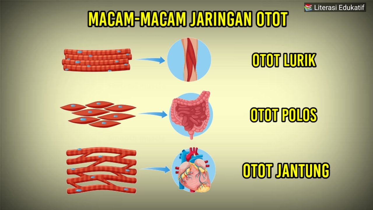
APA BEDANYA OTOT LURIK OTOT POLOS DAN OTOT JANTUNG?

Cellule musculaire : organisation - SVT - SANTÉ Term spé #7 - Mathrix
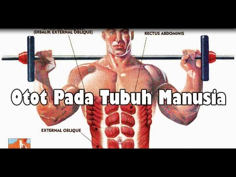
Otot Pada Tubuh Manusia

Types of Tissue Part 3: Muscle Tissue
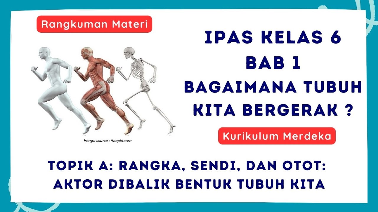
Rangkuman IPAS KELAS 6 BAB 1: Bagaimana Tubuh Kita Bergerak?. Topik A: Rangka, sendi, dan otot
5.0 / 5 (0 votes)
