PET vs SPECT | The basics (Updated video)
Summary
TLDRThis video explains the key differences between PET and SPECT scans, two major molecular imaging techniques used in nuclear medicine. While both detect biochemical changes, PET scans use positron-emitting radioisotopes like fluorine-18 to identify cancer cells and assess metabolic activity, detecting abnormalities months earlier than CT scans. SPECT scans, on the other hand, use gamma-emitting radioisotopes like technetium. The video highlights how both techniques involve radioactive isotopes that function as tracers, allowing doctors to track and visualize organ function, commonly combined with CT scans for both anatomical and metabolic information.
Takeaways
- 😀 PET and SPECT are the two main molecular imaging modalities in nuclear medicine.
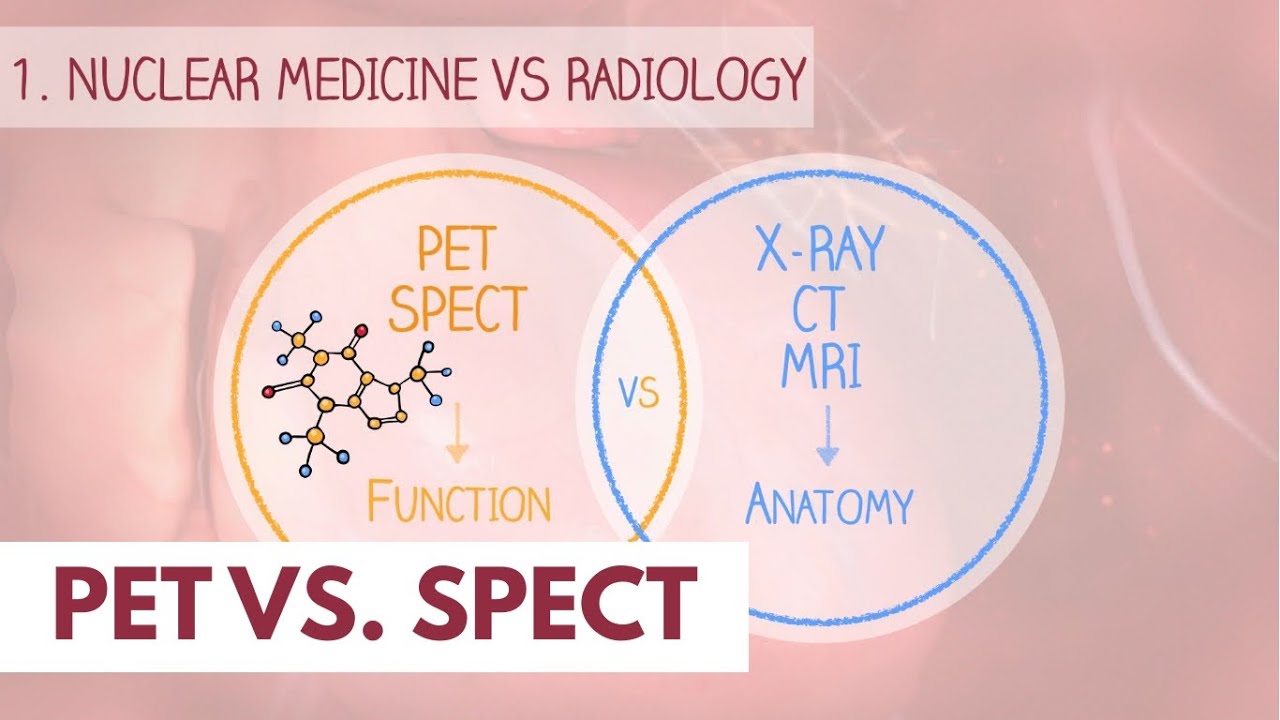
- 😀 Unlike X-ray, CT, or MRI, PET and SPECT visualize organ function rather than anatomy.
- 😀 PET scans can detect cancer about 6 months earlier than CT scans by identifying biochemical changes.
- 😀 PET stands for Positron Emission Tomography and uses positron-emitting isotopes like fluorine-18.
- 😀 SPECT stands for Single Photon Emission Computed Tomography and uses gamma-emitting isotopes like technetium.
- 😀 Radioactive isotopes are attached to compounds or molecules used by the body, such as glucose, to create radiopharmaceuticals.
- 😀 Fluorodeoxyglucose (FDG) is a commonly used radiopharmaceutical for PET scans that mimics glucose and accumulates in metabolically active areas like cancer cells.
- 😀 The level of glucose uptake by cancer cells often correlates with the aggressiveness of the cancer.
- 😀 PET and SPECT both use radiation to create images, but PET detects pairs of gamma rays emitted by positrons, while SPECT detects single photons.
- 😀 PET scanners use hundreds of detectors in rings around the patient, while SPECT scanners typically have two large rectangular detectors.
- 😀 PET and SPECT scans are often combined with CT scans to provide both anatomical and metabolic information.
Q & A
What is the main difference between PET and SPECT imaging techniques?
-The main difference is that PET (Positron Emission Tomography) uses positron-emitting radioisotopes, while SPECT (Single Photon Emission Computed Tomography) uses gamma-emitting radioisotopes. Both are used in nuclear medicine to visualize organ function, but they do so using different types of radiation and detection methods.
How do PET and SPECT differ from other imaging modalities like CT and MRI?
-PET and SPECT focus on visualizing organ function, whereas CT and MRI assess anatomical structures. This functional imaging is crucial for diagnosing and staging conditions like cancer, as PET and SPECT can detect biochemical changes before anatomical changes are visible on CT or MRI.
How much earlier can PET scans detect cancer compared to CT scans?
-PET scans can detect cancer on average six months earlier than CT scans, because PET can identify biochemical changes in tissues that may precede visible anatomical changes.
What does the term 'radiopharmaceutical' refer to?
-A radiopharmaceutical is a compound consisting of a radioactive isotope and a tracer molecule that is used in nuclear medicine to track biological processes in the body. For example, in PET imaging, fluorine-18 is attached to glucose to form FDG (fluorodeoxyglucose).
How does fluorine-18 in a PET scan help detect cancer?
-Fluorine-18 is attached to glucose to form FDG. Since cancer cells often have a higher rate of glucose metabolism, FDG accumulates in these cells, allowing PET scans to detect and measure the activity of cancerous tissue.
What is the significance of the rate of glucose uptake in cancer diagnosis?
-The rate of glucose uptake in tissues is a key indicator of their metabolic activity. Most cancers have higher glucose uptake compared to normal tissues, making this process useful in detecting and evaluating the aggressiveness of cancer.
What are the key differences in the way PET and SPECT generate images?
-In PET, fluorine-18 emits positrons that annihilate with electrons, producing two gamma rays detected by the scanner. In SPECT, gamma-emitting isotopes like technetium release single photons that are detected by the camera. PET scanners detect paired gamma rays, while SPECT detects single photons.
How are the gamma rays detected in PET and SPECT imaging?
-In PET, the gamma rays are detected when they travel in opposite directions after the annihilation of positrons and electrons. In SPECT, the camera detects the single photon emitted by the radioisotope as it exits the body.
How do the detectors in PET and SPECT scanners differ?
-PET scanners typically use hundreds of detectors arranged in rings around the patient, while SPECT scanners generally use two large rectangular detectors that rotate around the patient to capture the emitted radiation.
Why are PET and SPECT scans often combined with CT scans?
-PET and SPECT scans are often combined with CT scans to provide both metabolic (functional) and anatomical information. This combination allows for more accurate diagnosis and localization of diseases, including cancer.
Outlines

此内容仅限付费用户访问。 请升级后访问。
立即升级Mindmap

此内容仅限付费用户访问。 请升级后访问。
立即升级Keywords

此内容仅限付费用户访问。 请升级后访问。
立即升级Highlights

此内容仅限付费用户访问。 请升级后访问。
立即升级Transcripts

此内容仅限付费用户访问。 请升级后访问。
立即升级浏览更多相关视频

PET vs. SPECT scan | Dr. Paulien Moyaert

Kedokteran Nuklir itu apa sih? Simak Penjelasannya Disini

Nuclear Chemistry Medical Applications

Cara kerja Siklotron untuk produksi radiofarmaka di Kedokteran Nuklir| Nuclear Medicine #3

How does a PET scan work? | Nuclear medicine

Teknologi Nuklir Untuk Kesehatan
5.0 / 5 (0 votes)
