Gamma camera | Components & Function l Visual explanation
Summary
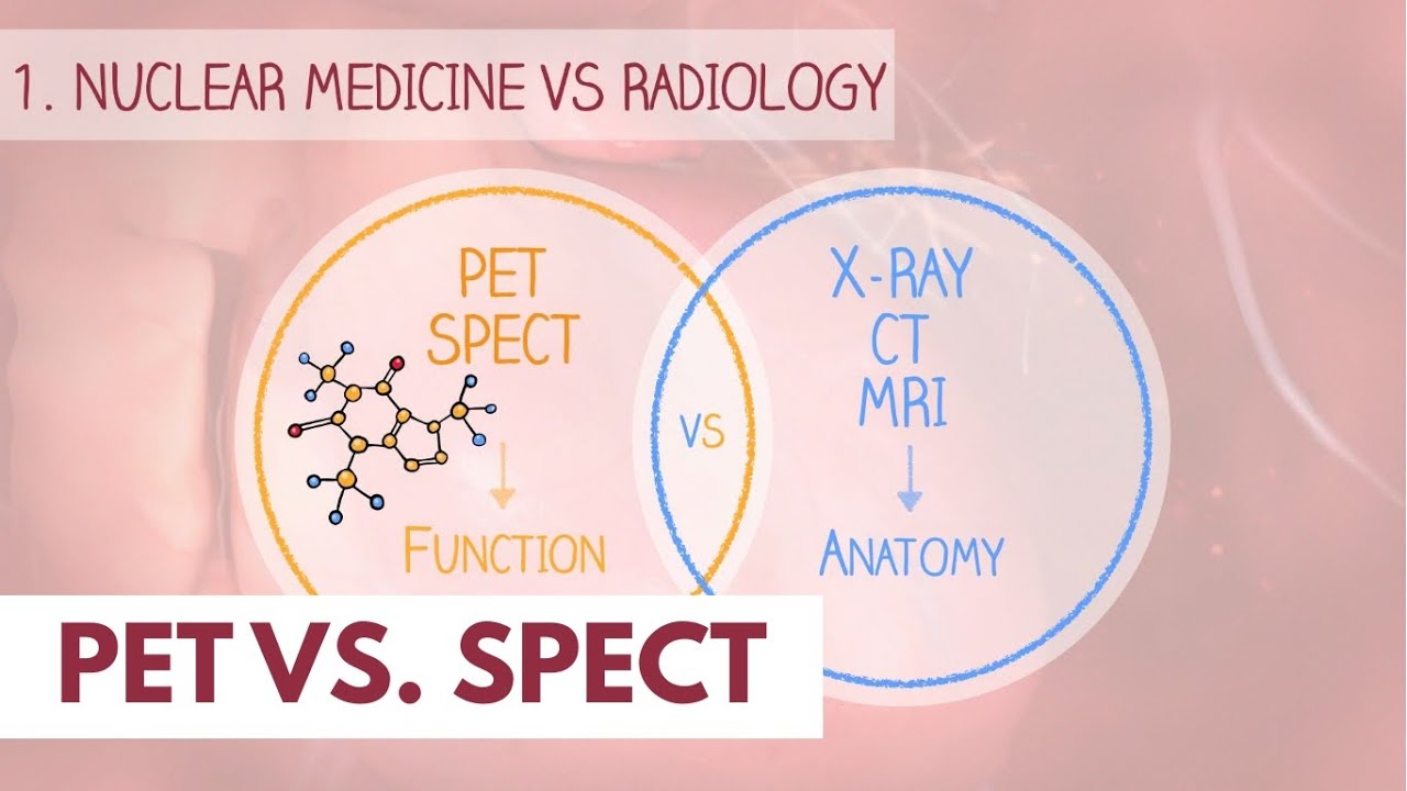
TLDRThe gamma camera is an imaging device used to capture gamma-emitting radioisotopes. It provides two-dimensional projections like X-rays but can also create 3D images through SPECT, a rotating imaging technique. The camera detects gamma photons using a collimator and scintillation crystals. Collimators, either pinhole or multihole, control photon direction, impacting image resolution and sensitivity. Photons absorbed by the crystal emit light, which photomultiplier tubes amplify to form images. The thickness of the crystal affects image clarity, balancing gamma ray absorption and spatial resolution. The data is processed by a computer to produce detailed images.
Takeaways
- 📷 The gamma camera is used to image gamma-emitting radioisotopes, similar to how X-rays capture 2D projections.
- 🧭 3D images can be produced using SPECT by rotating the gamma camera around the patient.
- 💡 The gamma camera detects gamma photons using crystals and consists of a collimator, crystal plane, and photomultiplier tubes connected to a computer.
- 🔍 The collimator functions like a photographic lens, controlling which photons reach the detector and preventing indistinct images.
- 🔒 Photons that are not parallel to the collimator holes are absorbed by the septa, reducing the sensitivity of the camera system.
- 🎯 High-resolution collimators have small or long holes and thicker septa, but they reduce sensitivity, requiring more radiation for imaging.
- 🔄 Pinhole collimators invert the image and are used for small organs like the thyroid but have low sensitivity due to the limited amount of radiation passing through the pinhole.
- 🌀 Multihole collimators can be parallel, diverging, or converging, with the parallel-hole type being the most common.
- 💥 The crystal absorbs gamma rays and emits flashes of light, known as scintillation, which is proportional to the absorbed energy.
- ⚡ Photomultiplier tubes convert scintillation light into measurable electrical signals that are processed by a computer to form an image.
Q & A
What is the primary function of a gamma camera?
-A gamma camera is used to image gamma-emitting radioisotopes, providing two-dimensional projections of three-dimensional objects. It can also produce 3D images using the SPECT technique.
How does SPECT enhance the imaging capabilities of a gamma camera?
-SPECT enhances imaging by rotating the camera around the patient, allowing for the creation of 3D images, including frontal, transversal, and sagittal cuts.
What role does the collimator play in a gamma camera?
-The collimator is the first device to encounter the emitted photons. It functions like a lens, controlling which photons reach the detector by allowing only photons traveling parallel to its holes to pass through.
Why is the collimator essential for image clarity?
-Without the collimator, photons would be emitted in all directions, resulting in a blurry, indistinct image. The collimator's holes help ensure only specific photons contribute to the final image.
What is the difference between high-resolution and high-sensitivity collimators?
-High-resolution collimators have small or long holes with thicker septa, providing detailed images but low sensitivity. High-sensitivity collimators have larger holes, allowing more photons to reach the detector but with lower resolution.
What is the primary use of pinhole collimators?
-Pinhole collimators are used for high-resolution images of small organs, such as the thyroid. However, they have poor sensitivity due to the small amount of radiation allowed to pass through the pinhole.
How does the scintillation crystal in the gamma camera work?
-The scintillation crystal absorbs gamma photons and emits the absorbed energy as flashes of light. The intensity of the light is proportional to the energy absorbed by the crystal.
What trade-off occurs with thicker scintillation crystals?
-Thicker scintillation crystals absorb more gamma rays, improving sensitivity, but they also increase photon scattering, leading to poorer spatial resolution.
How do photomultiplier tubes (PMTs) contribute to image formation in a gamma camera?
-PMTs detect the weak light flashes from the scintillation crystal and convert them into electrical signals. These signals are amplified and processed to form the final image.
What is the purpose of dynodes in a photomultiplier tube?
-Dynodes amplify the electrical signal by causing an avalanche multiplication process, where electrons strike the dynodes, releasing more electrons that are accelerated toward the next dynode, ultimately producing a measurable electrical charge.
Outlines

此内容仅限付费用户访问。 请升级后访问。
立即升级Mindmap

此内容仅限付费用户访问。 请升级后访问。
立即升级Keywords

此内容仅限付费用户访问。 请升级后访问。
立即升级Highlights

此内容仅限付费用户访问。 请升级后访问。
立即升级Transcripts

此内容仅限付费用户访问。 请升级后访问。
立即升级


5.0 / 5 (0 votes)
