Virtual tour | Nuclear medicine

Royal Papworth Hospital NHS Foundation Trust
8 Apr 202205:14

Summary

TLDRKatie, a radiographer, introduces a hybrid SPECT/CT gamma camera used for cardiac stress imaging to assess blood flow in the heart. The camera captures radiation from a radioactive isotope injected into the patient, while the CT scanner provides anatomical details. The process involves monitoring the patient's heartbeat and creating a 3D reconstruction of the heart's contraction. Katie also explains the safety measures taken to minimize radiation exposure for both patients and staff.

Takeaways

  • šŸ„ Katie introduces herself as a radiographer and welcomes the viewer to the nuclear medicine department.
  • šŸ“ˆ The hybrid SPECT CT gamma camera is used for cardiac and stress imaging to examine blood flow to the heart muscle.
  • šŸ” The gamma camera detects radiation from a radioactive isotope given to patients an hour before the scan.
  • šŸ“Ž The CT scanner provides anatomical information to enhance the SPECT image of the heart.
  • šŸ›ļø Patients lie on a table during the scan with ECG leads attached to monitor their heart rate.
  • šŸ’“ The scan measures the length of each heartbeat and creates a 3D reconstruction of the heart's contraction.
  • šŸ“Š Real-time images of the heart are displayed on a screen for both the patient and the medical staff to observe.
  • šŸ’‰ Patients are injected with a radioactive isotope, which circulates in the body and is absorbed by the heart muscle.
  • šŸ§Ŗ The radioisotope lab is where radioactive isotopes are prepared and stored for injection into patients.
  • šŸ›”ļø Radiographers are closely monitored for radiation exposure with finger and whole-body dosimeters.
  • šŸ’‰ Shielding and safety measures are taken when handling radioactive isotopes to minimize radiation exposure.

Q & A

  • What is the primary purpose of the hybrid SPECT CT gamma camera?

    -The primary purpose of the hybrid SPECT CT gamma camera is to perform cardiac stress imaging to observe the blood flow to the heart muscle under stress and resting conditions.

  • How does the gamma camera part of the SPECT CT system work?

    -The gamma camera part, which consists of two scanners at the front, picks up radiation from the radioactive material given to the patient an hour prior to the scan, collecting information emitted from the patient.

  • What role does the CT scanner play in the hybrid SPECT CT system?

    -The CT scanner, resembling a giant donut at the back, provides anatomical information that helps enhance the SPECT image of the heart.

  • What is the purpose of the ECG leads attached to the patient during the scan?

    -The ECG leads are used to monitor the patient's heart rate during the scan, allowing the scan to measure the length of each heartbeat.

  • How does the scan create a 3D reconstruction of the heart?

    -The scan breaks down the heartbeats into sections and creates a 3D reconstruction, which allows for the observation of how the heart is contracting.

  • What is the typical timing for injecting the radioactive isotope into the patient?

    -The radioactive isotope is injected into the patient an hour prior to imaging to allow time for it to circulate and absorb into the heart muscle.

  • What part of the heart does the radioactive injection highlight in the images?

    -The radioactive injection highlights the left ventricle of the heart in the images.

  • What is the purpose of the radioisotope lab mentioned in the script?

    -The radioisotope lab is where radioactive isotopes are prepared for injection into patients and where sealed sources for quality control checks on calibrators and the gamma camera are stored.

  • How is the staff's radiation exposure monitored?

    -Staff members are closely monitored for radiation exposure through the use of finger dose rings and whole body dosimeters, which are changed every two months and provide a yearly total of exposure.

  • What safety measures are taken when handling radioactive isotopes?

    -Safety measures include using lead-shielded vials and syringes, tongs to maximize distance from radiation, and a lead glass screen to protect from high doses during the drawing up process.

  • How is the radioactive contamination checked before staff members touch other objects?

    -A mini scintillation monitor is used to check for radioactive contamination on hands before touching other objects, with counts per second indicating background radiation or contamination levels.

Outlines

00:00

šŸ„ Introduction to Nuclear Medicine and Equipment

Katie, a radiographer, welcomes viewers to the nuclear medicine department, specifically focusing on cardiac stress imaging. She introduces the hybrid SPECT CT gamma camera used for examining blood flow to the heart muscle under stress and resting conditions. The gamma camera captures radiation from radioactive isotopes given to patients, while the CT scanner provides anatomical information to enhance the SPECT image. Katie explains the process of attaching ECG leads to monitor the patient's heart rate during the scan, which is later used to create a 3D reconstruction of the heart's contraction. The images are displayed in real-time for both the medical staff and the patient to observe.

Mindmap

Keywords

šŸ’”Radiographers

Radiographers are healthcare professionals who specialize in imaging technology to diagnose and treat illnesses. In the context of the video, Katie, a radiographer, is introducing the nuclear medicine department and explaining the processes involved in cardiac imaging. Radiographers play a crucial role in operating the specialized equipment and ensuring that patients receive accurate and safe scans.

šŸ’”Nuclear Medicine

Nuclear medicine is a branch of medical imaging that uses small amounts of radioactive material to diagnose or treat diseases. The video script describes a nuclear medicine procedure where a hybrid SPECT/CT gamma camera is used to examine blood flow to the heart muscle. This technique is vital for diagnosing heart conditions and assessing the effectiveness of treatments.

šŸ’”Hybrid SPECT/CT Gamma Camera

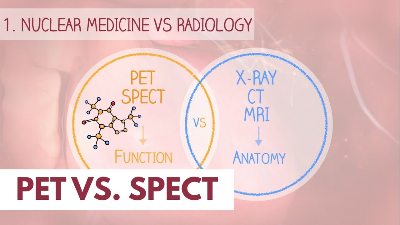
A hybrid SPECT/CT gamma camera is a medical imaging device that combines two technologies: Single Photon Emission Computed Tomography (SPECT) and Computed Tomography (CT). The SPECT part detects radiation from radioactive tracers, while the CT part provides detailed anatomical images. The script mentions this camera being used for cardiac stress imaging, allowing doctors to visualize both the function and structure of the heart.

šŸ’”Cardiac Stress Imaging

Cardiac stress imaging is a non-invasive diagnostic test used to assess the blood flow to the heart muscle under stress and at rest. The video explains that this procedure helps to identify potential heart problems by monitoring how the heart reacts to increased demand during stress. It is a critical tool for diagnosing coronary artery disease.

šŸ’”Gamma Camera

A gamma camera is a device used in nuclear medicine to detect and measure gamma rays emitted by radioactive tracers in the body. The script describes the gamma camera as having two scanners at the front that pick up radiation given to the patient, which is essential for collecting data on the heart's function.

šŸ’”ECG Leads

ECG leads are wires attached to electrodes that are placed on the patient's chest to monitor the electrical activity of the heart. In the video, Katie mentions attaching four ECG leads to the patient to track their heart rate during the scan. This data is crucial for synchronizing the imaging data with the heartbeats to create accurate 3D reconstructions of the heart's contractions.

šŸ’”3D Reconstruction

3D reconstruction is a process that creates a three-dimensional model from a series of two-dimensional images. The script explains that the data from the heartbeats is broken down into sections and used to create a 3D model of the heart's contraction. This reconstruction allows medical professionals to visualize the heart's function in detail.

šŸ’”Radioactive Isotope

A radioactive isotope is a variant of a chemical element that has an unstable nucleus and emits radiation. In the video, Katie discusses injecting a radioactive isotope into the patient an hour before imaging to allow it to circulate and be absorbed by the heart muscle. This is essential for visualizing the heart's function in the nuclear medicine scan.

šŸ’”Myocardium

The myocardium is the muscular layer of the heart that contracts to pump blood. The video script mentions that the radioactive injection is taken up within the myocardium, which allows the imaging to focus on the left ventricle of the heart. Understanding the health of the myocardium is critical for diagnosing and treating heart conditions.

šŸ’”Dose Calibrator

A dose calibrator is a device used to measure the exact amount of radioactive material in a sample. In the script, Katie describes using a dose calibrator to ensure that the radioactive isotope drawn up for injection is within the recommended dose range based on the patient's weight. This device is essential for patient safety and accurate imaging results.

šŸ’”Radiation Safety

Radiation safety refers to the measures taken to protect individuals from the harmful effects of ionizing radiation. The video script mentions the use of lead shielding, finger dosimeters, and whole-body dosimeters to monitor and minimize radiation exposure for staff. These practices are crucial in a nuclear medicine setting to ensure the safety of both patients and healthcare workers.

Highlights

Introduction to nuclear medicine and hybrid SPECT/CT gamma camera

Explanation of cardiac stress imaging to assess blood flow to the heart muscle

Description of the gamma camera's role in detecting radiation from patients

Details on the CT scanner's contribution to providing anatomical information

Process of attaching ECG leads to monitor heart rate during scans

How the scan measures heartbeats and creates 3D reconstructions

Real-time display of images on the screen for both staff and patients

Timing of radioactive isotope injection relative to imaging

Visualization of the left ventricle using radioactive injections

Importance of gating on the scanner for measuring heartbeats

Introduction to the radioisotope lab where isotopes are prepared

Storage of sealed sources for quality control on calibrators and gamma camera

Demonstration of drawing up radioactive isotope

Explanation of radiation exposure monitoring for staff

Use of finger and whole body dosimeters to track radiation exposure

Procedure for drawing up isotopes with lead shielding to protect staff

Measurement of isotope activity in a dose calibrator

Ensuring the correct dose of isotope based on patient weight

Use of tongs and lead shielding to handle isotopes safely

Checking for contamination with a mini scintillation monitor

Explanation of radiation detection levels with the scintillation monitor

Transcripts

play00:03

Hi. My name's Katie. I'm one of theĀ  radiographers. Welcome to nuclearĀ medicine.

play00:06

This is our hybrid spec ct gammaĀ  camera and this is where we do mainly cardiacĀ Ā 

play00:13

stress imaging to have a look at the blood flowĀ  to the heart muscle under stress and under restingĀ Ā 

play00:19

conditions. As you can see here we have the gammaĀ  camera part which is these two scanners at theĀ Ā 

play00:25

front so this will pick up the radiation that weĀ  give to the patient an hour prior to their scanĀ Ā 

play00:31

so it's collecting information coming off them andĀ  at the end at the back with a bit that looks likeĀ Ā 

play00:36

the giant donut is our ct scanner and that givesĀ  us anatomical information and it helps enhanceĀ Ā 

play00:42

the spect image of the heart before. As we do theĀ  scan our patient will be laying on the table hereĀ Ā 

play00:48

and we attach four little ecg leads andĀ  this basically monitors the patient'sĀ Ā 

play00:53

heart rate whilst we're doing the scan so our scanĀ  basically can measure the length of each heartbeatĀ Ā 

play00:59

and then when we do the reconstruction of theĀ  images later it breaks all of the heartbeatsĀ Ā 

play01:04

down into little sections and then it createsĀ  a 3d reconstruction allowing us to look at howĀ Ā 

play01:09

the heart is contracting. The images are displayedĀ  on the screen here so we can see it in real-timeĀ Ā 

play01:15

and the patient can also watch it as we're doingĀ  their scan. Normally the patient is injected withĀ Ā 

play01:21

a radioactive isotope an hour prior to theĀ  imaging because it needs an hour for it toĀ Ā 

play01:26

circulate round into the body and absorb intoĀ  the heart muscle and allow us to see the images.

play01:33

So what you're seeing here isĀ  the basically the left ventricleĀ Ā 

play01:37

so the radioactive injection that weĀ  give gets taken up within the myocardiumĀ Ā 

play01:42

and this is showing you the left ventricle asĀ  if it was squashed flat so this is the apexĀ Ā 

play01:47

of the heart and then this is the lateral sideĀ  and this would be the right side of the heartĀ Ā 

play01:52

and with as through the gating on the scannerĀ  those heartbeats that it's measuring allows usĀ Ā 

play01:57

to get a real-time image of how the heartĀ  has been contracting through the scan.

play02:06

So this is our radio radioisotope lab. ThisĀ is where we draw up all of the radioactiveĀ Ā 

play02:11

isotopes that we inject into the patients. We alsoĀ store all our sealed sources which are used to doĀ Ā 

play02:16

controller quality control on our calibratorsĀ  and the gamma camera and if you follow me we'reĀ Ā 

play02:21

going to go and have a look at the screenĀ  and you'll see me draw up some radar isotope.

play02:27

So as staff members, we're all monitored veryĀ  closely for our radiation exposure.Ā Ā 

play02:32

I have a ring underneath my glove that basicallyĀ  picks up the finger dose that i'm gettingĀ Ā 

play02:37

and i also have a whole body dosimeterĀ  which measures the dose i get to my bodyĀ Ā 

play02:42

and these are changed every two months and weĀ  get a yearly total of what we've been exposed to.

play02:50

So this is a radio isotope pot. So they just come in a little, small glass vials.

play02:56

The radio active isotope is just a clear, scentless liquid. You wouldn't know any different to water really,

play03:03

but these are all quite heavy. They're lead shielded to protect our fingers

play03:07

and contain everything with inside

play03:10

and then our syringe should have a syringe shield with about 6mm of lead around them

play03:16

and this again reduces our finger dose when we are injecting our patients.

play03:21

So to draw up, we would just, hold it in the pot, and draw.

play03:36

Our injections are very small.

play03:38

They're approximately one mil in volume, sometimes smaller,

play03:49

and then once this has been drawn up, it goes into a dose calibrator where we measure exactly how much

play03:55

activity has been measured up

play04:02

and then we are ensuring that the dose we haveĀ  drawn up is suitable based on the patient's weightĀ Ā 

play04:07

and it's within their recommended rangeĀ  for the dose levels for our investigations.

play04:12

So it's important when we handle the isotopesĀ  that we use tongs to basically maximize theĀ Ā 

play04:17

distance for the radiation away from ourĀ  hands until it's back into its shielded coverĀ Ā 

play04:25

and this glass screen in front of me is also madeĀ  of thick lead so this protects any high dose thatĀ Ā 

play04:31

I may get from drawing up. Once completelyĀ  drawn up we should always monitor our handsĀ Ā 

play04:38

to make sure that there's no contaminationĀ  on them before we go and touch something elseĀ Ā 

play04:42

so we have a monitor right here whichĀ  is called a mini scintillation monitorĀ Ā 

play04:47

and it will make a littleĀ  clicking sound as i turn it on

play04:52

and then we can put our hands in front of itĀ  to make sure that it's not detecting anything.

play04:57

So on here it measures in counts per second soĀ  anywhere between 10 and 20 counts per second isĀ Ā 

play05:03

like background radiation and anything aboveĀ  that would be radioactive or contaminated.

Rate This
ā˜…
ā˜…
ā˜…
ā˜…
ā˜…

5.0 / 5 (0 votes)

Related Tags
Nuclear MedicineHybrid CameraCardiac ImagingStress ImagingGamma CameraCT ScannerRadioisotopeRadiation SafetyIsotope HandlingMedical Imaging