Structure of a skeletal muscle - Muscle Physiology Animations || USMLE videos

Dr.G Bhanu Prakash Animated Medical Videos
4 Apr 202105:06

Summary

TLDRThis video explains the structure and function of skeletal muscle, emphasizing the layers of connective tissue (endomysium, perimysium, epimysium, deep fascia) that cover muscle fibers, fascicles, and muscles. It describes the organization of muscle fibers, highlighting thick filaments made of myosin and thin filaments made of actin, along with proteins like tropomyosin and troponin that regulate contraction. The video also explains the role of sarcomeres, sarcoplasmic reticulum, and transverse tubules in muscle contraction, focusing on the calcium ions and the triad structure that initiates the process.

Takeaways

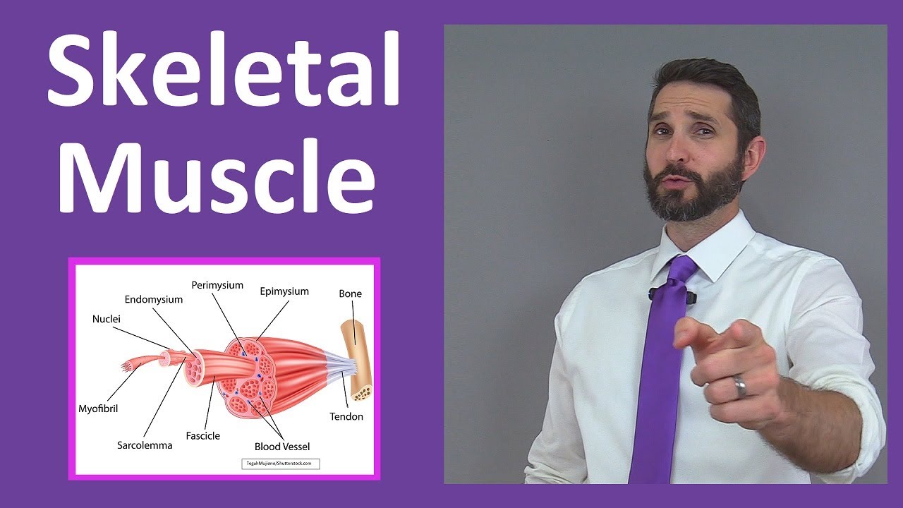
  • 💪 Skeletal muscles are organs made up of muscle fibers, connective tissue, blood vessels, and nerves.
  • 🧵 Each muscle fiber is wrapped in a thin layer of connective tissue called endomysium.
  • 📚 Muscle fibers are grouped into fascicles, each covered by a second connective tissue layer called perimysium.
  • 🛡️ Fascicles are bundled together to form muscles, which are covered by epimysium and an outer layer of deep fascia.
  • 🔗 The deep fascia can extend past the muscle to attach it to bones, cartilage, or other muscles.
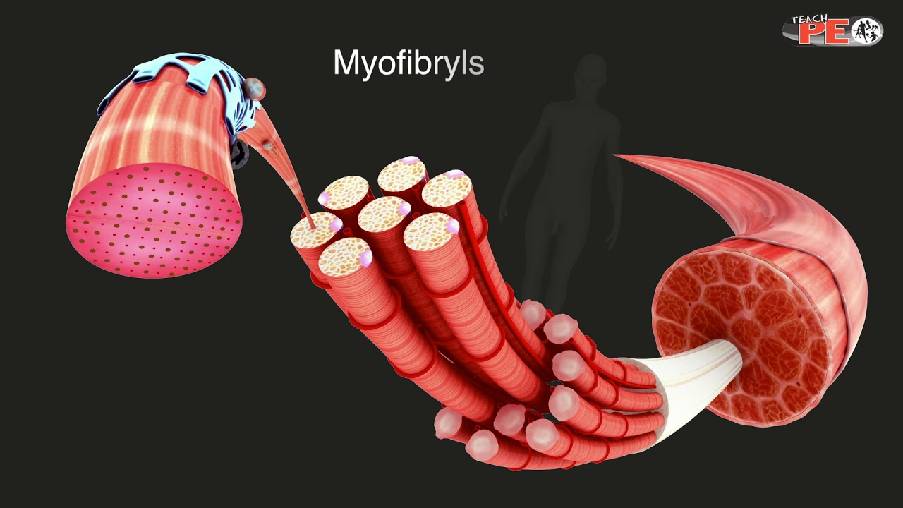
  • 🧬 Muscle fibers contain myofibrils, composed of thick myosin and thin actin protein filaments.
  • 🎯 Troponin and tropomyosin proteins regulate the interaction between actin and myosin during muscle contraction.
  • ⚙️ Striations in skeletal muscles are caused by the arrangement of thick and thin filaments within the myofibrils.
  • 🔋 Sarcoplasmic reticulum surrounds myofibrils and stores calcium ions, which play a key role in muscle contraction.
  • 🔗 Transverse tubules are membranous channels that help transmit signals deep into muscle cells and are part of the triad with sarcoplasmic reticulum.

Q & A

  • What are the main components of a skeletal muscle?

    -A skeletal muscle consists of muscle fibers, connective tissue coverings, blood vessels, and nerve fibers.

  • What is the role of endomysium in skeletal muscle?

    -The endomysium is a thin, delicate layer of connective tissue that wraps each individual muscle fiber in a skeletal muscle.

  • What is a fascicle and how is it formed?

    -A fascicle is a bundle of muscle fibers. Multiple muscle fibers are grouped together and wrapped in connective tissue called perimysium to form a fascicle.

  • How does the epimysium differ from the deep fascia in a skeletal muscle?

    -The epimysium is a dense fibrous layer that covers the entire skeletal muscle, while the deep fascia is a tougher layer of connective tissue that may extend beyond the muscle to attach it to bones, cartilage, or other muscles.

  • What are myofibrils and what proteins are they composed of?

    -Myofibrils are long, thin structures within muscle fibers composed of thick and thin protein filaments. Thick filaments are primarily made of myosin, while thin filaments are mainly composed of actin.

  • What is the function of tropomyosin and troponin in muscle contraction?

    -Tropomyosin stabilizes the actin filament, while troponin binds to actin, tropomyosin, and calcium ions, controlling the interaction between actin and myosin during muscle contraction.

  • What causes the striated appearance of skeletal muscle fibers?

    -The striated appearance is due to the arrangement of thick and thin filaments within the myofibrils, with alternating dark (A bands) and light (I bands) areas.

  • What is a sarcomere and where is it located?

    -A sarcomere is the functional unit of a myofibril and is defined as the segment between two Z lines within a myofibril.

  • What role does the sarcoplasmic reticulum play in muscle contraction?

    -The sarcoplasmic reticulum stores calcium ions and releases them into the sarcoplasm when stimulated by a muscle impulse, which is crucial for initiating muscle contraction.

  • What is a triad in skeletal muscle structure?

    -A triad refers to the structure formed by a transverse tubule flanked by two sarcoplasmic reticulum cisternae, near the area where actin and myosin filaments overlap. It plays a key role in muscle contraction activation.

Outlines

00:00

💪 Structure and Organization of Skeletal Muscles

Skeletal muscles are composed of muscle fibers, connective tissue, blood vessels, and nerves. Each muscle fiber is wrapped in a delicate connective tissue called endomysium, bundled into fascicles covered by perimysium. Multiple fascicles combine to form a muscle, which is wrapped in a dense layer called epimysium. The entire muscle is further surrounded by a tough, fibrous layer called deep fascia, which can extend to attach muscles to bones, cartilage, or other muscles.

🧬 Composition and Function of Muscle Fibers

Muscle fibers are long, thin cells made of myofibrils, which contain thick (myosin) and thin (actin) protein filaments. Myosin filaments have rod-like tails and globular heads (cross bridges) that bind with actin during muscle contraction. Thin actin filaments are helical and stabilized by tropomyosin, while troponin regulates the interaction of actin and myosin by binding to actin, tropomyosin, and calcium ions. These proteins coordinate the muscle contraction process.

🔍 Striations and Sarcomere Structure

Muscle striations arise from the arrangement of thick and thin filaments within the myofibrils. The A band (dark area) shows where thick and thin filaments overlap, while the I band (light area) contains only thin filaments. Myofibrils are divided into sarcomeres, the functional units of muscle contraction, which are separated by Z lines. This segmentation is essential for the muscle's ability to contract efficiently.

⚙️ Sarcoplasmic Reticulum and Calcium Ion Regulation

The sarcoplasmic reticulum, a membranous network surrounding each myofibril, stores and regulates calcium ions, critical for muscle contraction. It maintains a high concentration of calcium ions through active transport. When a muscle impulse triggers the sarcoplasmic reticulum, calcium ions are released into the sarcoplasm, initiating contraction.

🌀 Role of Transverse Tubules and the Triad in Contraction

Transverse tubules, continuous with the muscle cell membrane (sarcolemma), extend deep into the muscle fiber, facilitating the spread of muscle impulses. These tubules are filled with extracellular fluid and work closely with the sarcoplasmic reticulum, particularly in areas where actin and myosin filaments overlap. Together, they form a structure called the triad, crucial for activating the muscle contraction mechanism.

Mindmap

Keywords

💡Skeletal Muscle

Skeletal muscle is a type of muscle tissue that is attached to bones and enables voluntary movements. In the video, it is described as an organ made up of muscle fibers, connective tissue, blood vessels, and nerves, highlighting its complex structure. The skeletal muscle's ability to contract is central to the body's movement.

💡Endomysium

Endomysium is the thin, delicate layer of connective tissue that wraps around each individual muscle fiber. This layer provides support and plays a key role in protecting and separating muscle fibers within the larger muscle structure, making it critical for muscle function and movement.

💡Fascicles

Fascicles are bundles of muscle fibers wrapped in a second layer of connective tissue known as perimysium. This grouping of muscle fibers into fascicles helps organize the muscle and plays an important role in muscle contraction and strength.

💡Epimysium

Epimysium is the dense, fibrous connective tissue that covers the entire skeletal muscle. This outermost layer provides structural support, protects the muscle, and maintains its shape, allowing it to function efficiently during contractions.

💡Myofibrils

Myofibrils are the long, thin structures inside muscle fibers, made up of thick and thin protein filaments. These filaments, primarily myosin and actin, are responsible for the muscle’s ability to contract, which is fundamental to movement. Myofibrils are the core of muscle contraction.

💡Myosin

Myosin is the protein that forms the thick filaments in myofibrils. It has rod-like tails and two globular heads, which act as cross-bridges that interact with actin filaments to facilitate muscle contraction. Myosin’s role is central to generating the force needed for muscle movement.

💡Actin

Actin is the protein that makes up the thin filaments in myofibrils. Its helical structure resembles twisted strands of pearls. Actin works in conjunction with myosin to produce muscle contraction, making it a critical element of muscle movement.

💡Sarcoplasmic Reticulum

The sarcoplasmic reticulum is a membranous organelle that surrounds myofibrils and is involved in regulating calcium ion concentrations, which are essential for muscle contraction. The video explains how this network releases calcium into the muscle cell when stimulated, activating the contraction process.

💡Transverse Tubules

Transverse tubules, or T-tubules, are channels that extend from the muscle cell membrane deep into the muscle fiber. These tubules carry extracellular fluid and help transmit the electrical impulses that trigger muscle contraction by interacting with the sarcoplasmic reticulum.

💡Sarcomere

A sarcomere is the basic functional unit of a muscle fiber. It is the segment of the myofibril that is responsible for muscle contraction, consisting of overlapping thick and thin filaments (myosin and actin). Sarcomeres are arranged end-to-end and give skeletal muscle its striated appearance.

Highlights

Skeletal muscle is an organ made up of muscle fibers, connective tissue coverings, blood vessels, and nerve fibers.

Each muscle fiber is wrapped in a delicate layer of connective tissue called endomysium.

Fascicles, which are groups of muscle fibers, are wrapped in a second layer of connective tissue called perimysium.

A skeletal muscle consists of multiple fascicles wrapped in dense fibrous connective tissue called epimysium.

Skeletal muscles are also covered by deep fascia, a tough fibrous layer that can attach muscles to bones, cartilage, or other muscles.

Muscle fibers are long, thin cells composed of myofibrils, which are in turn made up of thick (myosin) and thin (actin) protein filaments.

Myosin filaments have a rod-like tail and two globular heads, which interact with actin filaments during muscle contraction.

Actin filaments resemble twisted strands of pearls and are stabilized by tropomyosin, a rod-shaped protein.

Troponin, a complex of polypeptides, binds to actin, tropomyosin, and calcium ions, playing a role in muscle contraction regulation.

Striations in skeletal muscles are caused by the arrangement of thick and thin filaments in myofibrils.

The dark A band contains overlapping thick and thin filaments, while the light I band contains only thin filaments.

Sarcomeres, the functional units of muscle fibers, are divided by Z lines and contain the key components involved in contraction.

Sarcoplasmic reticulum, a specialized membranous organelle, surrounds each myofibril and stores a high concentration of calcium ions.

Transverse tubules, continuous with the muscle cell membrane, extend deep into the muscle fiber, carrying extracellular fluid.

The sarcoplasmic reticulum and transverse tubules work together to trigger muscle contraction by releasing calcium ions when stimulated.

Transcripts

play00:00

[Music]

play00:14

do

play00:17

[Music]

play00:24

skeletal muscle each skeletal muscle

play00:28

is an organ made up of skeletal muscle

play00:31

fibers

play00:32

connective tissue coverings blood

play00:34

vessels

play00:35

and nerve fibers structure

play00:39

of a skeletal muscle connective tissue

play00:42

coverings

play00:44

each muscle fiber of the skeletal muscle

play00:47

is wrapped in a thin

play00:49

delicate layer of connective tissue

play00:51

called endomysium

play00:54

many muscle fibers are bundled together

play00:56

into groups

play00:58

called fascicles each fascicle

play01:01

is wrapped in a second layer of

play01:03

connective tissue

play01:04

made of collagen called perimysium

play01:08

many fascicles are bundled together to

play01:11

form

play01:11

a skeletal muscle each skeletal muscle

play01:15

is covered by a third layer of dense

play01:18

fibrous connective tissue called

play01:20

epimysium

play01:23

each skeletal muscle is then covered by

play01:25

a fourth

play01:26

very tough fibrous layer of connective

play01:29

tissue

play01:29

called deep fascia the deep fascia may

play01:33

extend

play01:33

past the length of the muscle and attach

play01:36

that muscle to a bone

play01:38

cartilage or muscle

play01:42

skeletal muscle fibers a muscle fiber

play01:46

is a long thin cell each muscle fiber is

play01:50

composed of myofibrils

play01:52

each myofibril is composed of two

play01:55

protein filaments

play01:58

thick filaments are primarily composed

play02:00

of the protein

play02:02

myosin they have a rod like tail

play02:05

that terminates into two globular heads

play02:08

or cross bridges these cross bridges

play02:12

interact with active sites on thin

play02:14

filaments

play02:15

thin filaments which are primarily

play02:18

composed of the protein actin

play02:20

they are coiled helical structures that

play02:23

resemble twisted strands of pearls

play02:27

tropomycin is the rod shaped protein

play02:30

spiraling around the backbone of actin

play02:33

to stabilize it troponin

play02:36

is a complex of polypeptides

play02:39

one binds to actin one that binds to

play02:43

tropomyosin

play02:44

and one that binds to calcium ions

play02:48

both tropomycin and troponin help

play02:50

control

play02:51

actin's interaction with myosin during

play02:54

contraction

play02:56

striations are caused by the arrangement

play02:59

of

play02:59

thick and thin filaments within the

play03:01

myofibrils

play03:03

a band also known as dark area

play03:06

in which there is overlapping of thick

play03:09

and thin

play03:09

filaments eye band also known as light

play03:13

area has thin filaments alone

play03:16

the length of each myofibril is divided

play03:19

into sarcomeres

play03:21

sarcomeres meet one another at an area

play03:24

called the z line within the sarcoplasm

play03:28

of a muscle fiber

play03:29

there are two specialized membranous

play03:32

organelles

play03:33

sarcoplasmic reticulum it is a network

play03:37

of membranous channels that surrounds

play03:39

each myofibril and runs parallel to it

play03:43

it is the same as endoplasmic reticulum

play03:45

in other cells

play03:47

sarcoplasmic reticulum has a high

play03:49

concentration

play03:51

of calcium ions compared to the

play03:53

sarcoplasm

play03:54

which is maintained by active transport

play03:56

calcium pump

play03:58

when stimulated by muscle impulse

play04:01

membranes become more

play04:02

permeable to calcium ions and calcium

play04:05

diffuses

play04:06

out of the sarcoplasmic reticulum into

play04:09

the sarcoplasm

play04:11

transverse tubules they are a set of

play04:14

membranous channels that

play04:16

extend into the sarcoplasm as

play04:19

invaginations

play04:20

continuous with muscle cell membrane the

play04:22

sarcolemma

play04:24

transverse tubules are filled with

play04:26

extracellular fluid

play04:28

and extend deep into the cell

play04:31

each transverse tubule runs between two

play04:34

enlarged portions of sarcoplasmic

play04:37

reticulum called

play04:38

cisternae these structures form a triad

play04:42

near the region where actin and myosin

play04:45

overlap

play04:47

sarcoplasmic reticulum and transverse

play04:49

tubules are

play04:50

involved in activating the muscle

play04:52

contraction mechanism

play04:54

because one transverse tubule is

play04:57

associated with two sarcoplasmic

play04:59

reticulum

play05:00

they are termed as the triad

Rate This

5.0 / 5 (0 votes)

相关标签
Skeletal MuscleMyofibrilsMuscle ContractionConnective TissueCalcium IonsTroponinTropomyosinSarcomereSarcoplasmic ReticulumMuscle Anatomy
您是否需要英文摘要?