FETAL SKULL | Bones | Sutures | Diameters | Fontanels | Anatomy | OBG Lecture | The Nurses Station
Summary
TLDRThis video discusses the anatomy of the fetal skull, focusing on its bones, regions, sutures, fontanels, and diameters. It explains the skull's significance during childbirth, highlighting how its bony structure can pose challenges while also being somewhat compressible. The video details the various areas of the skull, including the vertex, face, occiput, and base, as well as the key sutures and fontanels. Finally, it covers the different diameters of the skull, which play crucial roles in the birthing process depending on the baby's head position.
Takeaways
- 💡 The fetal skull is larger in proportion to the body and is the most challenging part to pass through the mother's pelvis due to its hard bony nature, though it is somewhat compressible.
- 🔍 The skull is divided into zones of obstetrical importance: vertex, face, brow, and occiput, each with specific boundaries and landmarks.
- 🦴 The skull bones include two frontal bones, two parietal bones, one occipital bone, and two temporal bones, each with unique features and functions.
- 🤕 The sutures of the skull are non-fused membranes that allow for the gliding movement of bones during childbirth, a process known as molding.
- 🔗 The obstetrically important sutures include the frontal, sagittal, coronal, lambdoid, and squamosal sutures, which facilitate the molding of the skull during delivery.
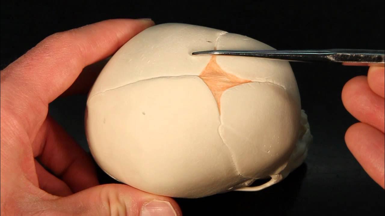
- 💠 Fontanels are wide gaps in the suture lines, with the anterior fontanelle (bregma) and posterior fontanelle (lambda) being of particular obstetric importance.
- 💎 The anterior fontanelle is diamond-shaped with approximate diameters of three centimeters, while the posterior fontanelle is triangular and about 1.2 centimeters in size.
- 🧠 The fetal skull's engaging diameter depends on the degree of head flexion, with different diameters engaging in various presentations such as vertex, face, or brow.
- 📏 The suboccipitobregmatic diameter is crucial for complete head flexion in vertex presentation, measuring 9.5 centimeters.
- ⏳ The anterior fontanelle ossifies by 18 months after birth, and failure to do so by 24 months is considered pathological.
Q & A
What is the significance of the fetal skull's shape and size during childbirth?
-The fetal skull is larger in proportion to the fetal body and is the most difficult part of the baby to pass through the mother's pelvic canal due to its hard, bony nature. However, it is compressible to some extent, which is crucial during childbirth.
How is the skull divided into zones of obstetrical importance?
-The skull is divided into several zones: the vertex, the anterior fontanelle (bregma), the posterior fontanelle (lambda), the occiput, and the base. Each zone has specific boundaries defined by sutures and bony landmarks.
What are the key bones that form the fetal skull?
-The key bones include two frontal bones, two parietal bones, one occipital bone, and two temporal bones. Each plays a role in the structure and function of the fetal skull.
What is the role of sutures in the fetal skull during childbirth?
-Sutures are the immovable fibrous joints between the bones of the skull. They allow for some degree of compression and gliding of one bone over another, known as molding, which facilitates the passage of the skull through the birth canal.
What are fontanels and why are they important in obstetrics?
-Fontanels are wide gaps in the suture lines that allow for the bones of the skull to move and overlap during childbirth. The anterior and posterior fontanels are of particular obstetric importance.
What is the shape and size of the anterior fontanelle?
-The anterior fontanelle is diamond-shaped and is formed by the joining of four sutures. Its anteroposterior and transverse diameters measure approximately three centimeters each.
How does the posterior fontanelle differ from the anterior fontanelle in shape and size?
-The posterior fontanelle is triangular in shape and is formed by the junction of three suture lines. It measures about 1.2 by 1.2 centimeters, which is smaller than the anterior fontanelle.
What are the different diameters of the fetal skull and their significance during labor?
-The fetal skull's diameters include the suboccipitobregmatic, submentovertical, occipitofrontal, mentovertical, and submentovertical. These diameters determine the ease of the fetal head's passage through the birth canal depending on the degree of head flexion and presentation.
What is the suboccipitobregmatic diameter and its measurement?
-The suboccipitobregmatic diameter extends from the nape of the neck to the center of the bregma and measures 9.5 centimeters. It is the engaging diameter in case of complete flexion of the head and vertex presentation.
What is the biparietal diameter and its significance in labor?
-The biparietal diameter measures 9.5 centimeters and extends between two parietal eminences. It nearly always engages during labor, regardless of the position of the head.
Outlines
💠 Fetal Skull Anatomy and Obstetric Significance
This paragraph delves into the intricate details of the fetal skull, emphasizing its unique structure and obstetric importance. The skull is described as being disproportionately large relative to the fetal body and poses a significant challenge during childbirth due to its hard, bony composition. However, it retains some degree of compressibility. The skull is divided into distinct zones including the vertex, brow, and occiput, each with specific boundaries and characteristics. The bones of the skull are highlighted, with the frontal bones forming the brow and the parietal bones above, while the occipital bone is at the back. Sutures, which are the fibrous joints between the bones, are crucial for the skull's flexibility during labor, allowing for 'molding.' Fontanelles, the soft spots on the skull, are also discussed, with the anterior and posterior fontanelles being of particular obstetric interest. Lastly, the paragraph covers the various diameters of the fetal skull, which are critical for determining the ease of passage through the birth canal.
🔍 Detailed Examination of Fetal Skull Diameters
The second paragraph continues the exploration of the fetal skull, focusing on the specific diameters that are essential for understanding the mechanics of labor and delivery. The engaging diameters are detailed, including the suboccipitobregmatic, submentovertical, and the transverse diameters, each with its unique measurement and relevance to different stages and presentations of fetal head during childbirth. The paragraph explains how these diameters are engaged based on the degree of flexion or extension of the fetal head, which is pivotal for a successful vaginal delivery. The biparietal and bitemporal diameters are also mentioned, providing a comprehensive overview of the dimensions that medical professionals consider when assessing the feasibility of a vaginal birth.
Mindmap
Keywords
💡Fetal Skull
💡Sutures
💡Fontanels
💡Anterior Fontanel
💡Posterior Fontanel
💡Molding
💡Engaging Diameter
💡Suboccipitobregmatic Diameter
💡Vertex Presentation
💡Biparietal Diameter
Highlights
The fetal skull is larger in proportion to the body and can be challenging to pass through the mother's pelvis.
The skull is compressible to some extent, which is crucial for childbirth.
The skull is divided into zones of obstetrical importance: vertex, face, and occiput.
The vertex is a quadrangular area defined by specific sutures and eminences.
The face area, or since put, is bounded by the anterior fontanelle and the root of the nose.
The occipital region is limited to the occipital bone, which also contributes to the skull's base.
The bones of the skull include two frontal bones, two parietal bones, one occipital bone, and two temporal bones.
Sutures are non-fused membranes that unite the bones of the skull vault.
Obstetrically important sutures include the frontal, sagittal, coronal, lambdoid, and squamosal sutures.
Sutures allow for the gliding movement of bones during childbirth, known as molding.
Fontanels are wide gaps in the suture lines, with the anterior and posterior fontanels being of obstetric importance.
The anterior fontanelle is diamond-shaped and measures approximately three centimeters in diameter.
The posterior fontanelle is triangular and measures about 1.2 by 1.2 centimeters.
The engaging diameter of the fetal skull depends on the degree of head flexion.
Suboccipitobregmatic is the engaging diameter in case of complete head flexion and vertex presentation.
Submentovertical is the engaging diameter when there is partial extension of the fetal head.
Transverse diameters, such as biparietal and bitemporal, are crucial in the mechanism of labor.
Transcripts
foreign
[Music]
we will talk about the fetal skull
its Bones the areas of the skull sutures
fontanels and diameters
fetal skull is all shaped at term it is
larger in proportion to the fetal body
and in comparison with the true pelvis
the fetal skull is the most difficult
part of the baby to pass through the
mother's pelvic Canal due to the hard
bony nature of the skull however it is
compressible to some extent first we
will talk about the areas of skull the
skull is arbitrarily divided into
several zones of obstetrical importance
these are the vertex it is a
quadrangular area bounded anteriorly by
the bregma and coronal sutures behind by
the Lambda and lambdoid sutures and
laterally by the lines passing through
the parietal eminences bro or since put
is an area
bounded on one side by the anterior
fontanelle and coronal sutures and on
the other side by the root of nose and
supraorbital Rays of either side
pace is an area bounded on one side by
the root of nose and supraorbital Ridge
and on the other side by the junction of
the floor of the mouth with the neck
the occipital region or the occiput is
limited to the occipital bone
this region between the phase and the
occiput is known as the base
regarding the bones there are two
frontal bones anteriorly forming since
put already broke at the center of each
frontal bone is a frontal Eminence these
are the sides of ossification of the
frontal bone during embryological
development although may not be the
first sight frontal bones used to form
only one frontal bone by eight years of
age
there are two parietal Bones superiorly
the ossification centers of parietal
bones are called parietal eminences one
occipital bone lying at the back of the
head and forms the region of the occiput
part of the occipital bone contributes
to the base of the skull as it contains
the foramen magnum which protects the
spinal cord as it leaves the skull
literally there are two temporal bones
talking about the sutures plaid bones of
the Vault are united together by
non-is-fired membranes attached to the
margins of Bones these are called the
sutures and the fontanels the sutures of
obstetric importance are the frontal
suture lies between two frontal Bones
the sagittal or the longitudinal suture
which lies between two parietal Bones
the coronal sutures between parietal and
frontal Bones on either side the
lambdoid sutures between occipital bone
and two parietal bones
and the secret muscle suture between the
temporal bone and the parietal bone
the sutures permit gliding moment of one
bone over the other during childbirth
known as molding talking about the
fontanels a wide Gap in the suture line
is called a fontanel there are almost
six fontanels but only two have the
obstetric importance
first the anterior fountain or blackma
and the posterior Fontanel or Lambda the
anterior fontanelle is formed by the
joining of four sutures the sutures are
anteriorly frontal posteriorly sagittal
and on either side coronal the shape of
the anterior fontanelle is like a
diamond its anteroposterior and
transverse diameters measure
approximately three centimeters each
the floor of the anterior fontanelle is
formed by a membrane and it becomes
ossified 18 months after the birth it
becomes pathological if it fails to
osify even after 24 months posterior
fontanelle is found by Junction of three
suture lines sagittal suture anteriorly
and lambdoid sutures on either sides it
is triangular in shape and measures
about 1.2 by 1.2 centimeters its floor
is membranous but becomes bony at tone
lastly we will discuss about the
diameters of fetal skull the engaging
diameter of the fetal skull depends on
the degree of flexion present the
anterior posterior diameters of head
which may engage are suboxito pragmatic
which extends from the nape of neck to
the center of the bregma this measures
9.5 centimeters and it is the engaging
diameter in case of complete flexion of
the head and vertex presentation
sub occipital frontal which extends from
the nape of the neck to the anterior and
of the anterior fontanelle or center of
the sync support it measures 10
centimeters
and it is the engaging diameter in case
of incomplete flexion and vertex
presentation
occipital frontal extends from the
occipital Eminence to the root of the
nose measuring 11.5 centimeters it is
the engaging diameter in case of marked
deflection of the head and vertex
presentation mental vertical extends
from the midpoint of the chin to the
highest point on the subject and suture
it is 14 centimeters long and it is the
engaging diameter in case of partial
extension of the head and bro
presentation
submanto vertical this diameter extends
from junction of the floor of the mouth
and neck to the highest point on the
sagittal suture it measures 11.5
centimeters and it is the engaging
diameter when there is incomplete
extension of the fetal head and there is
Phase presentation
pragmatic this extends from the junction
of floor of the mouth and neck to the
center of the bagma measures 9.5
centimeters and it is the engaging
diameter in case of complete extension
of the fetal head and face presentation
the transverse diameters which are
concerned in the mechanism of Labor
include biparita diameter measures 9.5
centimeters it extends between two
parietal eminences whatever may be the
position of the head this diameter
nearly always engages
by temporal diameter this measures 8
centimeters it is the distance between
the anterior inferior ends of the
coronal suture or the farthest points on
the coronal suture
so this completes the fetal skull in
this we discussed about the fetal skull
its regions bones sutures fontanels and
the diameters
5.0 / 5 (0 votes)