Premature Ventricular Contraction - causes, symptoms, diagnosis, treatment, pathology
Summary
TLDRThis script explains premature ventricular contractions (PVCs), abnormal heartbeats originating in the ventricles. It details the normal cardiac cycle, the role of pacemaker cells, and how PVCs disrupt it. The script describes how PVCs appear on an ECG, their potential causes, and the effects on the heart's rhythm. It also discusses the significance of compensatory pauses, different types of PVCs, and treatment options, emphasizing that while most PVCs are harmless, some may indicate a serious underlying condition.
Takeaways
- 💓 A premature ventricular contraction (PVC) is an early heartbeat that originates in the ventricles, disrupting the normal cardiac cycle.
- 🔌 The sinoatrial (SA) node normally sends electrical signals that coordinate the heartbeat, but in PVC, signals from the ventricles interrupt this sequence.
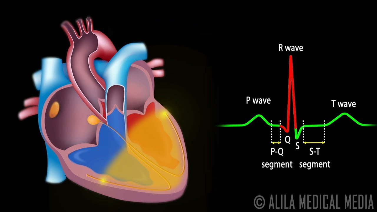
- 📊 On an electrocardiogram (ECG), atrial contractions appear as P-waves, ventricular contractions as QRS complexes, and ventricular repolarizations as T-waves.
- 🌊 The QRS complex, which indicates ventricular contraction, is typically less than 100 milliseconds and consists of three deflections: Q, R, and S waves.
- 🛑 Latent pacemakers such as the atrioventricular (AV) node, Bundle of His, and Purkinje fibers can generate electrical signals, but at a slower rate than the SA node.
- 🚫 Causes of PVCs include stressors like electrolyte imbalances, drugs, ischemic damage, or increased sympathetic activity.
- 🔄 Reentrant loops, where a depolarization wave circles around non-depolarizing tissue, can lead to continuous ectopic beats.
- 🔄 The direction of the ectopic focus determines the appearance of the QRS complex on ECG, with left or right bundle branch block patterns.
- ⏲️ Compensatory pauses occur when PVCs reset the SA node's timing, causing a longer interval between ventricular contractions.
- 🏥 PVCs are usually asymptomatic, but if frequent or symptomatic, they may be treated with medications or procedures like radiofrequency ablation.
Q & A
What is a premature ventricular contraction (PVC)?
-A premature ventricular contraction is an abnormal heartbeat that occurs when the ventricles contract earlier than normal in the cardiac cycle due to an abnormal contraction signal originating from within the ventricles rather than the sinoatrial (SA) node.
How does a normal cardiac cycle initiate contractions?
-In a normal cardiac cycle, the sinoatrial (SA) node sends an electrical signal that propagates through the atria, causing them to contract. This signal then moves to the atrioventricular (AV) node, where it is briefly delayed before traveling down to the ventricles, causing them to contract.
What is the significance of the PR segment on an ECG?
-The PR segment on an ECG corresponds to the pause in the atrioventricular (AV) node, which is the brief delay between the atrial depolarization and the ventricular depolarization.
What does the QRS complex represent on an ECG?
-The QRS complex on an ECG represents the ventricular depolarization, which is the electrical activity causing the ventricles to contract.
What are the three smaller waves that make up the QRS complex?
-The QRS complex is typically made up of three smaller waves: a Q wave (if the first wave after the P-wave is downwards), an R wave (the next upward deflection), and an S wave (any downward deflection after the R wave).
What are latent pacemakers and why are they significant?
-Latent pacemakers are cells in the AV node, Bundle of His, and Purkinje fibers that can generate an electrical potential and take over the pacemaker function if the SA node fails. They have slower depolarization rates and are significant as backup pacemakers for the heart.
What causes a premature ventricular contraction (PVC)?
-PVCs can be caused by enhanced automaticity in latent pacemaker cells or cardiac muscle cells due to factors such as electrolyte imbalances, drug use, ischemic damage, or increased sympathetic activity.
What is an early-afterdepolarization and a delayed-afterdepolarization?
-An early-afterdepolarization is a depolarization event that occurs during ventricular repolarization, while a delayed-afterdepolarization happens after repolarization is complete. Both can lead to ectopic beats.
What is a reentrant loop in the context of cardiac arrhythmias?
-A reentrant loop is a type of ventricular ectopic focus where a depolarization wave encounters non-depolarizing tissue, such as scar tissue, and circles around it, repeatedly sending depolarization waves to the heart tissue.
How can the origin of a PVC be determined from an ECG?
-The origin of a PVC can be determined by observing the QRS complex on an ECG. If the ectopic focus originates in the right ventricle, the QRS complex resembles a left bundle branch block, while if it originates in the left ventricle, it resembles a right bundle branch block.
What is a compensatory pause and how does it differ from a noncompensatory pause?
-A compensatory pause is a pause in the cardiac rhythm where a normal sinus complex lands exactly two times the normal sinus interval, following a PVC. A noncompensatory pause occurs when the sinus complex lands in less than twice the normal sinus interval after a PVC, indicating the PVC did not fully reset the sinus node's timing.
Outlines
💓 Understanding Premature Ventricular Contractions (PVCs)
This paragraph explains the concept of premature ventricular contractions (PVCs), which occur when the ventricles of the heart contract earlier than they should in the cardiac cycle. It details the normal electrical conduction system of the heart, starting from the sinoatrial (SA) node and moving through the atrioventricular (AV) node, bundle of His, and Purkinje fibers. The paragraph also describes how an ECG represents the electrical activity of the heart, with P-waves, QRS complexes, and T-waves corresponding to different stages of the cardiac cycle. It further discusses latent pacemakers and how they can lead to PVCs due to factors such as stress, drug use, or heart damage. The paragraph concludes by explaining how ectopic foci within the ventricles can cause early depolarization waves, leading to PVCs.
🔍 ECG Patterns of Premature Ventricular Contractions
The second paragraph delves into how PVCs manifest on an electrocardiogram (ECG) and the different scenarios that can occur. It describes how the origin of an ectopic focus in either the right or left ventricle affects the appearance of the QRS complex, resembling a left or right bundle branch block, respectively. The paragraph also explains how the V1 lead on an ECG can indicate the origin of the ectopic focus. It discusses the concept of compensatory and noncompensatory pauses following a PVC, which are identified by the timing of the subsequent P wave in relation to the normal sinus interval. Additionally, it covers various points in the cardiac cycle where PVCs can occur and their corresponding ECG appearances, including during the P-wave, PR segment, and ST segment. The paragraph concludes with a discussion of more serious arrhythmias that can be triggered by PVCs.
🩺 Diagnosing and Treating Premature Ventricular Contractions
The final paragraph focuses on the diagnosis and treatment of PVCs. It emphasizes the importance of the ECG in identifying PVCs and mentions the use of Holter monitors for continuous monitoring over several days. The paragraph outlines that while most PVCs do not require treatment, if they persist or cause symptoms, medications like beta blockers or calcium channel blockers may be prescribed. In cases where an ectopic focus is causing arrhythmias, radiofrequency ablation may be performed to eliminate the problematic tissue. The paragraph concludes with a recap of PVCs, their potential causes, and the necessity of treatment in certain circumstances.
Mindmap
Keywords
💡Ventricles
💡Depolarization
💡Sinoatrial (SA) Node
💡Atrioventricular (AV) Node
💡Bundle of His
💡Purkinje Fibers
💡Electrocardiogram (ECG)
💡Premature Ventricular Contraction (PVC)
💡Compensatory Pause
💡Latent Pacemaker
💡Reentrant Loop
Highlights
Premature ventricular contractions (PVCs) are abnormal contractions originating in the ventricles.
PVCs occur when the ventricles contract earlier than normal in the cardiac cycle.
An abnormal depolarization signal from the ventricles, rather than the pacemaker cells, triggers PVCs.
The sinoatrial (SA) node normally sends the first electrical signal in the heart's conduction system.
The atrioventricular (AV) node delays the signal before it reaches the ventricles.
The bundle of His and Purkinje fibers facilitate the signal's propagation to the ventricles.
On an ECG, atrial depolarization appears as a P-wave, and ventricular contraction as a QRS complex.
The PR segment on an ECG corresponds to the delay in the AV node.
The ST segment represents the interval between ventricular depolarization and repolarization.
The TP segment indicates the heart's quiet time after repolarization.
The QRS complex, typically less than 100 milliseconds, consists of three smaller waves.
A downward first wave after the P-wave is called a Q wave, while an upward wave is called an R wave.
A reentrant loop in the ventricles can cause a PVC due to a wave encountering non-depolarizing tissue.
Ectopic foci in the right ventricle produce a QRS complex resembling a left bundle branch block.
Lead V1 on an ECG captures depolarization waves moving towards the right ventricle.
A compensatory pause follows a PVC, with a normal sinus complex landing twice the normal sinus interval.
A noncompensatory pause occurs when the sinus complex lands less than twice the normal sinus interval after a PVC.
PVCs can be asymptomatic or cause lightheadedness due to reduced blood delivery to the brain.
In rare cases, PVCs can trigger more serious arrhythmias like ventricular tachycardia or fibrillation.
Diagnosis of PVCs is typically done via ECG, sometimes requiring a Holter monitor for continuous monitoring.
Treatment for PVCs may include beta blockers, calcium channel blockers, or radiofrequency ablation if necessary.
Premature ventricular contractions are usually benign but may indicate an underlying disorder requiring treatment.
Transcripts
The heart has two lower chambers, called the ventricles, so a premature ventricular contraction
is when the ventricles contract earlier than normal in the cardiac cycle.
This happens because an abnormal contraction signal, called a depolarization, originates
from somewhere in the ventricles rather than coming from the pacemaker cells.
So, if we simplify this heart a little bit, normally, the sinoatrial node or SA node sends
an electrical signal that propagates out through the walls of the heart and contracts both
upper chambers.
Then that signal moves to the atrioventricular node or AV node, where the signal is delayed
for a split second, and then goes down into the ventricles or lower chambers where it
moves down the bundle of His and into the left and right bundle branches and into each
ventricle’s Purkinje fibers, causing them to contract as well.
So, in a healthy heart the upper chambers contract first, then shortly after, the lower
chambers contract.
On an electrocardiogram or ECG which measures the electrical activity of the heart via electrodes
that are placed on the skin.
The atrial depolarization, and therefore it’s contraction, is seen as a p-wave, the ventricular
contraction is seen as a QRS complex, and the ventricular repolarization, and therefore
it’s relaxation, is seen as a T-wave.
This empty space here is called the PR segment, and it corresponds to the pause in the AV
node, and this one is called the ST segment, and it corresponds to the interval between
ventricular depolarization and repolarization, and this one is called the TP segment, which
represents the heart’s quiet time when the cells are finished repolarizing and are ready
for another signal.
Now, if we just look at the QRS complex, which normally lasts less than 100 milliseconds
or 2-and-a-half little boxes, it’s usually made up of three smaller waves, also called
deflections.
If the first wave after the p-wave is downwards, or negative, it’s called a Q wave - which
you can remember by the letter Q having a downward tail.
If the next deflection is upward, or positive, then it’s called the R wave.
If, though, the first wave after the p-wave is upwards, instead, or positive, you basically
skip the Q and just called it an R wave.
Finally, any downward deflection after the R wave is called the S wave.
Now, the interesting thing is that in addition to the pacemaker cells in the SA node, cells
in the AV node, Bundle of His, and the Purkinje fibers, all have the ability to generate an
electrical potential.
Those last three are called latent pacemakers, and they have slower depolarization rates—which
is the rate at which they fire off electrical signals—and they get slower as you move
further down.
Let’s use this bar to visualize the SA node’s depolarization rate, which is the fastest,
and then each one below is slightly slower.
Notice that each time the SA node fires, it resets all the slower ones, and this is exactly
how it works.
If, for example, the SA node stopped altogether, then the AV node would take over at it’s
slightly slower pace.
Now, though, let’s say you have a ventricular ectopic focus somewhere in the ventricles,
which is a cell or area of tissue that sends off an early depolarization wave, before even
the SA node gets to fire.
This is what leads to a premature ventricular contraction, or PVC.
One cause of a latent pacemaker cell or cardiac muscle cell depolarizing early is it gets
enhanced automaticity which might result from irritating stressors like electrolyte imbalances,
drugs like cocaine or methamphetamines, ischemic damage like a heart attack, or anything that
increases sympathetic activity, like anxiety.
Ectopic beats can also have “triggered activity” which is where cells depolarize early.
The exact mechanism here, though, is unclear, but it might be due an ion channel dysfunction
that leads to an unexpected change in the membrane potential during or right after repolarization.
When a cell depolarization happens during ventricular repolarization, it’s called
an early-afterdepolarization, and if it happens after repolarization is finished, it’s called
a delayed-afterdepolarization.
A final type of ventricular ectopic focus is a reentrant loop, where a depolarization
wave encounters tissue that doesn’t depolarize - which can be something like scar tissue
after a heart attack - and as a result the wave starts going around and around that tissue
- forming what’s called a reentrant loop.
A reentrant loop basically starts sending out depolarization waves to the rest of the
heart tissue each time the wave goes around.
If the ectopic focus originates in the right ventricle, the wave will depolarize the right
ventricle first and then the left ventricle, and this produces a QRS complex that looks
like a left bundle branch block.
If the ectopic focus originates in the left ventricle, the wave will depolarizing the
left ventricle first and then the right ventricle, which produces a QRS complex that looks like
a right bundle branch block.
Now, lead V1 on an ECG measures a depolarization wave that moves towards the right ventricle.
So, when an ectopic focus originates in the left ventricle, and moves towards the right
ventricle, the V1 lead shows a large positive complex, with a dominating R wave.
When an ectopic focus originates in the right ventricle, and then moves towards the left
ventricle, the V1 lead shows a large negative complex, with a dominating S wave.
Regardless of the originating ventricle, a premature ventricular contraction often has
an abnormal T wave since the timing and direction of repolarization will be abnormal as well.
Alright, so let’s say this heart’s cruising along at about 60 bpm, which means that there’s
1 second between p waves and QRS complexes.
All the sudden an ectopic focus in the ventricles fires off, which contracts the ventricles,
and that wave of depolarization tries to travel up to the atrium, but since this happens so
close to the previous depolarization, the atrium’s still in its refractory period
so the wave gets stopped at the AV node.
A split second later, the ventricle enters its refractory period.
Since the sinus node’s going at 1 second per beat, and it’s out of refractory, it
fires off a signal and the atria contracts and you get another P wave, which is right
on schedule.
But this time the opposite thing happens, and the ventricle’s in refractory so it
doesn’t contract!
K, now both relax and come out of refractory, and exactly one second later, the sinus node
sends another signal and the atrium contracts as it should, and then everything carries
on as per usual.
This situation is called a compensatory pause, which is defined as having a normal sinus
complex landing exactly 2 times the normal sinus interval, which was 1 second, so 1 times
2 equals 2 seconds.
What ends up happening here is there’s this big long pause between ventricular contractions,
greater than the sinus interval.
Longer time between contractions means more ventricular filling, which means the heart
contracts with greater strength, which can be felt as a palpitation.
Alright, now let’s run a slightly different scenario, one where the PVC comes a little
bit later, after the atrium comes out of refractory.
In this case the wave of depolarization makes it into the atrium and depolarizes the atrium,
including the sinus node, so essentially the sinus node resets about 4.4 boxes from the
last, or 0.88 seconds.
In this case, after being depolarized the sinus node waits its 1 second, and then sends
another signal and again we carry on as normal.
This time we call this a noncompensatory pause, because the sinus complex lands less than
2x the normal sinus interval.
And this is how you could tell if the PVC depolarized the atrium or not.
A ventricular ectopic focus can fire at different points in the cardiac cycle, which we can
look at using the ECG.
First off, it might happen during a P-wave, which can get completely lost in the QRS complex,
but can sometimes be seen if you search for it.
Next, the ectopic beat could happen during the PR segment, and because the ectopic depolarization
happens relatively slowly, oftentimes, it will combine with the normal depolarization
wave coming down the ventricular conduction system, resulting in a ventricular fusion
beat.
These can appear lots of different ways depending on where the two depolarization waves meet
each other.
Early after-depolarizations might start during the ST segment or even during the T-wave,
which is called R-on-T phenomenon.
This used to be considered an ominous sign of an impending dangerous arrhythmia, but
recent studies show that this is a bit more unclear than once thought.
Sometimes premature ventricular contractions can keep happening rather than being isolated
events.
For example, ventricular bigeminy is when a premature ventricular contraction consistently
comes after each normal cardiac cycle.
Ventricular trigeminy, on the other hand, is when one consistently comes after every
two normal cardiac cycles.
Also, you might have multiple ectopic foci PVCs, producing different appearing QRS complexes
on a single rhythm strip.
Now, most people with premature ventricular contractions don’t notice them, but if they
keep occurring they can cause lightheadedness because of less blood getting delivered to
the brain.
In rare situations, an ectopic focus can trigger ventricular tachycardia or even ventricular
fibrillation, which are more serious arrhythmias where the ventricles beat too quickly to fill
up with an adequate amount of blood.
The diagnosis of a premature ventricular contraction is based on the ECG, but sometimes a Holter
monitor - which is like a continuous ECG monitor used over a few days - is needed to capture
the event.
Typically, premature ventricular contractions don’t need treatment, and if there is an
obvious cause like a medication or use of a substance, then stopping that typically
resolves the issue.
If the premature ventricular contractions keep happening or cause palpitations, they
can be treated with beta blockers or calcium channel blockers, both of which prevent the
heart from beating too strongly.
If an ectopic focus is triggering ventricular arrhythmias, radiofrequency ablation can be
done, which is where radiofrequency waves are used to destroy the tissue that’s causing
the ectopic heartbeat.
Alright, as a quick recap - Premature ventricular contractions are abnormal heartbeats that
originate in the ventricles, and show up as tall and wide QRS complexes on an ECG.
They can be caused by abnormal automaticity, reentry, or a triggered afterdepolarization.
They’re usually asymptomatic, but when they’re a sign of an underlying disorder, treatment
including a radiofrequency ablation may be necessary.
浏览更多相关视频
Cardiac Conduction System and Understanding ECG, Animation.
Cardiac Arrhythmias, Animation
Guyton and Hall Medical Physiology (Chapter 11) REVIEW The Normal Electrocardiogram || Study This!
Potencial de ação cardíaco e contração do coração – Fisiologia Humana
Cardiac Cycle | Body Fluids and Circulation | Class 11 | NEET
The Heart, Part 2 - Heart Throbs: Crash Course Anatomy & Physiology #26
5.0 / 5 (0 votes)