Heart Conduction System & ECG (EKG)

Siebert Science
29 Apr 202417:45

Summary

TLDRThis educational video script delves into the intricacies of the human heart's cardiac conduction system, explaining how it coordinates heartbeats. It introduces the sinoatrial (SA) node as the heart's pacemaker, the role of the atrioventricular (AV) node in delaying signals for organized contractions, and the function of the bundle of His and Purkinje fibers in rapid signal transmission. The script also correlates these processes with an ECG (EKG), describing the significance of the P-wave, QRS complex, and T-wave, and how they reflect atrial and ventricular activity. Realistic anatomical visuals from Anatomage enhance the learning experience.

Takeaways

  • 💓 The human heart has been beating continuously since the first heartbeat as a fetus, thanks to the cardiac conduction system.
  • 🔍 The cardiac conduction system is a network of specialized tissue that coordinates heartbeats, including the sinoatrial (SA) node, atrioventricular (AV) node, and the bundle of His.
  • 📍 The SA node, located in the right atrium, acts as the heart's pacemaker, initiating the heartbeat by sending electrical signals.
  • 🚦 The AV node introduces a delay to ensure the atria contract before the ventricles, allowing for efficient blood transfer.
  • 🛤️ The bundle of His and bundle branches rapidly transmit the electrical signal from the AV node to the ventricles, preparing them for contraction.
  • 🏭 The Purkinje fibers spread the signal throughout the ventricles, ensuring synchronized contraction for effective blood pumping.
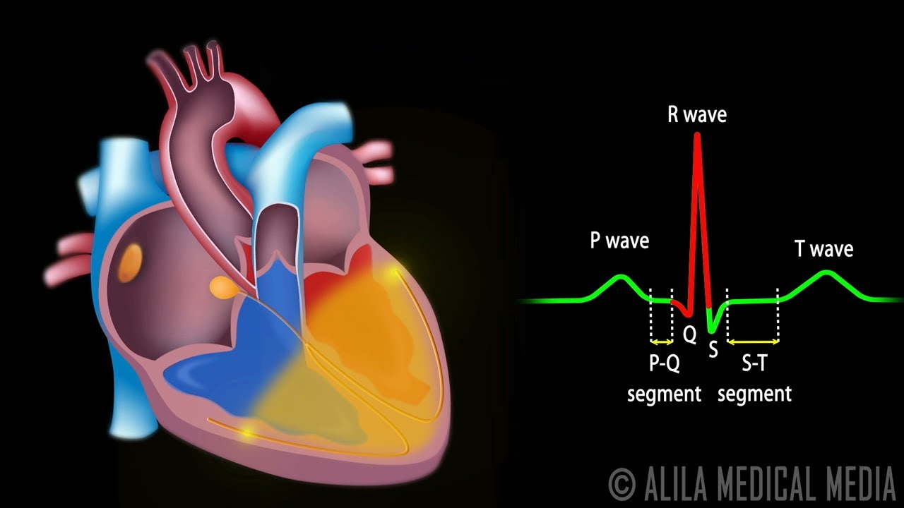
  • 📊 An ECG (electrocardiogram) measures the heart's electrical activity, with the P-wave, QRS complex, and T-wave corresponding to atrial depolarization, ventricular depolarization, and repolarization, respectively.
  • 🩺 The P-wave on an ECG represents atrial depolarization, which leads to atrial contraction and the subsequent pushing of blood into the ventricles.
  • 🏋️‍♂️ The QRS complex indicates ventricular depolarization, which is the signal for the ventricles to contract and pump blood out of the heart.
  • 🔙 The T-wave signifies the repolarization of the ventricles, marking the end of their contraction phase and the beginning of relaxation.
  • 🎥 Anatomage provides 3D models and virtual dissection tables that help visualize the complex structures of the heart in a three-dimensional space.

Q & A

  • What is the primary function of the sinoatrial (SA) node?

    -The SA node, also known as the pacemaker of the heart, is responsible for initiating the heartbeat by sending out electrical signals that trigger the contraction of the heart muscle.

  • How does the cardiac conduction system differ from cardiac muscle tissue?

    -The cardiac conduction system is non-contractile and functions like nervous tissue, conducting electrical signals rapidly throughout the heart. In contrast, cardiac muscle tissue is contractile and pumps blood but conducts signals more slowly.

  • What is the purpose of the delay in the atrioventricular (AV) node?

    -The delay in the AV node ensures that the atria contract and push blood into the ventricles before the ventricles contract. This sequence prevents the atria and ventricles from contracting simultaneously, allowing for efficient blood flow.

  • Why is the blood color in diagrams often depicted as blue for the right side of the heart and red for the left?

    -In diagrams, blue is used to represent the right side of the heart, which contains deoxygenated blood, and red is used for the left side, which contains oxygenated blood. However, it's important to note that blood is always red; these are just conventional colors used for illustration.

  • What is the role of the interatrial pathway in the heart's electrical conduction?

    -The interatrial pathway facilitates the rapid transmission of electrical signals from the SA node to the left atrium, ensuring that both atria contract in unison to efficiently move blood into the ventricles.

  • How does the bundle of His contribute to the heart's electrical activity?

    -The bundle of His, also known as the atrioventricular bundle, receives the signal from the AV node and rapidly transmits it through the left and right bundle branches, which then stimulate the ventricles to contract.

  • What is the significance of the QRS complex in an ECG?

    -The QRS complex in an ECG represents the ventricular depolarization, which is the electrical signal passing through the ventricles causing them to contract and pump blood to the body and lungs.

  • What does the T-wave indicate on an ECG?

    -The T-wave on an ECG signifies the repolarization of the ventricles, which is the return to the resting state after contraction, marking the end of the cardiac cycle's electrical activity.

  • How does the heart sound relate to the ECG?

    -The first heart sound corresponds to the end of the QRS complex when the tricuspid and mitral valves close, while the second heart sound occurs at the end of the T-wave when the aortic and pulmonary valves close.

  • What is the role of the interatrial and internodal pathways in the heart's electrical conduction system?

    -The interatrial pathway rapidly transmits the signal from the SA node to the left atrium, while the internodal pathways conduct the signal through the right atrium. Together, they ensure synchronized atrial contraction.

Outlines

00:00

🫀 Introduction to the Cardiac Conduction System

This paragraph introduces the concept of the cardiac conduction system, which is responsible for coordinating heartbeats. It explains that the heart has been beating continuously since fetal development, thanks to a small mass of cardiac tissue known as the sinoatrial (SA) node. The SA node, along with a complex conduction system, ensures the blood is pumped through the arteries. The video aims to educate viewers about the cardiac conduction system and how it relates to ECG (electrocardiogram) readings. Real human cadavers and anatomage images are used to provide a three-dimensional understanding of the heart's structure. The script also covers the basic structure of the heart, including the atria and ventricles, and the flow of blood through these chambers.

05:00

🚀 The Cardiac Conduction System and ECG Waves

This section delves into the three types of tissue in the heart: the cardiac conduction system, cardiac muscle tissue, and non-conductive fibrous tissue. The SA node's role as the heart's pacemaker is emphasized, explaining how it generates electrical signals that initiate each heartbeat. The script describes the pathway of these signals from the SA node through the internodal pathway to the atrioventricular (AV) node, and the purpose of the delay in the AV node to ensure the atria contract before the ventricles. It also discusses the bundle of His and the bundle branches that facilitate rapid signal transmission, leading to the contraction of the ventricles. The paragraph concludes with an overview of how these processes relate to the different waves of an ECG, specifically the P-wave, QRS complex, and T-wave.

10:01

🔬 Detailed Explanation of ECG Intervals and Waves

The paragraph provides a detailed explanation of the different intervals and waves seen in an ECG, correlating them with the electrical activity in the heart. It describes the P-wave, which signifies atrial depolarization, the PR interval during which the atria contract and push blood into the ventricles, and the QRS complex that indicates ventricular depolarization. The script also explains the ST interval, which corresponds to ventricular contraction and the first heart sound, and the T-wave, which represents ventricular repolarization and the second heart sound. The paragraph emphasizes the importance of these intervals and waves for understanding the heart's electrical activity and function.

15:02

📚 Recap and Visualization of Cardiac Processes

In this final paragraph, the script recaps the key points about the cardiac conduction system and the corresponding ECG readings. It summarizes the role of the SA node, the pathways of the electrical signals through the heart, and the significance of the ECG's P-wave, PR interval, QRS complex, ST interval, and T-wave. The paragraph also highlights the importance of practice in understanding these concepts. The video includes real-time visualizations of the heart's contractions and the electrical signals that drive them, provided by Anatomage, to help viewers grasp the complexity of the cardiac conduction system. The script ends with a call to action for viewers to practice labeling the parts of the cardiac conduction system and to explore more videos on the topic.

Mindmap

Keywords

💡Cardiac Conduction System

The Cardiac Conduction System is a network of specialized heart cells that generate and coordinate electrical impulses to regulate the heartbeat. In the video, it is described as the orchestrator of the heart's rhythmic contractions, ensuring that the atria and ventricles contract in a coordinated manner. The script mentions the SA node, AV node, and the bundle of His as key components of this system.

💡Sinoatrial (SA) Node

The Sinoatrial (SA) Node is often referred to as the heart's natural pacemaker. It is an autorhythmic cell cluster that initiates the heartbeat by sending electrical signals throughout the heart. The video script describes how the SA node's self-rhythmic nature ensures a consistent heartbeat, even without external input, by increasing the permeability to sodium and calcium ions.

💡Atrial Node

The Atrial Node, also part of the Cardiac Conduction System, is mentioned in the script as a secondary pacemaker that can take over if the SA node fails. It is located in the right atrium and is involved in the transmission of electrical signals from the SA node to the ventricles.

💡Atrioventricular (AV) Node

The Atrioventricular (AV) Node is a critical component of the Cardiac Conduction System that receives signals from the atria and transmits them to the ventricles, but with a slight delay. This delay, as explained in the script, ensures that the atria contract and push blood into the ventricles before the ventricles contract.

💡Bundle of His

The Bundle of His is a part of the Cardiac Conduction System that receives signals from the AV node and quickly transmits them to the ventricles. The script describes how this structure branches into the left and right bundle branches, which are essential for the rapid depolarization of the ventricles.

💡Depolarization

Depolarization is the process by which cardiac cells lose their electrical potential, leading to muscle contraction. The script explains that the SA node's depolarization initiates a heartbeat and that this process occurs in both the atria and ventricles, though at different times to ensure proper blood flow.

💡Repolarization

Repolarization is the return of cardiac cells to their resting state after depolarization. It is highlighted in the script as the process that follows the contraction of the heart muscle, allowing the muscle to relax and prepare for the next contraction cycle.

💡ECG/EKG

An Electrocardiogram (ECG or EKG) is a graphical representation of the heart's electrical activity. The video script uses the ECG to illustrate the different phases of the heartbeat, including the P-wave, QRS complex, and T-wave, each corresponding to specific events in the Cardiac Conduction System.

💡P-Wave

The P-Wave on an ECG represents atrial depolarization. The script explains that the P-wave corresponds to the electrical signal traveling through the atria, causing them to contract and push blood into the ventricles.

💡QRS Complex

The QRS Complex on an ECG indicates ventricular depolarization. As described in the script, the QRS complex is a significant spike that represents the rapid spread of electrical signals through the ventricles via the Bundle of His and Purkinje fibers, leading to their contraction.

💡T-Wave

The T-Wave on an ECG represents ventricular repolarization. The script mentions that the T-wave corresponds to the ventricles returning to their resting state after contraction, which is the final electrical event in a heartbeat cycle.

Highlights

The heart's rhythm is maintained by a small mass of cardiac tissue called the sinoatrial (SA) node.

The cardiac conduction system coordinates heartbeats through a complex network within the heart.

An ECG (Electrocardiogram) or EKG represents the electrical activity of the heart.

The heart is divided into right and left sides, each with an atrium and ventricle, colored to represent oxygen levels.

Blood flows through specific pathways, from the vena cava to the right atrium, then to the right ventricle and out through the pulmonary artery.

The left side of the heart receives oxygenated blood from the lungs and pumps it through the aorta to the rest of the body.

The cardiac conduction system is non-contractile and functions like nervous tissue to conduct signals.

Cardiac muscle tissue is contractile and also plays a role in signal conduction.

Fibrous tissue in the heart is non-conductive, ensuring the atria and ventricles do not contract simultaneously.

The SA node, or the heart's pacemaker, sends signals for each heartbeat without external input.

The internodal pathway transmits the signal from the SA node to the AV node through three branches in the right atrium.

The interatrial pathway quickly conducts the signal from the SA node to the left atrium.

The AV node introduces a delay to ensure the atria contract before the ventricles.

The bundle of His and bundle branches quickly transmit the signal down the heart's septum.

Purkinje fibers extend from the bundle branches to distribute the signal throughout the ventricles.

The ECG's P-wave corresponds to atrial depolarization, the QRS complex to ventricular depolarization, and the T-wave to ventricular repolarization.

The PR interval represents the time from atrial depolarization to the beginning of ventricular depolarization.

The ST interval is when the ventricles are contracting, pushing blood through the aorta and pulmonary artery.

The first and second heart sounds correspond to the closing of the heart valves during different stages of the cardiac cycle.

Transcripts

play00:00

your heart has beat continuously for

play00:02

your entire life from that very first

play00:04

heartbeat back when you were a fetus

play00:05

developing in the uterus until this very

play00:07

day that constant Rhythm that keeps the

play00:09

blood pumping through your arteries is

play00:10

thanks to a small mass of cardiac tissue

play00:12

called the CYO atrial node along with a

play00:15

complex cardiac conduction system that

play00:16

runs through your heart coordinating

play00:18

your heartbeat in this video we're going

play00:19

to build out the cardiac conduction

play00:21

system piece by piece learn how it all

play00:23

works and then use that to understand

play00:25

the different parts of an ECG or EKG and

play00:27

throughout the video we'll look at real

play00:28

human cavers and other fac images

play00:30

provided by anatomage the creator of the

play00:32

world's first virtual dissection table

play00:34

so you can see all these structures are

play00:36

arranged three-dimensionally in the body

play00:38

and by the end of this video you're

play00:39

going to know this whole process by

play00:40

heart well by brain because that's where

play00:43

memories are stored but you know what I

play00:44

mean let's jump to the Whiteboard and

play00:46

get started so let's start by drawing

play00:47

out the heart here we have an outline of

play00:49

the main structure of the heart on the

play00:51

cardiac muscle we've got the right

play00:52

atrium and the right ventricle and the

play00:54

left atrium and the left ventricle and

play00:56

of course we have the right side in blue

play00:58

because it's low oxygen blood the left

play01:00

side in red because it's high oxygen

play01:02

blood that's just come from the lungs

play01:04

and remember our blood is always red

play01:06

never blue that's just the colors we use

play01:07

in the diagrams blood is going to come

play01:09

into the right side through the superior

play01:10

and inferior venne cavis into the right

play01:12

atrium it'll pass through the tricuspid

play01:14

valve into the right ventricle and the

play01:16

right ventricle is going to pump it out

play01:17

through the pulmonary artery to go to

play01:19

the lungs to receive some oxygen the

play01:21

blood is going to get back to the heart

play01:22

through the pulmonary veins and go into

play01:24

the left atrium from the left atrium

play01:26

it'll pass through the bicuspid or mitol

play01:28

valve into the left ventricle and then

play01:29

the the left ventricle is going to pump

play01:31

it very forcefully out through the aorta

play01:33

so it can travel throughout the rest of

play01:34

the body and deliver the oxygen and

play01:36

nutrients and hormones and all the other

play01:37

stuff that's in our blood in between the

play01:39

left side and the right side of the

play01:40

heart we have the septum that really

play01:43

divides the heart into those two halves

play01:45

and here we can see in the anatomage

play01:46

models the right atrium and the right

play01:48

ventricle sitting sort of anterior to

play01:51

the left side where we have the left

play01:52

atrium and the left ventrical now for

play01:54

the rest of the video I'm going to

play01:55

assume you know those structures pretty

play01:56

well but if you want to refresh your a

play01:58

whole lesson on all that check out my

play02:00

pathway of blood Through the Heart video

play02:01

links down below for that now in the

play02:03

heart there's three different types of

play02:05

tissue that we're concerned about in

play02:06

this video first we have the cardiac

play02:08

conduction system itself that's going to

play02:10

be in yellow on this diagram and that's

play02:12

non-contractile cardiac tissue in other

play02:14

words these aren't muscle cells that are

play02:15

Contracting they're going to be more

play02:17

like nervous tissue that's going to be

play02:18

conducting signals throughout the heart

play02:20

second we have the cardiac muscle tissue

play02:22

this is going to be contractile tissue

play02:24

it's going to be tissue that contracts

play02:25

and and pumps blood but it also conducts

play02:28

signals as well the card conduction

play02:30

system which is just about 1% of the

play02:32

total kind of cells in here that's going

play02:34

to conduct the signals very quickly

play02:36

whereas the cardiac muscle it will take

play02:38

a little bit longer for the signals to

play02:39

pass through the muscle tissue itself

play02:41

and the cardiac muscle tissue that's

play02:43

going to be the vast majority of tissue

play02:44

in the heart now there is some

play02:46

non-conductive tissue in the heart and

play02:48

that's going to be the fibrous tissue

play02:50

and that's going to run from the atrial

play02:52

floor between the atrium and the

play02:54

ventricles now the fact that this

play02:55

fibrous tissue right there is

play02:56

non-conductive is very important we

play02:58

don't want the Atria and the ventricles

play03:00

to be contracting at the same time we

play03:02

want the Atria to contract and push the

play03:04

blood into the ventricles and then the

play03:06

ventricles to contract and push all that

play03:07

blood out of the heart if this tissue

play03:09

right here was conductive then we would

play03:11

have the Atria and ventricles

play03:12

Contracting at the same time wouldn't be

play03:14

good and the only way that signal can

play03:16

pass through here is through the cardiac

play03:18

conduction system through this yellow

play03:19

section right there that's the only part

play03:22

where the signal travels through that

play03:24

fibrous connective tissue so that

play03:25

fibrous connective tissue separates the

play03:27

Atria from the ventricles and into two

play03:30

sections that we call the atrial sensium

play03:33

as well as the ventricular sensium a

play03:35

sensium is just a big sort of hardto

play03:38

pronounce word that means a group of

play03:40

cells that are all electrically

play03:42

connected to each other so all of the

play03:44

cardiac muscle in this atrial sensium

play03:46

are connected electrically meaning that

play03:48

if one of the cells depolarizes it

play03:50

depolarizes the next cardiac muscle cell

play03:52

and that depolarizes the next one and

play03:54

eventually they'll all be depolarized

play03:56

cuz they're all electrically connected

play03:58

same thing in the ventricular sens when

play04:00

one of those cells depolarizes that'll

play04:02

depolarize the next cell and the next

play04:04

cell and the next cell until it's all

play04:05

depolarized now the signal passing

play04:07

between cardiac muscle cells is sort of

play04:10

slow I kind of mentioned that earlier

play04:11

and that's why we need this cardiac

play04:12

conduction system to conduct those

play04:14

signals very quickly so that the Atria

play04:17

can contract as one unit and the

play04:19

ventricles especially can contract as

play04:21

one unit but again the atrial cisum will

play04:23

depolarize first Contracting the Atria

play04:25

and then the ventricular sensium will

play04:27

depolarize Contracting the ventri Les

play04:30

now let's take a look at the individual

play04:32

parts of the cardiac conduction system

play04:34

first here we have the Sino atrial node

play04:36

now the Sino atrial node is autorhythmic

play04:38

meaning that it's going to be sending

play04:40

pulses by itself even without input from

play04:43

some other source autorhythmic meaning

play04:45

self- rhythmic in other words that saay

play04:47

note is the pacemaker of the heart it

play04:49

sends a signal every time our heart

play04:51

beats now there will be input from the

play04:54

brain from the cardiac regions of the

play04:56

brain they'll be sending signals to the

play04:57

heart to speed up that saay node or to

play05:00

slow down that SA node but even without

play05:03

input from the brain the SA node is

play05:05

going to be sending signals itself

play05:07

causing our heartbeat rhythm this

play05:10

consistent Rhythm happens by increasing

play05:12

permeability in the SA node of sodium

play05:14

ions and calcium ions so those sodium

play05:17

and calcium ions are slowly entering

play05:18

into the SA node and it prevents

play05:20

pottassium from leaving the cells in the

play05:22

SA node so there's a slow buildup of

play05:25

positive charge over time as sodium and

play05:27

calcium come into the SA node and then

play05:29

as soon as reaches a threshold membrane

play05:31

potential the SA node will send an

play05:33

action potential now the signals will

play05:34

eventually get to another node called

play05:36

the atrio ventricular or AV node more on

play05:39

that in just a moment but next let's

play05:40

talk about the internodal pathway this

play05:42

is going to be how the signal transmits

play05:44

from the SA node to the AV node and if

play05:48

you look at that internodal pathway

play05:49

there's three branches of it and they're

play05:51

all passing through the right atrium now

play05:54

on my diagram they look sort of planer

play05:56

or flat with each other but if we look

play05:58

on the anatomage images we'll see that

play06:00

these are actually three-dimensional um

play06:02

running through that right atrium which

play06:04

we can see right there so the SA node

play06:06

depolarizes sends a signal through the

play06:08

internal Pathway to the AV node and

play06:10

that's going to depolarize the right

play06:12

atrium now eventually that

play06:13

depolarization would make itself over to

play06:15

the left atrium but that's going to take

play06:17

a long time without the interatrial

play06:19

pathway which is going to run from the

play06:21

SA node over into the left atrium that

play06:24

interatrial pathway will conduct the

play06:26

signal very quickly into the left atrium

play06:28

and deol ize it most of the diagrams I

play06:31

looked up have the interatrial pathway

play06:32

coming directly from the SA node but one

play06:35

thing I noticed in the anatomage images

play06:37

is that the interatrial pathway is

play06:39

actually branching off of one of the

play06:41

internodal branches even though most of

play06:43

the diagrams you look up on this show it

play06:45

coming from the SA node directly great

play06:47

so the signal comes from the SA node

play06:49

it's going to travel through the

play06:49

interatrial pathway as well as the

play06:51

internodal pathway depolarizing both

play06:54

Atria so that they can contract that

play06:56

signal then is going to make it to the

play06:57

AV node now we've talked about the

play06:59

cardiac conduction system needing to

play07:00

send these signals very quickly but the

play07:02

AV node sort of does the opposite

play07:04

there's going to be a delay in the AV

play07:06

node now what would be the benefit of

play07:08

that well like I said earlier we want

play07:09

the Atria to contract before the

play07:12

ventricles contract so that delay is

play07:14

going to really separate the atrial

play07:15

contraction from the ventricular

play07:17

contraction that way we can get all the

play07:19

blood from the Atria to the ventricles

play07:21

and then the ventricles can pump it all

play07:23

out from The Av node the signal is going

play07:25

to pass into the bundle of hiss also

play07:27

known as the atrio ventricular bundle

play07:29

that bundle is immediately going to

play07:31

separate into the right and left bundle

play07:33

branches now unlike the AV node which

play07:35

passes the signal very slowly the bundle

play07:37

of His and the bundle branches are going

play07:39

to transmit that signal very quickly

play07:41

down the septum of the heart also as the

play07:43

signal is traveling through the septum

play07:45

it's not going to be stimulating the

play07:46

ventricles to contract just yet the

play07:49

ventricles will be stimulated to

play07:50

contract when the signal is passing its

play07:52

way back up that's going to allow the

play07:53

pumping to happen from the apex of the

play07:55

heart on the way back up to kind of

play07:57

force the blood out through the

play07:59

pulmonary artery and the aorta this way

play08:01

the tricuspid and mitro valves will also

play08:03

snap shut during this time to prevent

play08:05

the blood from back flowing into the

play08:07

Atria now extending out of the left and

play08:09

right bundle branches we have something

play08:10

called the Peri fibers and the pingi

play08:13

fibers are going to take that signal

play08:14

traveling through the bundle branches

play08:16

and spread it out throughout the muscle

play08:18

of the right and left ventricles that's

play08:20

going to conduct that signal many many

play08:22

times faster than if we were only

play08:23

relying on the ventricular Sensi or the

play08:26

connections between all of the cardiac

play08:28

cells so quick recap of all that the SA

play08:31

node or the pacemaker of the heart will

play08:32

send out a signal that'll pass through

play08:34

the interatrial pathway to stimulate the

play08:36

left atrium it'll also pass through the

play08:39

internodal pathways to stimulate the

play08:41

right atrium the signal will make it to

play08:42

the AV node where it's going to pass

play08:44

very slowly to cause a delay before the

play08:47

ventricles will contract the signal will

play08:48

pass through the bundle of hiss and the

play08:50

left and right bundle branches on the

play08:52

way back up they'll pass through the

play08:54

pingi fibers that's going to stimulate

play08:56

the cardiac muscle and the ventricles to

play08:57

contract and pump the blood out through

play09:00

the pulmonary artery as well as the

play09:02

aorta now let's take a look at an ECG or

play09:05

an EKG this is the thing that you've

play09:06

seen in like doctor movies and stuff

play09:08

where you see the beep beep and if it

play09:10

stops you hear it go beep because the

play09:12

heart has stopped beating but it's a

play09:13

measure of the electrical activity

play09:15

happening in the heart and it's got

play09:17

three regions here it's got the p-wave

play09:18

the QRS complex as well as the t-wave

play09:22

and these three sections correspond to

play09:24

different things happening in the

play09:25

cardiac conduction pathway so again we

play09:27

have the p-wave the Q complex in the

play09:30

t-wave you can see that happening one

play09:31

more time there I just really like that

play09:33

animation so what we're going to do is

play09:35

we're going to connect this to the

play09:36

cardiac conduction pathway looking at

play09:38

the p-wave QRS complex and the t-wave

play09:40

we're going to start with the p-wave the

play09:42

p-wave is going to correspond to the

play09:44

depolarization of the Atria so we've got

play09:46

the depolarization happening and you can

play09:48

see those signals traveling through

play09:50

those different Pathways causing

play09:52

depolarization of the Atria so that's

play09:54

the main thing happening here in the

play09:55

p-wave the Atria will depolarize next we

play09:58

have what we call the pr interval the PR

play10:00

interval is going to start at the

play10:01

beginning of the p wve and last all the

play10:03

way really until the Q part right here

play10:06

we call it the PR interval I think

play10:07

because sometimes on ECGs the qwave

play10:10

might be hard to identify or might not

play10:11

show up so we refer to this as the PR

play10:14

interval you also might see something

play10:15

called the pr segment so just as a quick

play10:18

clarification the PR interval starts at

play10:20

the beginning of p and lasts until R

play10:22

whereas the pr segment is just from the

play10:25

end of P to the beginning of R so PR

play10:27

interval would be this PR PR segment

play10:29

would just be this now during the PR

play10:31

interval the Atria are going to contract

play10:34

and that's going to send blood from the

play10:35

right atrium Into The ventricle and the

play10:38

blood from the left atrium into the left

play10:40

ventricle basically any blood that was

play10:41

still left in the atrium is going to get

play10:43

squeezed out through the contraction of

play10:45

the Atria the Contracting of the Atria

play10:47

will really start early on in the p wve

play10:49

and last throughout this section right

play10:50

here also during this section of the PR

play10:52

interval which is the pr segment is

play10:54

where we have that AV noal delay

play10:56

happening because as soon as the QRS

play10:58

complex hits then we're going to be

play10:59

depolarizing the ventricles speaking of

play11:01

which let's move on to the QRS complex

play11:04

in the QRS complex you see this huge R

play11:06

Spike that's because of the signal

play11:08

passing through the bundle of His and

play11:09

the bundle branches and then through the

play11:11

bingi fibers stimulating all of this

play11:14

cardiac muscle that electrical activity

play11:16

is going to be much greater in the

play11:18

ventricles because the ventricles have

play11:19

more cardiac tissue they also have to

play11:21

pump the blood a lot farther the Atria

play11:24

just had to pump the blood from one

play11:25

chamber to another the ventricles have

play11:27

to pump the blood to the lungs and then

play11:29

also Al through the aorta throughout the

play11:30

whole body so they need a very strong

play11:32

contraction so we need a lot of

play11:34

electrical activity to cause that to

play11:35

happen so during the QRS complex that

play11:37

signal is going to pass through the left

play11:39

and right bundle branches and through

play11:40

theingi fibers that's going to

play11:42

depolarize the ventricles also the Atria

play11:45

are going to repolarize repolarization

play11:48

is the opposite of depolarization

play11:50

depolarization is when tissue becomes

play11:51

more positive and in this case causes it

play11:53

to contract repolarization is when it

play11:55

returns back to its resting membrane

play11:57

potential and that muscle is going to

play11:58

relax and stop Contracting so we have

play12:00

depolarization of the ventricles as well

play12:02

as repolarization of the Atria basically

play12:05

ventricles contract but the Atria will

play12:07

be stopping their contraction now that

play12:09

QRS complex that big spike in the r is

play12:12

caused by the ventricles depolarizing we

play12:15

can't really see the effect of the Atria

play12:17

repolarizing on the EKG because it's

play12:19

sort of hidden by that big

play12:21

depolarization of the ventricles but

play12:23

both of those things are happening

play12:24

during that QRS complex up next we have

play12:27

something called the St interval that St

play12:29

interval is going to start with s and

play12:31

last all the way to the end of the

play12:33

t-wave a subset of that is the ST

play12:36

segment which would just be this section

play12:37

in right there lasting until the

play12:39

beginning of the t-wave during the St

play12:41

interval we're going to have the

play12:43

ventricles Contracting that's going to

play12:45

cause blood to be pumped through the

play12:46

aorta as well as blood to be pumped

play12:48

through the pulmonary artery that very

play12:49

forceful contraction is going to be

play12:51

starting kind of at the end of the QRS

play12:53

complex and Lasting until the t-wave

play12:55

happens the t-wave is when we're going

play12:57

to be repolarizing the ventricles and

play12:59

stopping the contraction but those

play13:00

ventricles will be contracting during

play13:02

that St interval now when the ventricles

play13:05

contract that's where we're going to

play13:06

hear our first heart sound the love of

play13:08

the ldub ldub we represent that with S1

play13:12

for the first sound of the heart and

play13:14

that sound is caused by the tricuspid

play13:16

and mitol valves snapping shut right

play13:19

before the ventricles will contract and

play13:21

pump the blood out we need those valves

play13:22

to close of course because we don't want

play13:24

the blood to rush back into the Atria we

play13:26

want all that blood to be forced out

play13:27

through the aorta and pull AR artery so

play13:29

again that first heart sound is going to

play13:31

kind of happen right around the end of

play13:33

that QRS complex as the valves are

play13:35

snapping shut finally the last section

play13:36

of this is the t-wave and the t-wave is

play13:39

going to be the repolarization of the

play13:41

ventricles or in other words sort of the

play13:42

turning off of ventricular contraction

play13:45

after the ventricles are finished

play13:46

Contracting we're going to have the

play13:47

second heart sound the dub of ldb just

play13:50

like the first heart sound the second

play13:52

heart sound is going to be caused by

play13:53

Valve snapping shut but in this case

play13:55

that's going to be the pulmonary valve

play13:56

snapping shut as well as the aortic

play13:58

valve valve snapping shut so the

play14:00

ventricles are relaxing and we don't

play14:01

want the blood that's been pumped out of

play14:03

the ventricles to pass back into them

play14:05

through the aorta or pulmonary artery so

play14:07

we snap those valves shut to keep the

play14:09

blood out of the ventricles there and

play14:10

that second heart sound is going to be

play14:12

happening kind of right at the end of

play14:14

the t-wave somewhere right in there all

play14:15

right so a lot going on in that process

play14:17

let's do a quick recap we have the soo

play14:19

atrial node where the signal will start

play14:21

we have the interatrial pathway the

play14:23

signal will travel there to depolarize

play14:25

the left atrium we have the internal

play14:27

pathway the signal will travel through

play14:28

the noal Pathways to depolarize the

play14:31

right atrium the signal will travel to

play14:32

the AV node where it is slowed down or

play14:35

delayed so the Atria can finish

play14:36

Contracting before the ventricles get

play14:38

depolarized and contract we have the

play14:40

bundle of His which is going to separate

play14:42

into the left and right bundle branches

play14:44

the bundle and the branches are going to

play14:46

transmit the signal very quickly because

play14:47

we want the ventricles to contract as

play14:49

one contract all unit as quickly as

play14:51

possible signal passes down the septum

play14:53

and then on the way back up it's going

play14:54

to pass through pereni fibers which are

play14:56

going to distribute the signal

play14:58

throughout the heart muscle to help the

play14:59

ventricles contract all at once from the

play15:01

Apex up all of this electrical

play15:03

conduction is going to cause the ECG the

play15:06

electrocardiogram the ECG will start

play15:08

with the p-wave this is where the Atria

play15:10

are depolarizing up next we have the PR

play15:13

interval this is where the Atria are

play15:15

Contracting and it's going to be pushing

play15:16

blood from the right atrium to the right

play15:18

ventricle and from the left atrium to

play15:20

the left ventricle next is the QRS

play15:23

complex this is going to be where the

play15:24

ventricles are depolarizing and it's

play15:26

going to be where the Atria are

play15:28

repolarizing or sort of turning off once

play15:30

the ventricles are depolarized we move

play15:32

into the St interval and this is where

play15:34

the ventricles are going to be

play15:35

contracting causing blood to pass up

play15:37

through the aorta and pumping blood out

play15:39

through the pulmonary artery as well and

play15:41

at the beginning of that St interval is

play15:42

where we have the first heart sound

play15:44

which is caused by the tricuspid and

play15:45

mitro valves snapping shut up next we

play15:48

have the t-wave the t-wave is going to

play15:50

be where the ventricles are repolarizing

play15:52

or turning off or stopping their

play15:53

contraction and as those ventricles

play15:55

relax we're going to have the second

play15:57

heart sound which is the dub of love dub

play15:59

and that's caused by the aortic and the

play16:01

pulmonary semilunar valves snapping shut

play16:03

now let's take a look at some video from

play16:04

anatomize so we can see all of this

play16:06

pumping and Contracting and stuff

play16:08

happening in action so we have the

play16:09

signal starting in the SA node and we're

play16:12

going to see the depolarization of the

play16:14

Atria the Atria are going to be

play16:15

contracting and it's hard to see that

play16:17

Contracting of the Atria it's going to

play16:18

be much less forceful than the

play16:20

Contracting of the ventricles later on

play16:22

the signals passing through the Atria

play16:24

into the AV node where we have that AV

play16:26

noal delay and then the signals passing

play16:28

through the bundle branches and the

play16:29

pingi fibers which is going to cause

play16:31

that QRS complex and then we have the

play16:34

ventricles Contracting during the St

play16:36

interval and finally The ventricle is

play16:38

relaxing until we have another signal

play16:40

from the SA node and we get a new p wve

play16:42

and this process starts all over again

play16:44

and now let's watch that process

play16:45

happening in real time it's just a cool

play16:47

process imagine this is happening in

play16:49

your heart like every time it beats

play16:51

multiple times per second it's just wild

play16:54

now the only way to really learn this

play16:56

stuff is to practice yourself so here's

play16:58

the diagram that you can use pause the

play17:00

video test yourself see if you can label

play17:02

all the parts of the cardiac conduction

play17:03

system as well as explain what's

play17:05

happening through the different parts of

play17:06

the electroc cardiogram and here's all

play17:08

that information back so you can check

play17:10

and see how you did thanks again to

play17:12

anatomage for sponsoring this video they

play17:13

make these amazing virtual dissection

play17:15

tables they have a science table they

play17:17

also have anatomage lessons lots of

play17:18

awesome stuff go check those out in the

play17:20

website link below and special thanks to

play17:22

my supporters on patreon link in the

play17:23

description if you're interested in

play17:24

joining all my supporters on patreon get

play17:26

access to the diagrams both labeled and

play17:28

unlabeled from all my videos including

play17:30

this one thanks for learning about the

play17:31

heart in this video I've got more videos

play17:33

on the heart and cardiovascular system

play17:35

and other parts of the body so uh check

play17:37

those out on the channel if you're

play17:38

interested and may your Sino atrial node

play17:40

continue sending signals for years and

play17:42

years to come and I'll see you in the

play17:44

next video

Rate This

5.0 / 5 (0 votes)

Related Tags
Cardiac SystemHeart HealthMedical EducationAnatomy LessonElectrocardiogramConduction SystemSinoatrial NodeAtrioventricular NodeBundle of HisHeart ValvesCirculatory System